|
|
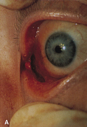
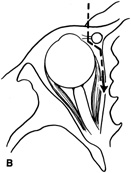
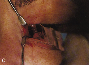
| Fig. 18. A. Incision for transcaruncular medial orbitotomy. The incision is placed just lateral to the caruncle and medial to the plica semilunaris. B. Axial diagrammatic scheme of route of dissection for transcaruncular orbitotomy. The incision in the medial fornix allows dissection to remain lateral to the lacrimal sac but medial to the globe and medial rectus muscle. Posterior to the sac, dissection is carried to the medial bony wall, where periosteum then can be incised and elevated posteriorly. C. The globe and medial rectus are drawn laterally by a malleable retractor, and the upper and lower lids are distracted to expose the medial extraperiosteal orbital space. |