|
|
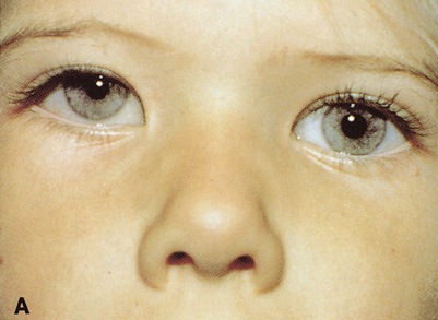
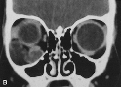
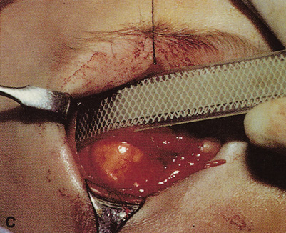
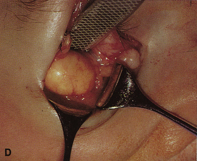
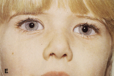
| Fig. 35. A. Patient with multiple cystic masses in the inferior and lateral orbit. These are causing supraplacement of the globe and expansion of the bony orbit as demonstrated on coronal CT scan (B). C. Approach is through a combined modified Berke lateral orbitotomy and transconjunctival inferior orbitotomy. After the lateral orbital rim is removed, wide exposure of the lateral and inferior orbits is afforded (D). E. Postoperative appearance of patient, revealing excellent scar camouflage afforded by the small lateral canthotomy incision and the hidden transconjunctival incision. |