|
|
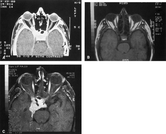
| Fig. 2. A. Axial orbital CT scan of right optic nerve meningioma. Note lack of detail in orbital apex. B. Axial orbital MR image, same patient. Note increased detail in orbital apex owing to lack of bone artifact. C. MRI with gadolinium contrast. Note extension into brain not easily appreciated with CT scanning or MRI without contrast. |