
|
|
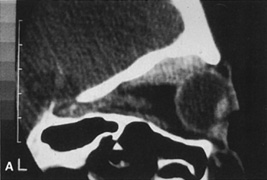
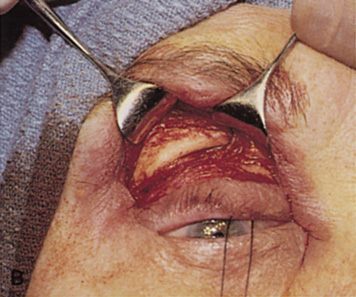
| Fig. 24. A. Sagittal CT scan of a lesion in the peripheral superior orbit behind the equator of the globe. B. Lid crease incision and superior dissection are used to expose the superior orbital rim. C. The superior orbital rim then can be burred away to provide greater access to the more posterior orbital lesion. |