Chapter 37
Vascular Lesions of the Orbit
Susan M Tucker
Main Menu   Table Of Contents

Search

ORBITAL VASCULAR HAMARTOMAS
NEOPLASMS
ORBITAL VASCULAR MALFORMATIONS
ARTERIOVENOUS FISTULA
OTHER VASCULOGENIC MASSES
REFERENCES

Vascular lesions of the orbit constitute approximately 11% of clinical orbital disease and 6% of orbital lesions that have undergone biopsy.1,2 Clinical presentation is determined by the effects of the resulting hemodynamic alterations on the tissues of the orbit. Features suspicious for a vascular lesion are intermittent exophthalmos, vascular engorgement with the Valsalva maneuver, orbital pulsation or bruit, and hemorrhage. The classification of vascular tumors of the orbit has been controversial1,3–5 but more recently has been better defined with advances in imaging, immunohistochemistry, and electron microscopic studies. In this chapter, vascular lesions of the orbit are classified as hamartomas, neoplasms, vascular malformations, arteriovenous fistulas, and “other vasculogenic masses.” Table 1 summarizes features of orbital vascular hamartomas (infantile capillary hemangioma and cavernous hemangioma) and neoplasms (e.g., hemangiopericytoma, malignant hemangioendothelioma, Kaposi's sarcoma, epithelioid hemangioma, Kimura's disease, vascular leiomyoma). Table 2 summarizes orbital vascular malformations according to the classification proposed by the Orbital Society in 1998.6 Other vasculogenic masses consist of ophthalmic artery aneurysm, orbital venous thrombosis, orbital hemorrhage, cholesterol granuloma, and epidermoid cholesteatoma.

TABLE 1. Vascular Tumors of the Orbit
Click Here to view Table 1.


TABLE 2. Orbital vascular malformations

No flow malformations
    Hemodynamically isolated
    Applicable to so-called lymphangiomas
Venous flow malformations
    Applicable to so-called primary varices
    Including distensible and nondistensible varieties
    Including mixed forms with venous and no flow components
         (grouped here to emphasize the clinical importance of the venous relationship)
Arterial flow malformations
    Arteriovenous malformations or arterialized veins distal to them
    Applicable to so-called secondary varices

From Harris GJ: Orbital vascular malformations: A consensus statement on terminology and its clinical implications. Am J Ophthalmol 127:453, 1999


Back to Top
ORBITAL VASCULAR HAMARTOMAS

INFANTILE CAPILLARY HEMANGIOMA

Hamartomas are composed of abnormal mixtures of tissue elements or an abnormal proportion of a single element that is usually present in that site. Benign hemangioendothelioma, strawberry hemangioma or nevus, and infantile or hemangioblastic hemangioma are additional terms used to describe this high-flow hamartomatous proliferation of primitive vasoformative tissues. Infantile capillary hemangioma is the most common orbital vascular tumor of childhood, estimated to occur in 1% to 2% of infants.7,8 It occurs predominantly in female infants, with a 3:2 female-to-male ratio most frequently reported.9,10 Although usually sporadic, rare heritable infantile hemangiomas have been described, and may perhaps be under-reported.11,12 Predisposing factors for the development of capillary hemangiomas include maternal chorionic villus sampling13 and prematurity.14

Clinical Features

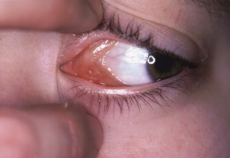
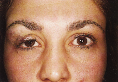
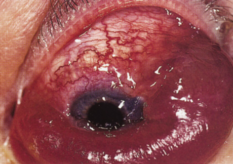
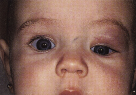
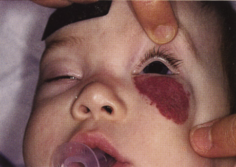
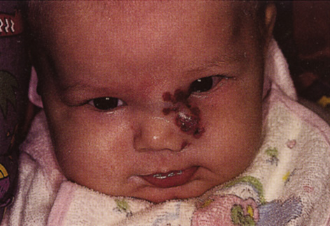
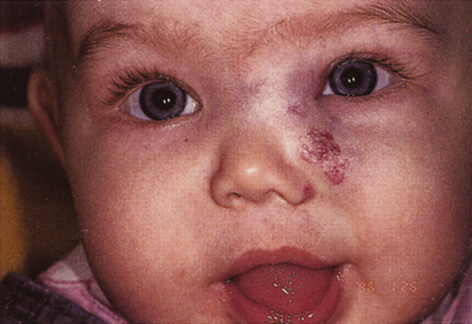
One-third of all orbital capillary hemangiomas are noted at birth, and virtually all are diagnosed by age 6 months.10 Most commonly a unilateral diffuse subcutaneous or circumscribed, dimpled, red dermal lesion (strawberry nevus) is noted on the upper eyelid (Figs. 1 and 2).9,10 In more than one-third of patients, the palpebral or fornical conjunctiva is involved.10 Commonly, this superficial lesion is associated with a component deep to the orbital septum resulting in variable degrees of proptosis in 38% of patients.10 More rarely, it can occur as an isolated lesion posterior to the orbital septum without a superficial component: In Haik and colleagues' large series of 101 infants with capillary hemangiomas, seven presented with proptosis alone.10 Forty-six percent of patients have an increase in lesion size with crying or Valsalva maneuver.10 Very rarely, hemangiomas can occur within the extraocular muscles.15 Early flat, circumscribed lesions rapidly expand into bulky, compressible masses over a period of weeks to months as the capillary lumina of the endotheliomatous hamartoma opens.16 This distinguishes the capillary hemangioma from the port-wine stain (nevus flammeus) associated with the Sturge-Weber and Klippel-Trenaunay syndromes, which remains flat to slightly thickened and is noncompressible. The infantile capillary hemangioma usually reaches its largest size within 6 months and then typically involutes.10,17 Fine, stellate areas of pale scarring (herald spots) appear during resolution (Fig. 3). Residual pigmentary skin changes and superficial cutaneous scarring may result.10 The most common ocular complication of adnexal capillary hemangiomas is visual loss, most often resulting from amblyopia or in rare cases optic atrophy. Amblyopia occurs in 44% to 64% of these infants and usually is a result of anisometropia, visual deprivation, or both.9,10,18 Anisometropia may result from axial myopia induced by the eyelid closure or astigmatism (plus cylinder axis points toward the tumor mass).19 Strabismus may be present in up to 34% of patients with periorbital infantile capillary hemangioma.18 Associated dermal or visceral hemangiomas throughout the body have been reported in 29% of infants with periorbital capillary hemangiomas.10 Laryngeal hemangiomas are the most frequent visceral vascular manifestation. In rare cases, hepatic, gastrointestinal, or intracerebral hemangiomas may be present.20 Sequestration of platelets and red blood cells leading to thrombocytopenia and bleeding diathesis (Kasabach-Merritt syndrome) can occur with large visceral lesions but are rare with isolated head and neck lesions.10,21

Fig. 1. Capillary hemangioma. Eight-month-old patient with a diffuse subcutaneous lesion of the left superior orbit, with a component deep to the orbital septum. Note the displacement of the globe inferiorly. (Courtesy of Dr. Nancy A. Tucker, Illinois Oculoplastic Associates)

Fig. 2. Capillary hemangioma. Six-month-old patient with a circumscribed, red, dimpled dermal “strawberry nevus” capillary hemangioma. This patient had resolution of the induced astigmatic anisometropic refractive error after one intralesional steroid injection.

Fig. 3. A. Patient with capillary hemangioma at age 2 months. B. Patient at age 5 months, 8 weeks after intralesional steroid injection. Note the stellate areas of pale scarring (herald spots) characteristic of the resolution phase of this lesion.

Investigations

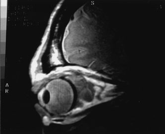
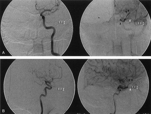
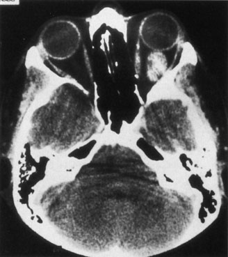
Most capillary hemangiomas can be diagnosed readily by clinical inspection. However, if delineation of the extent of deep orbital involvement is required, or the diagnosis is unclear, magnetic resonance imaging (MRI) with surface coils, gadolinium–pentetic acid enhancement, and fat suppression (to detect enhancement against the orbital fat) is indicated. The tumor is isointense to muscle and gray matter on T1- and hyperintense on T2-weighted images.22 The enhancement seen with intravenous contrast varies from moderate to intense and may be homogeneous or inhomogeneous.10 Lesions undergoing involution are less intense and more inhomogeneous. Major feeding vessels appear as black, serpiginous structures because of the “flow void” phenomenon.22 B-scan ultrasonography shows a smooth, lobular, or irregular mass with variable internal reflectivity that blends into surrounding orbital structures.23 A-scan ultrasonography shows alternating high internal reflectivity (high echo spikes) and low internal reflectivity (low echo spikes) resulting from the variable architecture and acoustic interfaces of vascular spaces, cellular areas, and septa, and moderate sound attenuation.24 High vascular flow may be demonstrated on Doppler echography. On computed tomography (CT) scan, the margins of deep infantile hemangiomas vary from moderately well defined to irregular.25 They can occur anywhere within the orbit and may be both intraconal and extraconal. There are no calcifications within these lesions. Occasionally, they indent the globe and are associated with bony orbital enlargement without erosion.10 Very rarely, angiography may be needed in the unusual situation where hazardous superselective embolization is used for life-threatening hemangiomas unresponsive to other therapies.20 These lesions frequently have multiple feeding vessels from both the internal and external carotid arteries.

Histopathology

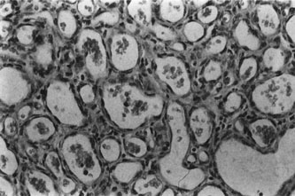
Infantile capillary hemangiomas have a characteristic histopathologic pattern with masses of plump proliferating endothelial cells organized into a network of basement membrane–lined vascular channels with small, irregular lumina (Fig. 4). No true capsule exists.10 A reticulin stain may help delineate primitive vascular structures in early stages of proliferation when there are few vascular spaces.10 During the proliferative phase or for poorly differentiated tumors, the localization of von Willebrand factor produced by the endothelial cells by either peroxidase or fluorescent antibody technique may prove useful.10 Lesions undergoing involution are characterized by fewer endothelial cells, larger and less numerous vascular channels, increasing collagen deposition, intralesional fat, and in some instances, inflammatory cell infiltrates. The late regression phase is dominated by fibrosis.10

Fig. 4. Infantile capillary hemangioma. Characteristic pattern of masses of plump endothelial cells organized into a network of basement membrane-lined vascular channels with small, irregular lumina. Red blood cells are present within these spaces (H&E, ×250).

Differential Diagnosis

The differential diagnosis of a rapidly expanding orbital mass in infancy and early childhood includes rhabdomyosarcoma, chloroma, neuroblastoma, vascular malformations, congenital hydrops of the nasolacrimal sac, dermoid cyst, and orbital cellulitis.

Management Natural History

Outlining the natural history of these lesions to the parents is extremely important in alleviating their concerns and anxieties and in justifying the usual decision for conservative management. The rate of complete resolution without treatment is 32% to 60% at 4 years of age, and 72% to 76% at 7 years of age; there is variable improvement in the remaining children until the age of 10 to 12 years.17

Treatment

Treatment is required for children with refractory amblyopia, threatened occlusion of the visual axis, compression of the optic nerve, or corneal exposure secondary to severe proptosis. Controversy exists about the most appropriate management of induced astigmatism and anisometropic refractive error, with some authors advocating early treatment to reverse severe refractive errors.10,26 Nonvisual indications for therapy include deep lesions that bleed frequently, secondary maceration and erosion of the epidermis, or severe disfigurement. Systemic indications for treatment are cardiovascular, hematologic, or obstructive complications.20 Intralesional corticosteroids have been the mainstay of initial therapy in lesions requiring treatment. Their therapeutic effectiveness results from arteriolar constriction and narrowing of precapillary sphincters.27 Combinations of short- and long-acting corticosteroids of various doses have been successful. Generally, the long-acting depot steroid is directed deep into the lesion to prevent deposits from being visible under the skin, and the more soluble short-acting corticosteroid is given subcutaneously around the periphery of the capillary hemangioma. Injections usually are given under mask inhalation; separate 1- to 3-mL syringes with 25- or 27-gauge needles are used for each substance. The following are two popular regimens: (a) 40 mg triamcinolone acetate and 6 mg betamethasone given separately into the lesion28 or (b) 40 mg methylprednisolone and 4 mg dexamethasone sodium phosphate given separately into the lesion.9 Some authors vary the dose according to the size of the capillary hemangioma.29,30 Involution of the tumor may begin several days after injection and usually is considerable within 2 to 4 weeks.26 Injections can be repeated at 6-week intervals, as needed.9,30 Intralesional corticosteroids are effective in inducing moderate to marked involution in 45% to 88% of cases,20,26,30 but recurrence and regrowth with diminution of the steroid dose is not infrequent. Potential complications of intralesional steroids include adrenal suppression, blindness secondary to retinal embolization of the corticosteroid, eyelid depigmentation, eyelid necrosis, and subcutaneous fat atrophy.31–34 Other treatment modalities include topical steroids, sub-Tenon infusion of steroids, systemic corticosteroids, laser therapy, interferon injections, selective arterial embolization, and surgery.20,35–37 Topical clobetasol propionate cream given twice daily for 2 weeks, with 1 week drug-free periods, may be an alternative mode of treatment in those patients whose parents decline intralesional steroids.37 Promising results without fibrosis or hypertrophic scarring have been reported with repeated treatments using the flashlamp pumped-pulsed dye lasers, particularly if initiated early, when lesions are relatively flat.38 Interferon alfa, which inhibits both endothelial cell and fibroblast proliferation, has been used for life-threatening hemangiomas resistant to corticosteroids.39,40 Hastings and colleagues examined 40 patients aged 2 to 36 months with life- or organ-threatening hemangiomas treated with daily subcutaneous interferon alfa-2b for 3 months, followed by taper or retreatment. An average 82% reduction in tumor volume occurred, with clinical response observed at an average of 6 weeks.40 Therapeutic arterial embolization is particularly hazardous and has yielded inconsistent results; it is therefore a last resort for life-threatening situations.20 Early surgical intervention in infants 2 to 20 months for selected hemangiomas without a significant cutaneous component has been reported with good results.25,35,41 Preoperative radiologic tests (magnetic resonance angiogram, arteriogram, or digital intravenous arteriogram) and blood typing are advisable for all patients. A true capsule is not present,35 making complete surgical excision of extensive infiltrating lesions very difficult. When surgery is performed in patients with Kasabach-Merritt syndrome (very large platelet-consuming lesions), systemic antifibrinolytic agents are needed, including aminocaproic acid or tranexamic acid. Cryotherapy, irradiation, and intralesional sclerosing agents have largely been abandoned because of inadequate effectiveness or unacceptable side effects

CAVERNOUS HEMANGIOMA

Cavernous hemangiomas are benign, noninfiltrative, low-flow hamartomas. Although often cited as the most common primary orbital tumor of adults,8,10 in the largest reported series from Moorfields Eye Hospital, cavernous hemangiomas constituted only 7% of 1,270 patients with primary orbital tumors and were outnumbered by venous flow malformations (primary orbital varices) by a ratio of 3:1.42

Clinical Features

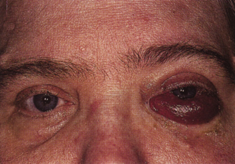
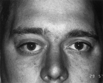
Patients typically present in the fourth and fifth decades, although the age range spans from occasional case reports in children younger than 18 years of age43 to an adult 78 years old.44 A 60% to 70% female preponderance is reported consistently.10,42,45 Seventy percent of patients present with gradually increasing painless proptosis, which usually is axial.10,45 Less common symptoms of orbital pain, eyelid swelling, diplopia, and gaze-induced amaurosis can occur.42,45–47 In a series of 162 cavernous hemangiomas (110 histologically proven), no episodes of acute or subacute hemorrhage were documented.48 Cavernous hemangiomas appear to enlarge by proliferation of capillaries,10 and local hemodynamic disturbances, hypoxia, and hormonal changes during pregnancy may stimulate their growth.10,44,49On clinical examination, other signs of mass effect may accompany the proptosis: In their series of 85 patients, McNab and Wright reported 32% with posterior pole choroidal striae and 16% with compressive optic neuropathy.42 Orbital cavernous hemangiomas usually are isolated lesions.45 Very rarely, they may be associated with multiple hemangiomas involving predominantly the arms and trunk (usually evident at birth) and visceral lesions commonly in the small intestine, leading to gastrointestinal bleeding and iron deficiency anemia (the blue rubber bleb nevus syndrome).50,51 This is usually a sporadic syndrome, although pedigrees with autosomal-dominant inheritance have been reported.52

Investigations

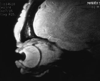
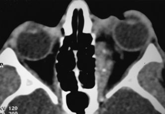
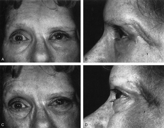
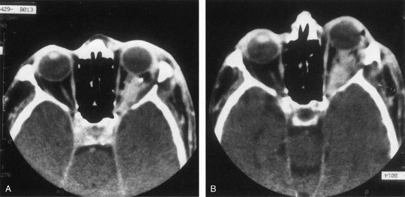
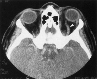
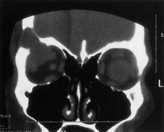
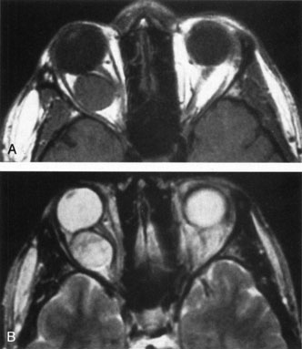
Contrast-enhanced CT of the orbits with direct and coronal cuts is the radiologic study of choice for these patients.45 A well-demarcated, encapsulated oval or rounded mass is revealed, which is typically intraconal in the lateral part of the middle third of the orbit (Fig. 5) but occasionally may extend to the extraconal space.42 Septa within the tumor may be apparent on high-resolution CT. The posterior pole of the globe frequently is indented by the rounded anterior margin of the tumor.42 The optic nerve typically is displaced rather than surrounded by the tumor. Subtle outward bowing of the lateral orbital wall or increase in orbital size may be present, consistent with a long-standing, slowly growing mass lesion.10,42 Enhancement with intravenous contrast occurs and may be homogeneous or inhomogeneous.10,45 Rarely, cavernous hemangiomas may occur as an intraosseous tumor within the orbital or facial bones.42,53–55 Although usually an isolated intraorbital lesion, multiple lesions in one orbit occurred in 8 of 164 (5%) patients of three combined large studies,10,42,45 and bilateral multiple cavernous hemangiomas also have been described.56 In contrast to patients with venous flow malformations in which phleboliths are common, phleboliths are rare in cavernous hemangiomas. Three large studies comprising 164 patients with cavernous hemangioma all reported that no calcification was detected within the tumor.10,42,45If the diagnosis is still unclear or if better definition of details and localization of the lesion is required, then MRI should be performed. Magnetic resonance imaging demonstrates nonspecific characteristics of a lesion isointense to muscle and gray matter on T1-weighted images and hyperintense on T2-weighted images (Fig. 6).57 The lesions show initial central patchy enhancement, which fills up homogeneously within 20 to 60 minutes.58 If ultrasonography is performed, B-scan ultrasonography shows a well-circumscribed mass with a sharply defined anterior acoustic border.23 A-scan ultrasonography shows high reflectivity of the echo signals resulting from the multiple blood-filled vascular channels, regular internal structure with a higher anterior and posterior spike marking the capsule, and moderate sound attenuation (angle of decrease of the echo spike within the lesion).24 Arteriography is not indicated.
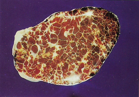
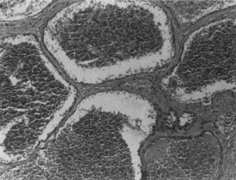
Fig. 5. Cavernous hemangioma. Contrast-enhanced axial computed tomography scan shows a well-demarcated, oval intraconal mass in the lateral part of the middle third of the orbit. Note the enhancement within the lesion, which in this instance is inhomogeneous, but can also be homogeneous.

Fig. 6. Cavernous hemangioma. Axial magnetic resonance imaging demonstrates a well-defined, homogeneous intraconal mass that is isointense to muscle and gray matter on T1-weighted image (A), and hyperintense on T2-weighted image (B). Note the displacement of the optic nerve and indented posterior globe.

Histopathology

Histopathology reveals large, endothelium-lined, blood-filled spaces separated by fibrous septa and surrounded by a fine capsule (Figs. 7 and 8). Abundant, loosely distributed smooth muscle is present in the vascular walls, and scattered inflammatory cells may be seen.10,44 Histopathologic confirmation of the low-flow character of this lesion is seen by the presence of menisci in some vascular spaces and evidence of decomposed blood.

Fig. 7. Cavernous hemangioma. Gross cut specimen showing large blood-filled spaces separated by fibrous septa and surrounded by a fine capsule. (Courtesy of Dr. Seymour Brownstein, University of Ottawa)

Fig. 8. Cavernous hemangioma. Histopathologic appearance of the widely dilated vascular spaces filled with red blood cells. Abundant, loosely distributed smooth muscle is present in the vascular walls, and scattered inflammatory cells can be seen (H&E, ´100).

Differential Diagnosis

The differential diagnosis of cavernous hemangioma includes neurofibroma, cystic schwannoma, fibrous histiocytoma, and vascular leiomyoma, which all can have identical clinical, CT, and MRI findings. Hemangiopericytoma, hemangioendothelioma, and cystic schwannoma can have similar ultrastructural features.45 Osseous hemangioma usually can be distinguished from meningioma by the lack of extensive hyperostotic changes and from osteosarcoma, multiple myeloma, and metastatic cancer by the absence of irregular bone destruction.53

Management Natural History

As the natural history of cavernous hemangiomas is to enlarge, some authors advise excision as soon as the diagnosis is established,1,59 whereas others believe that observation of asymptomatic cavernous hemangiomas is reasonable.10

Treatment

Treatment, when indicated, involves surgical excision of the lesion. Blunt dissection reveals a plump, nodular, plum-colored, encapsulated mass with vascular channels on its well-defined surface. Because of its low-flow character, a cavernous hemangioma can be punctured during surgery, leading to exsanguination and shrinkage of the tumor, which facilitates its removal.42 A cryoprobe may assist with extraction. If an apical vascular tag can be identified, it should be cauterized; otherwise the gush of blood when it is transected can be controlled with gentle tamponade. There is no evidence of recurrence from incompletely excised lesions.10 The risks of surgery depend on the location of the hemangioma within the orbit: Removal at the orbital apex can be challenging. Although the transcranial approach is optimal for tumors of the superomedial orbital apex, other apical locations can be approached transorbitally.60 Goldberg and colleagues reported no visual loss and no dysmotility in two patients with inferior and superolateral apical hemangiomas resected transorbitally.60 Two of four apical tumors operated on by Missori and colleagues via a transcranial approach developed “complete amaurosis.”61 Reports of postoperative complications from nonapical tumors vary greatly. Lumping together four apical and 21 nonapical intraconal hemangiomas, Missori and colleagues found no complications in the two patients operated on transorbitally, but permanent ptosis in 3/23 (13%), permanent “visual worsening” in 8/23 (62%), and ophthalmoplegia in 4/23 (17%) of patients operated on transcranially.61 Kersten and Kulwin described 37 surgeries for removal of orbital hemangiomas (location within orbit not given); 35 removed via orbitotomy and two via craniotomy. One patient suffered “reduction in visual acuity,” and limitation of extraocular motility occurred in two patients.62 Gdal-On and Gelfand reported on 12 patients with hemangiomas confined mainly to the middle third of the orbit (no apical tumors) undergoing cryosurgical extraction from a 360-degree conjunctiva peritomy. One patient lost vision because of intraoperative cilioretinal arterial occlusion; there were no complications in the remaining 11 patients.63 Pelton and Patel described five intraconal cavernous hemangiomas removed via a superomedial lid crease approach. One patient had transient vertical diplopia for 2 months, and two patients had transient ptosis for 2 and 6 weeks.64

Back to Top
NEOPLASMS

HEMANGIOPERICYTOMA

Hemangiopericytoma is a rare mesenchymal tumor of vascular origin. Its histogenesis remains uncertain, but is believed to originate either from the pericyte found in the walls of capillaries and venules or a pluripotential perivascular mesenchymal cell.65,66 Hemodynamically, hemangiopericytomas have a rapid circulation with a significant amount of shunting of blood. They may arise primarily in the orbit, secondarily invade the orbit from adjacent sinuses, or metastasize from a distant site, including the retroperitoneum and lower limbs.

Clinical Features

Patients usually present as middle-aged adults, but hemangiopericytoma has been diagnosed in patients from 20 months of age to 87 years.67–69 At the time of diagnosis, symptoms usually have been present for less than 1 year; however, there is wide variation from 1 month to 26 years. The major clinical features typically are painless nonaxial proptosis with downward displacement of the globe.68,69 A soft, compressible mass with ill-defined margins is often palpable.69 Rarely, hemangiopericytoma can originate within the lacrimal sac or medial canthal tissues.70,71 In 49 patients from three combined studies, proptosis was present in 86%, decreased vision in 24%, diplopia in 26%, swelling and ecchymosis of the eyelids (which may be intermittent) in 19%, and pain in 20%.67,69,72 Choroidal folds were present in two of 12 patients (17%) in one of these studies.69 Clinical evidence of active hemodynamics is rare but can be demonstrated by angiography.6

Investigations

Computed tomography and MRI characteristics are uniform but nonspecific. Computed tomography reveals a round or ovoid mass most commonly in the superior aspect of the orbit, with well-defined borders.67–69 In contrast to cavernous hemangiomas, no internal trabeculation is seen. The lesion usually is extraconal, but occasionally intraconal.69 Associated irregular calcification,68,73 bony orbital expansion,69 or erosion of adjacent orbital bone can occur.63,64,68 Intense early homogeneous enhancement (staghorn vessels) is seen with contrast injection. Magnetic resonance imaging shows a lesion isointense to gray matter on both T1- and T2-weighted images, which enhances heterogeneously with gadolinium.68,69 Hemangiopericytoma typically has large arterial feeding vessels,69 and carotid angiogram shows intense tumor blush without prominent arteriovenous shunting.69 Extension into the central nervous system, paranasal sinuses, and eyelids may be seen.72 A-scan echography shows low to medium internal reflectivity, regular to slightly irregular internal structure, and mild to moderate sound attenuation. Ultrasonographic findings of a hemangiopericytoma can be indistinguishable from schwannoma.

Histopathology

Currently, clinical and investigative diagnostic criteria do not allow a reliable preoperative diagnosis, and diagnosis depends on histologic confirmation. Histopathology reveals sheets of tightly aligned elongated cells with a vascular component often forming branching (staghorn) channels, and usually surrounded by a pseudocapsule.65,67,72 Silver stains show a network of reticulum fibers throughout the tumor.69 The pericyte is a spindle-shaped cell with a moderate amount of cytoplasm and an indistinct border; routine histopathologic differentiation from endothelial cells, histiocytes, and fibroblasts may be difficult. Ultrastructurally, the pericyte forms delicate interdigitating cytoplasmic processes, has a frequently multilayered basal lamina, possesses frequent pinocytotic vesicles, and has poorly developed desmosomes.69 Hemangiopericytomas can be divided into benign, intermediate, and malignant tumors on the basis of histopathologic criteria. The benign pattern shows minimal atypia with fewer than two to three mitotic figures per high-power field,69 whereas borderline and malignant patterns demonstrate increasing mitosis, compression of vascular spaces, pleomorphism, necrosis, hemorrhage, and infiltrative margins.67 Interestingly, there is no clear correlation between histologic features and clinical behavior;74 although hypercellularity, high mitotic activity, and necrosis are tumor features that may be more closely associated with local recurrence or metastatic spread to the lung, bone, or liver.65,66

Differential Diagnosis

The differential diagnosis of hemangiopericytoma includes fibrous histiocytoma, cavernous hemangioma, schwannoma, neurofibroma, vascular leiomyoma, cellular infantile capillary hemangiomas, hemangioendothelioma, mesenchymal chondrosarcoma, and angioblastic meningioma. Fibrous histiocytoma, cavernous hemangioma, schwannoma, neurofibroma, and vascular leiomyoma have different MRI characteristics of a lesion with a sharply circumscribed border that is isointense to gray matter and muscle on T1- and hyperintense on T2-weighted images.45 Histopathologically, fibrous histiocytomas are dominated by a storiform highly cellular pattern without a significant vascular component, and much less reticulin deposition compared with a hemangiopericytoma. Distinction on a cellular basis may be best achieved by electron microscopy to demonstrate the ultrastructural features of the pericyte within the hemangiopericytoma. An infantile capillary hemangioma usually can be distinguished with the reticulin stain, and mesenchymal chondrosarcomas contain areas of chondroid or cartilaginous tissue.66 Angioblastic meningiomas do not deposit a great deal of reticulin, stain with S100 protein, and are virtually nonexistent in the orbit. Orbital calcifications also can be seen with no flow and venous flow malformations and rarely with cavernous hemangiomas.

Management Natural History

In a study of 30 patients with orbital hemangiopericytomas, Croxatto and Font reported a 5-year actuarial survival rate of 89%.67 However, following incomplete excision, there is a 30% recurrence rate ranging from 1 month to 7 years after surgery, and a 10% to 15% risk of metastasis to the lung, bone, or liver up to 32 years later.67 Therefore, long-term follow-up is necessary to ensure a complete cure.

Treatment

When histologic confirmation of hemangiopericytoma is established, a metastatic survey, including chest radiograph, abdominal CT, bone scan, and lumbar puncture are required. If hemangiopericytoma is suspected, complete excision is recommended.67,69 When an incisional biopsy has been performed already, wide local excision with excision of the biopsy tract or exenteration should be carried out.69 At surgery most of these tumors are well-circumscribed, pinkish or violaceous masses, and may have large, draining, telangiectatic vessels; rarely, grossly infiltrating lesions have been noted. Radiation therapy and systemic chemotherapy have been used in too small a number to determine their efficacy. Orbital recurrence or aggressive local behavior may require exenteration.69,75

MALIGNANT HEMANGIOENDOTHELIOMA

Several terms are used interchangeably with this rare malignant, endothelial-derived vascular tumor: angiosarcoma, malignant angioma, malignant endothelioma, angioendothelioma, and hemangioendothelial sarcoma. The majority of cases involve the axial skeleton or extremities, fewer than a dozen cases involving the orbit have been reported.76–78They usually arise within orbital soft tissues, although rarely they arise in the orbital bones or involve the eyelids.76

Clinical Features

Most patients present in the pediatric age group with a range in age from 2 weeks to 68 years. The clinical presentation is often of rapidly developing proptosis, globe displacement, and painful ophthalmoplegia.76,77 Rarely, hemangioendothelioma may arise within the orbital bones.76 One-fourth of reported cases have associated local sensory (usually trigeminal nerve) or motor neurologic signs and symptoms.77 Hemangioendotheliomas may be locally aggressive and are capable of metastatic spread to parenchymal organs.78

Investigations

Computed tomography scan reveals one or several circumscribed or infiltrative masses usually associated with significant bone destruction.76 Enhancement occurs with contrast, and carotid angiography shows intense tumor blush. Hemangioendothelioma has a highly irregular ultrasonic outline.23

Histopathology

Histopathology reveals sheets of tightly aligned, elongated cells with a vascular component often forming branching “staghorn” channels, and usually surrounded by a pseudocapsule.76 The many-layered, endothelial-like cells are seen within the reticulin sheath. Ultrastructurally, endothelial features are noted: a basal lamina, luminal pinocytotic vesicles, tight junctions, cytofilaments, and Weibel-Palade bodies. Factor VIII–related antigen, an endothelial marker, and S100 protein may be noted.77 Wold and colleagues have divided hemangioendothelioma involving bones outside the orbit into three grades, with disease-free survival decreasing with increasing grade: grade 1, 95%; grade 2, 62%; and grade 3, 20%.79

  Grade 1: Lesions with abundant vascular spaces lined by cells with mild atypia and rare mitotic figures.
  Grade 2: Lesions showing intermediate anaplasia.
  Grade 3: Lesions that are less vasoformative, with marked nuclear anaplasia and numerous mitotic figures.

Differential Diagnosis

The differential diagnosis includes hemangiopericytoma, fibrous histiocytoma, cavernous hemangioma, Kaposi's sarcoma, schwannoma, neurofibroma, vascular leiomyoma, cellular capillary hemangioma, mesenchymal chondrosarcoma, and angioblastic meningioma. When occurring in children, the differential diagnosis includes entities presenting with a rapidly expanding orbital mass: rhabdomyosarcoma, vascular malformation, chloroma, neuroblastoma, congenital hydrops of the nasolacrimal sac, and orbital cellulitis.

Management Natural History

Mean survival times for hemangioendotheliomas of the face and scalp range from 10.7 to 20 months, and only 12% survive 5 years.80

Treatment

When the diagnosis of hemangioendothelioma is confirmed histologically, a metastatic survey, including a chest radiograph, abdominal CT, bone scan, and lumbar puncture is indicated.76 They are very aggressive tumors, and tend to recur readily and metastasize early. The optimal treatment is unknown, but wide surgical excision is recommended to prevent recurrence or metastases.76,77 Radiation and systemic chemotherapy have been used in too small a number to establish their efficacy

KAPOSI'S SARCOMA

Kaposi's sarcoma is a malignant mesenchymal tumor of the skin and mucous membranes that occurs predominantly in patients with AIDS, but also in elderly men and immunocompromised HIV-negative persons. Ocular involvement with Kaposi's sarcoma occurs in approximately 30% of patients with AIDS, and is the initial clinical manifestation in 4% of these patients.81

Clinical Features

The lesion appears as a painless, violaceous, flat patch, papule, or nodule on the eyelid. Less commonly, the conjunctiva (particularly of the lower fornix) harbor discrete to diffuse, raised, red-purple masses, or what appears as persistent subconjunctival hemorrhage.81 The globe itself is not involved. Ophthalmic Kaposi's sarcoma lesions generally are slowly progressive.81 Secondary orbital involvement may occur by direct extension of the tumor

Investigations

Fluorescein angiography may be useful in the relatively transparent conjunctiva for detecting the peripheral extent of the tumor.82

Histopathology

Stage I lesions consist of small, irregularly shaped, thin-walled vascular channels without slit spaces, and a moderate mononuclear cell infiltrate but no spindle cells. Stage II lesions have early slit vessels and a few foci of immature spindle cells. Stage III lesions are characterized by aggregates of densely packed spindle cells and occasional mitotic figures with a reduction in the inflammatory cell background.82

Differential Diagnosis

The clinical differential diagnosis of a conjunctival Kaposi's sarcoma includes hemorrhagic conjunctivitis or subconjunctival hemorrhage, pyogenic granuloma, foreign-body granuloma, hordeolum, arteriovenous fistula, capillary hemangioma and cavernous hemangioma

Management Natural History

Because ophthalmic Kaposi's sarcoma lesions usually are slow growing and rarely invasive, observation is often appropriate.

Treatment

Indications for treatment are discomfort, obstruction of vision by bulky lesions, and cosmetically disturbing lesions. Small localized ocular lesions may be treated with surgical excision; larger lesions can be treated with cryotherapy, injectable chemotherapeutic agents, or focal irradiation. Radiotherapy (2,000 to 3,000 cGy given in fractions over a 3-week period) often results in complete regression within 3 to 6 weeks.81 If the disease is multifocal or visceral involvement is present, systemic chemotherapy is given, which may lead to regression of the ophthalmic lesions. Chemotherapy may consist of either single-agent therapy or any combination therapy with the following: doxorubicin hydrochloride, bleomycin sulfate, vinblastine sulfate, and interferon-alfa or -beta. Recurrences after treatment occur in approximately half of patients, usually within weeks to several months after treatment.81 Further studies are needed for a more accurate comparison of treatment outcomes and recurrence rates

EPITHELIOID HEMANGIOMA

Epithelioid hemangioma is an uncommon benign neoplasm of endothelial cells characterized by subcutaneous nodules, and plaques and papules of the face, scalp, and ear. The cause of epithelioid hemangioma remains uncertain. Other terms that have been used to describe this condition include angiolymphoid hyperplasia with eosinophilia, pseudopyogenic granuloma, atypical pyogenic granuloma, and inflammatory angiomatous nodule.

Clinical Features

Epithelioid hemangiomas usually present in middle-aged Caucasian women with multiple violaceous lesions involving the canthus, eyelids, or anterior orbit. With orbital involvement, patients may present with proptosis, tearing, and blurred vision occurring for several weeks to years.83 Nonaxial proptosis may be present, typically with globe displacement inferiorly and a palpable mass. A small proportion may have regional lymphadenopathy and eosinophilia of the peripheral blood,83 but there are no associated systemic diseases.84

Histopathology

Histopathology reveals dermal or subcutaneous lymphocytic infiltrate with mild to marked eosinophilia. Endothelial cells have atypical nuclei with abundant cytoplasm and sometimes intracytoplasmic vacuoles.84

Differential Diagnosis

The differential diagnosis includes Kimura disease, pyogenic granuloma, hemangioendothelioma, granuloma faciale, nonspecific orbital inflammation, insect bite, and Kaposi's sarcoma.

Treatment

The treatment of choice is local excision. The recurrence rate after surgical excision is 33%.85 Radiotherapy has been advocated for recurrent or incompletely excised lesions. Cryotherapy, intralesional injection of corticosteroids, and chemotherapy also have been used, but their value is unclear.84

KIMURA'S DISEASE

Kimura's disease most likely represents an allergic or autoimmune response. Although previously used synonymously with epithelioid hemangioma or angiolymphoid hyperplasia with eosinophilia, distinct clinical and histopathologic characteristics exists between these two entities.83

Clinical Features

Kimura's disease occurs almost exclusively in Asian men. Patients present with lymphadenopathy involving preauricular, parotid, and submandibular lymph nodes. Increased serum immunoglobulin E and blood eosinophilia usually are present.83 Nephrotic syndrome with proteinuria may be present.85

Histopathology

The histopathology includes dermal or subcutaneous mild to marked lymphocytic infiltrate, commonly with lymphoid aggregates with prominent germinal centers and mild to marked eosinophilia. Unlike epithelial hemangioma, atypical endothelial cells are not seen in Kimura's disease.84

Differential Diagnosis

The differential diagnosis includes epithelial hemangioma and its differential diagnosis.

Treatment

Like epithelial hemangioma, Kimura's disease has an indolent nature, is self-limiting, and potentially is curable with complete excision.84 Irradiation, steroids and chemotherapy also have been used

VASCULAR LEIOMYOMA

Angiomyoma and hemangioleiomyoma are terms used interchangeably with vascular leiomyoma. Vascular leiomyoma is a tumor of uncertain histogenesis, but is believed to arise from vascular smooth muscle or possibly the pericyte.86

Clinical Features

The few cases described in the orbit have been slow-growing, encapsulated masses occurring chiefly in patients younger than 40 years of age.86,87 Patients usually present with gradual, painless proptosis, which may be associated with motility restriction and diplopia. Pain and increased proptosis may be noted when patients perform the Valsalva maneuver.

Investigations

Magnetic resonance imaging reveals a well-circumscribed mass, more commonly found intraconally rather than extraconally.87 The appearance can be identical to cavernous hemangioma, neurofibroma, schwannoma, and fibrous histiocytoma: a lesion isointense to gray matter on T1- and hyperintense on T2-weighted images, which enhances with gadolinium.61,87

Histopathology

Histopathology shows encapsulated fascicular interwoven bundles of spindle-shaped smooth muscle cells interspersed within abundant endothelial sinusoids or dilated capillaries.86 Reticulin stain shows an orderly deposition of reticulin between the cells.86 Densely vascular lesions may be confused with hemangiopericytoma or cavernous hemangioma.87 The diagnosis is substantiated by identification of smooth muscle origin by means of histochemistry or electron microscopy (cytoplasmic filaments with fusiform densities, marginal plasmalemmal densities, and basement membrane formation).87

Differential Diagnosis

The differential diagnosis includes cavernous hemangiomas, neurofibroma, schwannoma, fibrous histiocytoma, and hemangiopericytoma. Fibrous histiocytoma is not encapsulated, and hemangiopericytoma consists of more primitive mesenchymal cells rather than differentiated smooth muscle, as evidenced by electron microscopy.86,87

Treatment

The treatment of choice is complete excision. Incomplete removal may lead to local recurrence.

Back to Top
ORBITAL VASCULAR MALFORMATIONS
In 1998 the Orbital Society, consisting of oculoplastic and orbital surgeons from North America, Europe and Australia, devised a nomenclature of classifying orbital vascular malformations according to their hemodynamic relationships as no flow, venous flow, and arterial flow lesions (see Table 2).6 It was the consensus that this hemodynamic classification was more germane to clinical management then purely morphologic differentiation.

NO FLOW MALFORMATIONS

The term no flow malformation (NFM) is appropriate when hemodynamic isolation is demonstrated: that is, the lack of connection of the lesion to the arterial or venous system on imaging studies. These lesions, previously called lymphangiomas, presumably arise embryologically from misdirected vascular anlage tissue capable of differentiating into lymphatics, blood vessels, and other mesodermal elements.88 A spectrum of resulting lesions exists, with hybrid forms composed of combinations of lymphatic and vascular elements.89

Clinical Features

No flow malformations are more prevalent in women by a ratio of 1.4 to one.89 Sixty-five percent present in the first decade, and 92% by the end of the third decade.48 However, they may be noted at birth or as late as 73 years of age.89 The channels of an NFM are fully formed at birth, but the lesion can remain inconspicuous until it expands. Lesions may: (a) have purely superficial, multicystic conjunctival (Fig. 9) or eyelid involvement; (b) be localized deep within the orbit; or (c) have both superficial and deep components. Superficial lesions on the eyelid and conjunctiva transilluminate and may appear bluish because of xanthochromic or partially blood-filled cysts.90 Deep lesions can be massive, involving the intraconal, extraconal, preseptal, and postseptal spaces, often resulting in significant cosmetic disfigurement. There are no clinical features of either arterial or venous connection; that is, Valsalva maneuver and alteration of head position do not produce a change in lesion size, and there is no bruit or pulsation.89 Expansion during upper respiratory infections presumably results from lymphoid hyperplasia in response to immune challenge.4,91,92 Bleeding likely results from the rupture of fine blood vessels that course through the flimsy septa of the lymph channels. Clinically, these episodes of expansion manifest as acute proptosis, ptosis, restricted motility, periorbital ecchymosis and swelling, and subconjunctival hemorrhages.3,89 Shrinkage of blood cysts usually occurs over a period of several weeks to several months.89 The probability and timing of future orbital hemorrhage is highly unpredictable. In a study of 30 patients diagnosed with NFM, 40% of patients managed without surgery had additional hemorrhages 2 weeks to 11 years later; 52% of patients who underwent orbitotomy for debulking had additional hemorrhages 4 days to 12 years later.89 Visual loss may result from anisometropic amblyopia, optic nerve compression, or exposure keratopathy from extreme proptosis.89,92 Associated ipsilateral congenital ocular malformations also may be present, including congenital ptosis, congenital strabismus, microcornea, sectorial cataract, heterochromia irides, and abnormal iris vessels.89,90 Associated extraorbital malformations, most commonly the hard and soft palate and paranasal sinuses, were present in 17% of 30 patients studied by Harris and colleagues.89 Very rarely, direct intracranial extension of the NFM can result in cerebral hemorrhage.90

Fig. 9. No flow malformation of the orbit. Conjunctival mass with multicystic structures containing serous fluid and an admixture of blood and blood products. (Courtesy of Nancy A. Tucker MD, Illinois Oculoplastic Associates)

Investigations

If NFM is suspected, MRI should be ordered of the orbits and brain to exclude noncontiguous intracranial anomalies.93 Magnetic resonance imaging demonstrates the dilated cystic spaces with either a hypointense (lymphatic cyst) or hyperintense (hemorrhagic cyst) signal on T1-weighted images, and a hyperintense signal on T2-weighted images (Fig. 10, A&B).94Layering may be seen within the cysts if there has been a recent hemorrhage leaving unresorbed blood. The CT scans of patients with a deep NFM show low-density, poorly defined masses behind the orbital septum in the extraconal and intraconal spaces, which may indent the globe.3 Calcification within the mass (Fig. 11) and inhomogeneous enhancement of the rim and focal areas within the lesion may be seen that corresponds to abnormal endothelially lined channels.3 Enlargement of the bony orbit can occur, particularly with combined lesions.3,89 If the diagnosis is still unclear, ultrasonography can be performed. A cystic orbital mass is seen on B-scan. A-scan shows features of a solid, cellular tumor: low reflectivity, regular homogeneous internal structure, and marked sound attenuation through the mass.3,89 Standard pulsed Doppler ultrasound confirms no intrinsic flow within the lesions.

Fig. 10. A and B. No flow malformation of the orbit. Sagittal magnetic resonance imaging showing dilated cystic spaces hypointense to muscle on T1-weighted images (A) and hyperintense to muscle on T2-weighted images (B).

Fig. 11. Axial computed tomography scan of patient in Figure 9, showing poorly defined orbital mass with calcifications. (Courtesy of Nancy A. Tucker MD, Illinois Oculoplastic Associates)

Histopathology

Histopathology consists of nonencapsulated networks of thin-walled, dysplastic, serous-containing vessels.58 The absence of lymphocyte aggregates does not preclude the diagnosis.89 Smooth muscle cells and pericytes may be identified within the septa, suggesting that some of the channels are vascular in nature.

Differential Diagnosis

The main clinical and histologic differential diagnosis of an NFM is a venous flow malformation (VFM). Clinical and imaging evidence for a lack of continuity with the systemic vascular system is required to differentiate between the two entities. The superficial component of an NFM generally is more striking, with evidence of varying types of tortuous vascular channels, some of which contain blood, menisci, or clear fluid, compared with the larger, blood-filled channels of a VFM. No flow malformations may be associated with expansion during intercurrent illness, and similar lesions may be seen elsewhere in the skin and mucous membranes of the head and neck. Other causes of recurrent and intermittent exophthalmos are neurofibromatosis, ruptured dermoids, orbital hemorrhage, and ethmoiditis.

Management Natural History

No flow malformations tend to enlarge slowly until the patient reaches adulthood. Spurts of growth can occur during upper respiratory tract infections and hemorrhages. In Wright and colleagues' study of 158 patients with NFM and VFM, two-thirds of the lesions enlarged permanently, usually incrementally with pain and evidence of orbital hemorrhage.95 End-stage disease occurred in five of these patients requiring radical surgery to remove fibrous tissue and clot, and in one case partial exenteration for pain relief.95

Treatment

It is important to educate the patient about the nature of an NFM. It should be emphasized that a clean excision or “cure” is impossible, and that each hemorrhagic episode is managed on the basis of its functional compromise. If cosmetically unacceptable, superficial lesions can be removed with relative ease because of their limited location and small size. Otherwise, observation is advisable whenever possible because a poor final visual acuity appears to be associated with multiple surgical procedures.89,96 There is increasing support for conservative management even when acute hemorrhage results in visual compromise, as long as there is no afferent pupillary defect. Cautious observation of 11 episodes of acute hemorrhage in six children with loss of vision predominantly in the range of 20/30, but up to 20/200 in the absence of a pupillary defect, resulted in 20/20 acuity after 3 days to several months, but usually within several weeks.96 Urgent surgical management is required for deep lesions if acute orbital hemorrhage causes optic nerve compression, significant corneal compromise, severe discomfort, or the threat of amblyopia in the susceptible age group. The surgical aim is anterior debulking; complete excision of an extensive orbital NFM is an unrealistic goal and should not be attempted. Carbon dioxide or continuous-wave Nd:YAG laser can be used as adjuncts during surgery.94,97 For severe proptosis, bony orbital decompression also may be effective.98 Both injection of sclerosing agents and radiation therapy have been used in the past, but are no longer advocated.

VENOUS FLOW MALFORMATIONS

Venous flow malformations are tangled masses of abnormal venous channels that probably originate from a congenital weakness in the wall of one or more orbital veins or from a misdirected venous anlage and have no direct connection with the arterial side of the circulation. More rarely, segmental tortuous dilatations of previously normal channels arise secondary to an arteriovenous shunt, either intracranially or within the orbit.1 Venous flow malformations are almost always unilateral; bilateral orbital involvement is quite rare.99 These lesions can be divided into nondistensible and distensible lesions depending on their connection to the main venous circulation in the orbit. Nondistensible lesions have small functioning systemic venous connections that lack significant connection to the venous bed. Distensible lesions have more significant connections to the venous bed with proptosis and pain that increases with straining, bending forward, or the Valsalva maneuver.

Clinical Features

Congenital VFM were the most frequent vascular tumor in Wright's series of 67 patients with orbital vascular lesions, outnumbering cavernous hemangiomas by a ratio of 3:1.1 In Wright and colleagues' study of 158 patients with NFM and VFM, 43% presented younger than 6 years of age, 60% younger than 16 years of age, and only 15% were older than 36 years of age when first seen.95 The most common initial complaint is eyelid swelling or mass (42%), followed by hemorrhage (37%), proptosis (15%), and pain (4%).95 Rarely, proptosis from a VFM may be noted at birth.100 At rest, the affected eye may even be enophthalmic because of enlarged orbits and perhaps some fat atrophy (Fig. 12). Patients with a superficial component have swelling and disfigurement of the eyelids and conjunctiva (Fig. 13), most often in the lower fornix, and the lesions may extend into the deep orbit.1,101

Fig. 12. Orbital venous flow malformation. This 77-year-old woman presented with a 10-year history of left enophthalmos. A. and B. Hertel measurements were 12 mm on the right and 8 mm on the left at a base of 96. With Valsalva maneuver the patient demonstrated 4 mm of proptosis on the left side. C. and D. This proptosis resolved 10 to 15 seconds after the Valsalva maneuver ended. (Courtesy of Dr. John V. Linberg, University of West Virginia, Morgantown, WV)

Fig. 13. Venous flow malformation. This woman, who had a superficial component to her lesion, demonstrates the characteristically lumpy, violaceous mass causing swelling and disfigurement of the upper eyelid. (Courtesy of Dr. John V. Linberg, University of West Virginia, Morgantown, WV)

Venous flow malformations do not enlarge. The episodes of acute exacerbation result from hemorrhage or thrombosis within the lesion or distention from raised venous pressure.102 At some point in their course, 55% of Wright and colleagues' 158 patients had hemorrhage, and 43% experienced pain (usually associated with hemorrhage).95 Multiple hemorrhages occurred in 27/55 (49%) of patients who underwent surgery and 10/33 (30%) of patients who did not have surgery.95 Fourteen of these 158 patients developed compressive optic neuropathy from posterior hemorrhage. Six of these 14 patients were watched without intervention: four patients had complete recovery of visual function, the remaining two patients with optic atrophy at presentation had final acuities of 20/60 and 20/100. Eight patients underwent surgical decompression: five patients improved (two to 20/20); and three showed no improvement with final acuities of no light perception, 20/100 and 20/300 (95). In their study of 158 patients with NFM and VFM, Wright and colleagues found 18% of patients to have extraorbital vascular lesions, mostly involving the palate, oral and nasal cavities, and forehead, and rarely involving the body, intracranium, and skull.95 Katz and colleagues, however, reported a more frequent association between extensive orbital lesions and intracranial involvement.103 In a study of 25 patients with “combined venous lymphatic malformations,” they found that seven patients (28%) with extensive orbital involvement had associated noncontiguous intracranial vascular anomalies.103 All these patients had orbital extension into or through the superior orbital fissure; no intracranial anomalies were found in patients with orbital lesions without superior orbital fissure involvement.103 Very rarely, orbital VFM may be part of the Klippel-Trenaunay syndrome, and other orbital findings in these cases include conjunctival telangiectasia, retinal varicosities, and angiomas of the conjunctiva, sclera, and choroids. Associated cutaneous and visceral venous varicosities and hemangiomas, as well as ipsilateral bony and soft tissue hypertrophy of the involved limb or trunk, also may be present.1

Investigations

There are no pathognomonic ultrasonography, CT, or MRI findings. A-scan ultrasonography shows well-delineated regular structures with low internal reflectivity and minimal attenuation owing to the congested pools of blood in the dilated veins. Doppler demonstrates flow within the lesion.58 Computed tomography may show abnormally dilated, irregular veins with uniform contrast enhancement or, when there has been hemorrhage, multilobular lesions.58 As the VFM may not be visualized when collapsed, direct coronal scans should be obtained in the prone position or during the Valsalva maneuver to distend the lesions (Fig. 14). Spiral CT with a single breath-holding technique may be useful in confirming an increase in size of the lesion during the Valsalva maneuver.104 The majority of venous malformations typically occupy the superomedial orbit in the area of the superior ophthalmic vein, followed by the inferolateral quadrant.95,104 In their study of 158 patients with NFM and VFM, Wright and colleagues found 45% with phleboliths.95 Enlargement of the affected orbit also may be seen.95 Obtaining MRI scans before and during a Valsalva maneuver may show distensibility, and uniform enhancement occurs.58 Venography is seldom used now because of the availability of other imaging modalities. Carotid angiography may demonstrate the VFM and can detect venous enlargement consequent to arteriovenous shunting.

Fig. 14. Orbital venous flow malformation. Axial computed tomography scan showing an ill-defined, dense, inhomogeneous orbital lesion with the patient in a supine position (A). Note the phlebolith, which indicates venous stagnation. As the venous flow malformation distends with the patient in the prone position, the lesion enlarges (B). (Courtesy of Dr. John V. Linberg, University of West Virginia, Morgantown, WV)

Histopathology

Histologically it is difficult to ascertain whether the endothelium-lined space with lymphocytic infiltration of the adjacent tissue is a lymph vessel or a small vein. Dysmorphic venous and lymphatic (when combined lymphatic-venous lesions) channels are present with varying amounts of new and old blood.58

Differential Diagnosis

The main clinical and histologic differential diagnosis of a VFM is an NFM and its differential diagnosis.

Management Natural History

In their study of 158 patients with NFM and VFM, Wright and colleagues found that two-thirds of the lesions enlarged permanently, usually incrementally with pain and evidence of orbital hemorrhage.95 End-stage disease occurred in five of these patients requiring radical surgery to remove fibrous tissue and clot, and in one case partial exenteration for pain relief.95

Treatment

Intervention is indicated for extreme orbital pressure with functional deficit, severe cosmetic disfigurement, and optic nerve compression or unremitting pain. As with NFM, most episodes of hemorrhage or thrombosis, even with mild visual compromise, resolve in a satisfactory fashion without intervention. Because it is impossible to be certain of the etiology of VFM in a given case, and the venous malformation of the orbit also may serve as part of the venous drainage of the brain, it is essential to investigate patients angiographically before surgery is considered.102 Surgery generally should be confined to the anterior orbit; surgery in the posterior two-thirds of the orbit is both difficult and hazardous.95 Bleeding can be extensive, and careful control of bleeders with clips, ligation, or cautery is essential. Carbon dioxide and Nd:YAG lasers may facilitate removal of the superficial and subcutaneous VFM. Rootman has developed an approach using intraoperative venography and controlled embolization combined with intraoperative obstruction of the outflow followed by excision of the radiopaque cast.58

ARTERIAL FLOW MALFORMATIONS

Arteriovenous malformations (AVM) are developmental anomalies whereby arteries and veins anastomose without capillaries, leading to arterialized veins with high-pressure flow. Most commonly, cerebral arteriovenous malformations result in secondary orbital manifestations; true congenital arteriovenous malformations of the orbit are rare.5,105 Of 67 orbital vascular anomalies reported by Wright, only three were AVM.1 Because the venous drainage is anterograde in the orbital veins, signs and symptoms generally are less profound than in fistulas where the flow is retrograde because of distal obstruction and shunting.

Clinical Features

Although AVM are congenital lesions, they usually only become symptomatic during the second and third decades of life.106 Patients most commonly present with pulsating exophthalmos with ocular and episcleral venous congestion, but any of the symptoms and signs described for arteriovenous fistulas can be present. Although an unruptured brain AVM carries a 1% to 2% yearly risk of bleeding, with a mortality rate from hemorrhage estimated to be from 6% to 14%,106 hemorrhage from an orbital AVM is rare.107 Arteriovenous malformations may be isolated, or they may be associated with neurocutaneous syndromes. Cerebral, and more rarely orbital, arteriovenous malformations have been described with encephalofacial cavernous hemangiomatosis (Sturge-Weber syndrome); orbital and cerebral arteriovenous malformations with racemose hemangiomatosis (Wyburn-Mason syndrome); and orbital arteriovenous malformations with hereditary hemorrhagic telangiectasia (Osler-Weber-Rendu syndrome). These entities are described in detail elsewhere in these volumes (see the index)

Investigations

Contrast-enhanced CT scan shows irregular, rapidly enhancing masses with possible enlargement of the optic canal and bony orbit.58 Magnetic resonance imaging can detect the vascular malformation and color Doppler imaging shows pulsation and dynamic flow.58,108,109 Carotid angiography remains invaluable if the diagnosis is still in question or treatment is required. Arteriovenous malformations usually are supplied by branches of both the internal and external carotid arteries.

Histopathology

The tissue is composed of mature, hypertrophied arteries and veins with many microvascular connections between them.58 Secondary capillary angiomatosis may occur, presumably in response to chronically elevated capillary pressure.110

Differential Diagnosis

Pulsating exophthalmos also can result from an orbital arteriovenous fistula, ophthalmic artery aneurysm, orbital meningocele or encephalocele, or neurofibromatosis. Also included in the differential diagnosis of an arteriovenous malformation are idiopathic orbital inflammatory syndrome and thyroid orbitopathy.

Management Natural History

The natural course is a slow rate of progressive recruitment of additional arterial feeders and enlargement of arteries and veins. Increased growth can occur during menarche, pregnancy, and blunt trauma.11

Treatment

Conservative treatment usually is indicated; however, recurrent hemorrhages or persistent pain may require intervention. When possible, embolization of the nidus and all feeding arteries, followed by surgical excision of the entire vascular mass 48 hours later is advised.102,112–114 When embolization alone is performed, the risk of incomplete embolization, resulting from the multiplicity of feeding and draining vessels and the small caliber of orbital vessels, with continued growth of the lesion is significant.102,112 Surgery alone may be possible, but there is a risk of intraoperative hemorrhage.105,115 Selective embolization can be performed with detachable balloon catheters, minicoils, or various thrombogenic materials including n-butyl cyanoacrylate, iodized oil, and polyvinyl alcohol particles and gel foam particles.112,114 Temporary materials only should be used if surgery is subsequently planned. The route of embolization (transarterial versus direct percutaneous venous access) is chosen based on the angiographic architecture of the malformation.114 Evocative tests with the loss of visual acuity or the onset of scotomata after injection of amobarbital or lidocaine into a given artery can indicate that the artery supplies the retina and cannot be safely embolized.116 Complications of embolization include postoperative orbital hemorrhage, recanalization of the vascular lesion, occlusion of the blood supply to other structures, and dislodgement and migration of the embolus with potential severe visual dysfunction.113 The complications of surgery include the inability to excise the entire lesion, bleeding, and functional impairment of surrounding organs.

Back to Top
ARTERIOVENOUS FISTULA
An arteriovenous fistula (AVF) is an acquired abnormal shunt between a previously normal artery and vein. Most commonly the arteriovenous fistula is intracranial with secondary orbital manifestations, although primary orbital fistulas rarely occur in which the arteriovenous fistula is situated within the orbit. Hemodynamically, arteriovenous fistulas may be either high flow or low flow; however, there are no objective criteria for clinically differentiating between these. Therefore these lesions are best classified angiographically into direct and indirect fistulas, which provide an objective method for grouping them, determining prognosis, and planning therapy.

DIRECT ARTERIOVENOUS FISTULA

A direct arteriovenous fistula (carotid cavernous fistula) is an acquired abnormal communication between the main trunk of the internal carotid artery and the venous cavernous sinus. These lesions represent 79% to 90% of all arteriovenous fistulas.106 They are most often the posttraumatic result of major head trauma associated with basal skull fracture, but also can occur with surgical trauma, rupture of an intracavernous carotid aneurysm, fibromuscular dysplasia, or occur in patients with connective tissue disorders such as Ehlers-Danlos syndrome Type 1V.117,118 A direct fistula may drain anteriorly into the ophthalmic venous system, superiorly into the sphenoparietal sinus and middle cerebral vein and posteriorly via the petrosal sinuses to the ponto-mesencephalic veins and sigmoid sinus.119–121 Even a small fistula can cause severe orbital congestion if anterior venous drainage is predominantly into the ophthalmic venous system. These manifestations usually are unilateral and ipsilateral to the fistula but may be bilateral or even contralateral because of the connections between the two cavernous sinuses.106

Clinical Features

Although usually seen in adults, rare cases of direct arteriovenous fistulas occurring in children have been described.122 Orbital symptoms often are significant because of the high-flow characteristics of these shunts. The clinical signs from data of several studies are as follows:


Tortuous conjunctival and episcleral vessels87% of 108 patients (Figs. 15 and 16)120,123
Proptosis81% of 235 patients118,120,123
Bruit 79% of 122 patients118,120,123
Sixth nerve paresis61% of 108 patients120,123
Increased intraocular pressure48% of 108 patients120,123
Third nerve paresis 46% of 108 patients120,123
Chemosis45% of 108 patients120,123
Decreased vision31% of 235 patients118,120,123
Fatality4% of 127 patients118
Intracerebral hemorrhage3% of 127 patients118
Subarachnoid hemorrhage3% of 127 patients118


Fig. 15. Direct arteriovenous (carotid cavernous) fistula. This 48-year-old woman presented with a 7-month history of periorbital discomfort, proptosis, diplopia, chemosis, and increased lacrimation treated as idiopathic orbital inflammatory syndrome with systemic immunosuppressants. No bruit was present, and intraocular pressure was normal. Computed tomography scan showed diffuse thickening of the left rectus muscles and an enlarged superior ophthalmic vein. Magnetic resonance angiography was interpreted as normal. She was referred for consultation service when she developed gradual visual loss to 20/400. Cerebral angiography confirmed the presence of a left direct arteriovenous fistula between the internal carotid artery and cavernous sinus.

Fig. 16. Direct arteriovenous fistula. Magnified view showing tortuous, arterialized conjunctival and episcleral vessels with marked inferior chemosis.

Cranial palsies (usually third or sixth, or both) resulting in extraocular muscle paresis may result from pressure on the distended cavernous sinus or venous congestion of the extraocular muscles, or be a direct effect of trauma. Decreased vision may be secondary to glaucoma, anterior segment ischemia, and corneal decompensation from exposure, retinal involvement (cystoid macular edema, hemorrhage into the macular area, ischemic maculopathy or retinal artery occlusion), or optic neuropathy.120,123 The optic neuropathy is likely to be secondary to a “vascular steal” phenomenon, rather than to a compressive effect.120

Investigations

If the clinical suspicion is very high for an arteriovenous fistula, particularly if intervention is required, bilateral selective arteriography of the internal and external carotid arteries is indicated. This shows retrograde opacification of the cavernous sinus and the involved venous drainage system (Fig. 17). A difference in the ocular pulse amplitude (OPA; i.e., the difference between the maximum and minimum intraocular pressure during the cardiac cycle) between a patient's two eyes of more than 1.6 mm Hg may be useful to identify patients with either direct or indirect arteriovenous fistulas.124 This is performed by instilling a topical anesthetic drop into each eye, instructing the patient to look at the 20/400 “E” on a projected Snellen chart, and placing a pneumotonometer tip on the central cornea for 10 to 15 seconds. The difference in OPA between the two eyes then is calculated from the intraocular pressure curves. Ultrasound, CT, and MRI are neither sensitive nor specific. A-scan orbital ultrasonography and Doppler illustrate venous distention and dynamic flow. Color Doppler ultrasonography shows enlargement and arterialization of the venous flow (blood flow reversal), as well as enlarged extraocular muscles and may be useful in monitoring the clinical course of arteriovenous fistulas.109,125 Reversal of flow in the superior ophthalmic vein is not pathognomonic for arteriovenous fistula: It also has been described in carotid sinus thrombophlebitis, orbital apex tumors, and in normal persons.126 Computed tomography demonstrates enlargement of the superior ophthalmic vein and frequently enlargement of the extraocular muscles proportional to the degree of shunting (Fig. 18). There may be CT evidence of venous thrombosis in the form of a nonenhancing defect in the lumen of the superior ophthalmic vein or cavernous sinus. Magnetic resonance angiography (MRA) also may be used to evaluate these lesions, but false-negative results can occur.109

Fig. 17. Left internal carotid angiogram. The internal carotid artery within the cavernous sinus is seen in anteroposterior projection as a dark circle (A) and in lateral projection as an S-shaped structure (B). The fistula (black arrow) in this patient drained anteriorly into the superior ophthalmic vein (double arrow) and inferior ophthalmic vein (white arrow). In the absence of a history of trauma, the presumed etiology of the fistula in this patient was rupture of a preexisting intracavernous aneurysm.

Fig. 18. Direct arteriovenous fistula. Axial computed tomography scan of this patient showing the enlarged left superior ophthalmic vein (white arrow). The rectus muscles of the left orbit were all diffusely enlarged.

Differential Diagnosis

The differential diagnosis includes those entities listed for arteriovenous malformations.

Management Natural History

The natural history of a direct AVF is incompletely understood. Spontaneous closure has been reported and carotid compression therapy has been successful in closure of 17% of direct arteriovenous fistulas.127

Treatment

Treatment usually is indicated in direct high-flow direct arteriovenous fistulas owing to the frequency of clinical signs described earlier, and the low but devastating risks of intracranial and subarachnoid hemorrhages. Selective cerebral angiography is performed to delineate the lesion, and then the fistula is closed. Over 95% of direct fistulas can be closed with endovascular approaches.120,128,129 Percutaneous embolization using detachable balloons, minicoils, or various thrombogenic materials via the internal carotid artery or the inferior petrosal sinus are currently the treatments of choice because they offer safe occlusion of the fistula while preserving flow in the internal carotid artery in the majority of cases.121,129,130 If the arterial supply is primarily from the internal carotid artery or if access through the inferior petrosal sinus is impossible, then a direct transvenous approach to the cavernous sinus through the superior ophthalmic vein can be performed and embolization performed with detachable balloons or minicoils.129–132 Alternate treatment option should be planned, because ligation of the superior ophthalmic vein without successful closure of the fistula can result in severe vision loss and neovascular glaucoma.133 Cyanoacrylic embolization with a cut-down procedure over the supraorbital artery with surgical excision 4 days later also has been described with successful outcome.102 When all other routes are impossible, surgical exposure of the cavernous sinus and direct puncture with embolization or permanent occlusion of the internal carotid artery proximal to the fistula in conjunction with surgical ligation of the internal carotid artery distal to the fistula remain alternatives.128 Complications of detachable balloon catheters include transient and occasionally permanent oculomotor nerve palsy, hemiparesis, fistula reoccurrence, unplanned carotid occlusion, and permanent neurologic deficit.120,121 Complications of coils include coil migration, cavernous sinus compression syndrome with cranial nerve deficits, and cavernous sinus perforation.128 In a study of 32 patients with successful occlusion of their fistulas, Kupersmith and colleagues found the signs of orbital congestion improved in all within the first 24 hours and for many resolved completely; all bruits stopped immediately; and secondary glaucoma resolved in all patients.120 Extraocular eye muscle function improved in all cases without traumatic cranial neuropathy, although the full extent of recovery occurred over a period of months.120

INDIRECT ARTERIOVENOUS FISTULA

An indirect arteriovenous fistula (dural arteriovenous fistula) is an acquired communication between the dural veins in the vicinity of the carotid sinus and dural branches of the external carotid artery (middle meningeal artery, distal internal maxillary, ascending pharyngeal artery) or the internal carotid artery (meningohypophyseal trunk, inferior cavernous sinus artery).134 Predisposing factors include vascular disease (systemic hypertension, ruptured giant intracavernous aneurysm), trauma, intracranial surgery, collagen-vascular disease, straining, sinusitis, and pregnancy.118,134–137 It has been postulated that systemic hypertension, trauma, intracranial surgery, or straining may result in rupture of normal meningeal arteries. In these lesions, rather than a network of arteries, one sees only a single meningeal feeding artery with a small shunt into the cavernous sinus. Although the etiology of “spontaneous” indirect arteriovenous fistulas remains enigmatic, it is believed to result from a vein that has thrombosed.138 This thrombosis may arise in a dural sinus, the intradural segments of afferent veins, or the emissary veins or venous plexuses at the base of the skull. It is postulated that as the thrombosed sinus becomes recanalized, pre-existing microscopic arteries in the wall of the dural sinus become sufficiently enlarged to constitute a significant hemodynamic shunt.138

Clinical Features

Indirect arteriovenous fistulas occur most frequently in postmenopausal women, although they have been described in patients ranging from 8 to 80 years of age.139 Although usually unilateral, 15% are bilateral.139 As with direct fistulas, ocular signs may be contralateral to the fistula if spontaneous thrombosis of a venous channel occurs on the involved side. Indirect arteriovenous fistulas usually are low-flow shunts, and therefore manifest lesser signs and symptoms compared with direct fistulas. Reported clinical signs and symptoms, in decreasing frequency, are as follows.


Arterialization of conjunctival vessels 93% of 135 patients140
Chemosis 87% of 135 patients140
Proptosis 80% of 163 patients118,140
Diplopia68% of 135 patients140
Bruit49% of 135 patients140
Retroorbital headache34% of 135 patients140
Elevated intraocular pressure34% of 135 patients140
Diminished visual acuity 30% of 163 patients118,140
Intracerebral hemorrhage 0.6% of 163 patients118,140
Subarachnoid hemorrhage0.6% of 163 patients118,140


Investigations

Bilateral selective internal carotid and external carotid angiography is necessary for optimal delineation of the arterial contributions to these fistulas.134,135,139 Ocular pulse amplitude difference between both eyes, ultrasound, color Doppler, CT, and MRI findings are similar to those described for direct arteriovenous fistulas.

Differential Diagnosis

The differential diagnosis includes chronic conjunctivitis, scleritis, and the items listed under the differential diagnosis for direct arteriovenous fistulas.

Management Natural History

Approximately 50% of indirect arteriovenous fistulas undergo spontaneous resolution, and closure with carotid compression therapy has been successful in 30% of patients.118,122,127,134,135,139 Resolution may occur from thrombosis at areas of venostasis-induced, damaged vascular lining.141 Thrombosis also has been reported after injection of iodinated contrast media, possibly towing to direct damage to the vascular endothelium.134

Treatment

Indirect arteriovenous fistulas are rarely life threatening and tend to resolve spontaneously; therefore, treatment is not required except in the presence of impending visual loss, uncontrollable glaucoma, anterior segment hypoxia, severe exophthalmos with exposure, progressive ophthalmoplegia, or incapacitating headache or bruits.135,139 Transarterial embolization of the external carotid artery feeders has been the traditional treatment of choice;139 however, more and more surgeons are now considering embolization of the cavernous sinus or external carotid feeders with detachable balloons, minicoils, or various thrombogenic materials by the venous approach.128,140,142 The most common venous route is via the inferior petrosal sinus; other venous routes are the pterygoid plexus, superior petrosal sinus, and superior ophthalmic vein.121,129,132 If surgical exposure or percutaneous puncture of the superior ophthalmic vein still does not allow passage of a microcatheter to the cavernous sinus, direct puncture of the posterior superior ophthalmic vein behind the globe may afford access and obviate a craniotomy.143

Back to Top
OTHER VASCULOGENIC MASSES

OPHTHALMIC ARTERY ANEURYSM

Clinical Features

An intraorbital ophthalmic artery aneurysm is extremely rare and usually is an incidental finding on angiography done for other reasons.144 Aneurysms in the orbit rarely cause functional deficits, although proptosis (ranging from absent to very obvious), pulsation of the globe, bruit, congestion of epibulbar vessels, and periorbital and subconjunctival hemorrhage can result.145 Loss of vision or constriction of the visual field may occur as the aneurysm compresses the optic nerve.146 Rupture of an intraorbital ophthalmic aneurysm is very rare, but can result in total blindness, or sudden onset of headache and subarachnoid hemorrhage.145,147 Ophthalmic artery aneurysm also may rarely present with Horner's syndrome, or recurrent retinal artery obstruction.148 Associated intracranial arteriovenous malformations may be present.144

Investigations

Ultrasound, CT, MRA, and angiography may aid in the diagnosis. Although MRA has shown potential to reveal cerebral aneurysms as small as 3 mm, false-negative tests do occur; therefore, conventional angiography remains the gold standard for aneurysm detection.149 Three-dimensional rotational angiography may demonstrate an aneurysm not detected on conventional cerebral angiography.147

Treatment

Surgery (resection or clipping) of the aneurysm should be done to prevent the rare but visually devastating effects of a rupture.146

ORBITAL VENOUS THROMBOSIS

Clinical Features

Orbital venous thrombosis may result in proptosis, chemosis, and vascular injection. Thrombosis may occur in association with vascular malformation, arteriovenous fistula, orbital infection, or trauma.

Investigations

Computed tomography reveals an enlarged superior ophthalmic vein with enhancement of the vessel wall (vaso-vasorum) and nonenhancing central thrombus.150 These orbital findings are also demonstrable on standard or color Doppler orbital ultrasonography.109 In this situation, the clinician can use MRA to help confirm or exclude the presence of flow without resorting to invasive angiography.108 If an underlying orbital vascular anomaly is suspected, selective cerebral angiography may be indicated if symptoms persist or further treatment is needed.108

Differential Diagnosis

The differential diagnosis is orbital cellulitis.

Treatment

Treatment depends on the underlying etiology. There are insufficient data in the literature to support or denounce the role of anticoagulation in superior ophthalmic vein occlusion

ORBITAL HEMORRHAGES

Clinical Features

Orbital hemorrhage usually results from orbital trauma, but it also has been associated with underlying orbital vascular lesions, most commonly orbital vascular malformations and rarely ophthalmic artery aneurysm or arteriovenous fistula.151,152 start Hematologic abnormalities (e.g., hypertension, leukemia, hemophilia, uremia, scurvy, sickle cell disease, malaria); hemodynamic abnormalities (e.g., labor, straining); and orbital myositis and idiopathic orbital inflammatory syndrome have also been associated with orbital hemorrhage.152–154 Rarely, hemorrhage can occur 3 to 4 days after orbital surgery, possibly because of the dissolution of intravascular clots. Approximately 5% of patients with nontraumatic orbital hemorrhage have no predisposing factors.151 Most orbital hemorrhages are unilateral, although rare cases of bilateral superior subperiosteal hemorrhages have been reported.155–157 It is interesting to note that orbital hemorrhage has not been reported in association with a malignant orbital tumor. Patients most commonly present with acute painful proptosis, associated with eyelid swelling or a mass.151 A smaller number of patients present with a painless, space-occupying mass lesions evolving over a period of several months.158 Periorbital bruising, visual disturbance, limited extraocular motility with diplopia, and nausea and vomiting also may be present.151,152,159 In a study of 115 patients with nontraumatic orbital hemorrhages, visual acuity was reduced in 32% at presentation.151 Vision may range from unaffected to no light perception

Investigations

An MRI is indicated if orbital hemorrhage is suspected. The hemorrhage usually is intraorbital; less commonly superior or medial subperiosteal hemorrhage occurs.155 In young patients, the blood tends to be localized and encysted compared to the more diffuse distribution seen in elderly patients.151 Fresh oxygenated blood (hemorrhage not older than 1 day) has the same MRI characters as water, being hypointense in T1-weighted images and hyperintense in T2-weighted images.160 Acute hemorrhages (1 to 7 days), because of the paramagnetic effect of deoxyhemoglobin, have a low signal on T1- and T2-weighted sequences.161 With the progressive oxidation of deoxyhemoglobin to methemoglobin, hemorrhages older than 7 days become hyperintense on T1-weighted images; T2-weighted images may be low or high.162 Hemosiderin encountered in scars or organized hematomas cause low signal on both T1- and T2-weighted sequences.161 Computed tomography scan reveals a homogeneous, rounded orbital mass, which may displace and flatten the globe.163,164 A-scan shows medium reflectivity with moderate sound attenuation.

Management Natural History

The natural course of an orbital hematoma usually is gradual absorption within weeks;152 therefore, no surgical treatment is necessary unless optic nerve compromise is present or a localized lesion persists.151 In a study of nontraumatic orbital hemorrhages, Sullivan and colleagues reported that of 107 patients treated conservatively, complete spontaneous resolution occurred in 71 patients (66%) (usually over a period of 4 to 6 weeks), 31 (29%) had partial resolution, and five (5%) had no resolution.151 Thirty-seven patients (32%) presented with reduced visual acuity. Eight patients with optic nerve compression underwent surgery to drain the hemorrhage. Of the remaining 29 patients, 11 recovered vision fully, two partially, and 16 had no recovery of vision.151 The incidence of final visual loss was 24% in patients under 20 years of age, and 21% over 50 years. Eight patients lost 1 or 2 Snellen lines of acuity, seven lost 3 lines, one lost 4 lines, and seven patients had less than 6/60 vision in the affected eye.151

Treatment

Investigation for systemic coagulopathies should be done when there is no history of trauma, no evidence of a vascular anomaly, or when the clinical course is atypical. There is support for conservative management, even when acute hemorrhage results in visual compromise so long as no afferent pupillary defect exists.96 However, compressive visual loss requires intervention: emergent canthotomy and cantholysis, needle aspiration, corticosteroids, osmotics, or even occasionally bony orbital decompression.158,163,164 Cantholysis with disinsertion of at least the inferior crus of the lateral canthal tendon is important in maximizing intraocular pressure reduction.165 In Sullivan and colleagues' study of 115 patients with nontraumatic orbital hemorrhage, eight patients underwent surgical drainage of the blood collection for optic nerve compression. Three had complete recovery and five had no recovery of vision. In rare cases, uncontrolled recurrent orbital hemorrhage or a grossly disfiguring abnormality may require exenteration despite a benign process.75

CHOLESTEROL GRANULOMA

Less specific terms such as hematic cyst, chronic hematic cyst, and hematoma also have been used to describe cholesterol granuloma. Cholesterol granuloma is a rare expansile orbital lesion that presumably arises from organization of incompletely resorbed orbital hemorrhage caused by trauma, orbital surgery, foreign body, hemorrhagic diathesis, or vascular lesion, with subsequent granulomatous response to blood breakdown products, fibrous encapsulation, and recurrent hemorrhage into the cyst.166,167 An area of osteolysis usually commences in the diploe of the frontal bone above the lacrimal fossae in the frontal bone and extends to but does not transgress the frontozygomatic suture line.166–170 Its pathogenesis is unclear, as is its tendency to occur in the frontal bone.166

Clinical Features

This uncommon lesion usually affects middle-aged men (age range, 25 to 68 years).166,171,172 In two combined studies of 42 patients with cholesterol granuloma, 24% had a preceding history of trauma.166,171 Patients usually present with painless, progressive proptosis, periorbital swelling, and globe displacement occurring over a period of weeks to several years (Fig. 19). Restricted upgaze with associated diplopia is present in 93% of patients with cholesterol granuloma.166,167,169–171 One-third of patients have periorbital pain, generally a dull supraorbital ache.166,171 Choroidal folds are present in 50% of patients.166,170

Fig. 19. Cholesterol granuloma. This patient has progressive, painless right proptosis, with displacement of the globe inferiorly. Restriction and associated diplopia were present in upgaze. (Courtesy of Dr. Edsel Ing, Toronto, Canada)

Investigations

Ultrasound shows a solid mass with well-defined borders. A-scan reveals low internal reflectivity with a slightly irregular internal structure from large clumps of cholesterol debris. These lesions are noncompressible during kinetic echography.171 Cholesterol granuloma on CT scan usually appears as a homogenous, well-defined mass isodense with brain, with bony orbital roof expansion and thinning and a variable amount of sclerosis at the margin of the bony orbital defect (Fig. 20).166,167,171 The globe may be indented.170 Cholesterol granuloma does not enhance, although its pseudocapsule may.169 Magnetic resonance imaging has greater specificity, with features of blood breakdown products filling a cystic lesion with bone erosion.166,170,173

Fig. 20. Cholesterol granuloma. Coronal computed tomography scan shows a homogeneous mass isodense with brain, with erosion of the bony orbital roof. Note that the area of osteolysis in the frontal bone extends to, but does not transgress, the frontozygomatic suture line. (Courtesy of Dr. Edsel Ing, Toronto, Canada).

Histopathology

Histopathology reveals a dense, fibrous pseudocapsule without evidence of epithelial or endothelial lining encasing cholesterol clefts, surrounded by a granulomatous reaction with foreign-body giant cells and lipid-laden macrophages.166,169,170 The cholesterol crystals are the result of the breakdown products of blood or keratin lipids. Hemosiderin, both extracellularly and within histiocytes, is seen along with a variable amount of fibrosis in the wall of the lesion.166,167,169 Cholesterol granuloma is distinguished from an epidermoid cholesteatoma by the lack of squamous epithelium and keratin debris.171

Differential Diagnosis

The differential diagnosis of cholesterol granuloma includes invasive lacrimal gland carcinoma, mucocele, dermoid or epidermoid cysts, teratoma, epidermoid cholesteatoma, aneurysmal bone cysts, cystic ossifying fibroma, lipid granuloma of the frontal bone, eosinophilic granuloma, metastases, and lytic Paget's disease.174

Treatment

If not removed, a cholesterol granuloma gradually enlarges. Therefore, these lesions are managed by extraperiosteal orbitotomy with aspiration of the content and stripping of the lining.166,168 Complete removal of the granulomatous mass should be attempted in each case to prevent recurrence.172 Percutaneous endoscopic assisted curettage through the superior sin crease may avoid bone removal in larger lesions with a significant intracranial component.175 The lesions consist of thick, brown fluid containing aggregates of golden crystals and yellowish tan tissue.171

EPIDERMOID CHOLESTEATOMA

The origin of epidermoid cholesteatoma, a rare orbital lesion, is unclear. Its variable location and its communication with the sinuses, however, make it likely that the frontal and occasionally the ethmoid sinuses are the sources for the epithelium that has undergone squamous metaplasia.176

Clinical Features

Epidermoid cholesteatoma is equally prevalent in males and females.171 The mean age at diagnosis is 38 years (range, 17 to 65 years).171 A history of trauma is present in one-third of patients. In a study of 14 patients, proptosis was present in 92%, pain in 36%, and decreased visual acuity in 25%.171

Investigations

In a study of 14 patients, CT demonstrated the lesion in the superior orbit in seven patients, in the supertemporal orbit in five patients, and in the supernasal orbit in two patients.171

Histopathology

Histopathologic examination reveals an encapsulated lesion with contents similar to cholesterol granuloma, but with the addition of epithelial elements.

Treatment

Surgical excision is performed. Unlike cholesterol granulomas, epidermal cholesteatoma may recur in up to 29% of patients.171

Back to Top
REFERENCES

1. Wright JE: Orbital vascular anomalies. Trans Am Acad Ophthalmol Otolaryngol 78:606, 1974

2. Shields JA, Bakewell B, Augsburger JJ, et al: Classification and incidence of space-occupying lesions of the orbit. A survey of 645 biopsies. Arch Ophthalmol 102:1606, 1984

3. Rootman J, Hay E, Graeb D, et al: Orbital-adnexal lymphangiomas. A spectrum of hemodynamically isolated vascular hamartomas. Ophthalmology 93:1558, 1986

4. Iliff WJ, Green WR: Orbital lymphangiomas. Ophthalmology 86:914, 1979

5. Jakobiec FA, Jones IS:Vascular tumors, malformations and degenerations. In: Diseases of the Orbit. Hagerstown, MD:Harper & Row, 1979:269

6. Harris GJ: Orbital vascular malformations: A consensus statement on terminology and its clinical implications. Am J Ophthalmol 127:453, 1999

7. Shields JA, Shields CL: Ocular tumors of childhood. Pediatr Clin North Am 40:805, 1993

8. Ducrey N: Les tumeurs vasculaires de l'orbite. Klin Monatsbl Augenheilkd 206:391, 1995

9. Boyd MJ, Collin JRO: Capillary hemangiomas: an approach to their management. Br J Ophthalmol 75:298, 1991

10. Haik BG, Jakobiec FA, Ellsworth RM, et al: Capillary hemangioma of the lids and orbit: An analysis of the clinical features and therapeutic results in 101 cases. Ophthalmology 86:760, 1979

11. Shields CL, Shields JA, Minzter R, et al: Cutaneous capillary hemangiomas of the eyelid, scalp, and digits in premature triplets. Am J Ophthalmol 129:528, 2000

12. Blei F, Walter J, Orlow SJ, et al: Familial segregation of hemangiomas and vascular malformations as an autosomal dominant trait. Arch Dermatol 134:718, 1998

13. Burton BK, Schulz CJ, Angle B, et al: An increased incidence of hemangiomas in infants born following chorionic villus sampling (CVS). Prenatal Diagn 15:209, 1995

14. Amir J, Metzker A, Krikler R, et al: Strawberry hemangioma in preterm infants. Pediatr Dermatol 3:331, 1986

15. Kiratli H, BilgiÇ S, Çaglar M, et al: Intramuscular hemangiomas of extraocular muscles. Ophthalmology 110:564, 2003

16. Bartoshesky LE, Bull M, Feingold M: Corticosteroid treatment of cutaneous hemangiomas: How effective? Clin Pediatr 17:625, 1978

17. Bowers RE, Graham EA, Tomlinson K: The natural history of the strawberry nevus. Arch Dermatol 82:59, 1960

18. Stigmar G, Crawford JS, Ward CM, et al: Ophthalmic sequelae of infantile hemangiomas of the eyelids and orbit. Am J Ophthalmol 85:806, 1978

19. Hoyt CS, Stone RD, Fromer C, et al: Monocular axial myopia associated with neonatal eyelid closure in human infants. Am J Ophthalmol 91:197, 1981

20. Enjolras O, Riche MC, Merland JJ, et al: Management of alarming hemangiomas in infancy: A review of 25 cases. Pediatrics 85:491, 1990

21. Kasabach HH, Merritt KK: Capillary hemangioma with extensive purpura. Report of a case. Am J Dis Child 59:1063, 1940

22. Gelbert F, Riche MC, Reizine D, et al: MR imaging of head and neck vascular malformations. J Magn Reson Imaging 1:579, 1991

23. Coleman DJ, Jack RL, Franzen LA: Hemangiomas of the orbit. Arch Ophthalmol 88:368, 1972

24. Hasenfratz G: Orbital tumors: The importance of standardized echography. Acta Ophthalmol 204(suppl): 82, 1992

25. Slaughter K, Sullivan T, Boulton J, et al: Early surgical intervention as definitive treatment for ocular adnexal capillary hemangioma. Clin Exp Ophthalmol 31:418, 2003

26. Kushner BJ: Infantile orbital hemangiomas. Int Pediatr 5:249, 1990

27. Stüttgen G: Vasoconstriction in response to corticosteroids observed in human lips. Dermatologica 152:91, 1976

28. Kushner BJ: Intralesional corticosteroid injection for infantile adnexal hemangioma. Am J Ophthalmol 93:496, 1982

29. Nelson LB, Melick JE, Harley RD: Intralesional corticosteroid injections for infantile hemangiomas of the eyelid. Pediatrics 74:241, 1984

30. Sloan GM, Reinisch JF, Nichter L, et al: Intralesional corticosteroid therapy for infantile hemangiomas. Plast Reconstr Surg 83:459, 1989

31. Goyal R, Watts P, Lane CM, et al: Adrenal suppression and failure to thrive after steroid injections for periocular hemangioma. Ophthalmology 111:389, 2004

32. Townshend LM, Buckley EG: Linear subcutaneous fat atrophy after a single corticosteroid injection for ocular adnexal hemangioma. Am J Ophthalmol 109:102, 1990

33. Cogen MS, Elsas FJ: Eyelid depigmentation following corticosteroid injection for infantile ocular adnexal hemangioma. J Pediatr Ophthalmol Strabismus 26:35, 1989

34. Ruttum MS, Abrams GW, Harris GJ, et al: Bilateral retinal embolization associated with intralesional corticosteroid injection for capillary hemangioma of infancy. J Pediatr Ophthalmol Strabismus 30:4, 1993

35. Walker RS, Custer PL, Nerad JA: Surgical excision of periorbital capillary hemangiomas. Ophthalmology 101:1333, 1994

36. Coats DK, O'Neil JW, D'Elia VJ, et al: Subtenon infusion of steroids for treatment of orbital hemangiomas. Ophthalmology 110:1255, 2003

37. Cruz OA, Zarnegar SR, Myers SE: Treatment of periocular capillary hemangioma with topical clobetasol propionate. Ophthalmology 102:2012, 1995

38. Garden JM, Bakus AD, Paller AS: Treatment of cutaneous hemangiomas by the flashlamp-pumped pulsed dye laser: Prospective analysis. J Pediatr 120:555, 1992

39. Ezekowitz RAB, Mulliken JB, Folkman J: Interferon alfa-2a therapy for life-threatening hemangiomas of infancy. N Engl J Med 326:1456, 1992

40. Hastings MM, Milot J, Barsoum-Homsy M, et al: Recombinant interferon alfa-2b in the treatment of vision-threatening capillary hemangiomas in childhood. JAAPOS 1:226, 1997

41. Plager DA, Snyder SK: Resolution of astigmatism after surgical resection of capillary hemangiomas in infants. Ophthalmology 104:1102, 1997

42. McNab AA, Wright JE: Cavernous hemangiomas of the orbit. Aust NZ J Ophthalmol 17:337, 1989

43. Shields JA, Bakewell B, Augsburger JJ, et al: Space-occupying orbital masses in children. A review of 250 consecutive biopsies. Ophthalmology 93:379, 1986

44. Garner A: Cavernous hemangioma of the orbit. A consideration of its origin and development. Orbit 7:149, 1988

45. Ruchman MC, Flanagan J: Cavernous hemangiomas of the orbit. Ophthalmology 90:1328, 1983

46. Orcutt JC, Tucker WM, Mills RP, et al: Gaze-evoked amaurosis. Ophthalmology 94:213, 1987

47. Otto CS, Coppit GL, Mazzoli RA, et al: Gaze-evoked amaurosis. A report of five cases. Ophthalmology 110:322, 2003

48. Selva D, Strianese D, Bonavolonta G, et al: Orbital venous-lymphatic malformations (lymphangiomas) mimicking cavernous hemangiomas. Am J Ophthalmol 131:364, 2001

49. Zauberman H, Feinsod M: Orbital hemangioma growth during pregnancy. Acta Ophthalmol 48:929, 1970

50. McCannel CA, Hoenig J, Umlas J, et al: Orbital lesions in the blue rubber bleb nevus syndrome. Ophthalmology 103:933, 1996

51. Chang EL, Rubin PAD: Bilateral multifocal hemangiomas of the orbit in the blue rubber bleb nevus syndrome. Ophthalmology 109:537, 2002

52. Oranje AP: Blue rubber bleb nevus syndrome. Pediatr Dermatol 3:304, 1986

53. Sood S, Vashisht S, Betharia SM, et al: CT evaluation of orbital osseous hemangiomas. Australas Radiol 36:124, 1992

54. Colombo F, Cursiefen C, Hofmann-Rummelt C, et al: Primary intraosseous cavernous hemangioma of the orbit. Am J Ophthalmology 131:151, 2001

55. Dias GDR, Cruz AAV: Intraosseous hemangioma of the lateral orbital wall. Ophthal Pl Reconstr Surg 20:27, 2004

56. Sullivan TJ, Aylward GW, Wright J, et al: Bilateral multiple cavernous hemangiomas of the orbit. Br J Ophthalmol 76:627, 1992

57. Wilms G, Raat H, Dom R, et al: Orbital cavernous hemangioma: Findings on sequential Gd-enhanced MR1. J Comput Assist Tomogr 19:548, 1995

58. Rootman J: Vascular malformations of the orbit: Hemodynamic concepts. Orbit 22:103, 2003

59. Wolin MJ, Holds JB, Anderson RL, et al: Multiple orbital tumors were cavernous hemangiomas. Ann Ophthalmol 22:426, 1990

60. Goldberg RA, Shorr N, Arnold AC, et al: Deep transorbital approach to the apex and cavernous sinus. Ophthal Plast Reconstr Surg 14:336, 1998

61. Missori P, Tarantino R, Delfini R, et al: Surgical management of orbital cavernous angiomas: Prognosis for visual function after removal. Neurosurgery 35:34, 1994

62. Kersten RC, Kulwin DR: Surgical management of orbital cavernous angiomas: Prognosis for visual function after removal. Neurosurgery 36:1058, 1995

63. Gdal-On M, Gelfand YA: Surgical outcome of transconjunctival cryosurgical extraction of orbital cavernous hemangioma. Ophthal Surg Lasers 29:969, 1998

64. Pelton RW, Patel BCK: Superomedial lid crease approach to the medial intraconal space. A new technique for access to the optic nerve and central space. Ophthal Plast Reconstr Surg 17:241, 2001

65. Popoff NA, Malinin TI, Rosomoff H: Fine structure of intracranial hemangiopericytoma and angiomatous meningioma. Cancer 34:1187, 1974

66. Newland RC, Maxwell LE, Constance TJ, et al: Malignant hemangiopericytoma: Case report and ultrastructural study. Pathology 10:277, 1978

67. Croxatto JO, Font RL: Hemangiopericytoma of the orbit. A clinicopathologic study of 30 cases. Hum Pathol 13:210, 1982

68. Kikuchi K, Kowada M, Sageshima M: Orbital hemangiopericytoma: CT, MR, and angiographic findings. Comput Med Imaging Graph 18:217, 1994

69. Sullivan TJ, Wright JE, Wulc AE, et al: Haemangiopericytoma of the orbit. Austr NZ J Ophthalmol 20:325, 1992

70. Charles NC, Palu RN, Jagirdar JS: Hemangiopericytoma of the lacrimal sac. Arch Ophthalmol 116:1677, 1998

71. Sekundo W, Roggenkämper P, Tschubel K, et al: Hemangiopericytoma of the inner canthus. Am J Ophthalmol 121:445, 1996

72. Karcioglu ZA, Nasr AM, Haik BG: Orbital hemangiopericytoma: Clinical and morphologic features. Am J Ophthalmol 124:661, 1997

73. Tijl JWM, Koornneef L, Blank LECM: Recurrent hemangiopericytoma and brachytherapy. Doc Ophthalmol 82:103, 1992

74. Battifora H: Hemangiopericytoma: Ultrastructural study of five cases. Cancer 31:1418, 1973

75. Rose GE, Wright JE: Exenteration for benign orbital disease. Br J Ophthalmol 78:14, 1994

76. Lyon DB, Tang TT, Kidder T: Epithelioid hemangioendothelioma of the orbital bones. Ophthalmology 99:1773, 1992

77. Messmer EP, Font RL, McCrary JA, et al: Epithelioid angiosarcoma of the orbit presenting as Tolosa-Hunt syndrome. A clinicopathologic case report with review of the literature. Ophthalmology 90:1414, 1983

78. Weiss SW, Ishak KG, Dail DH, et al: Epithelioid hemangioendothelioma and related lesions. Semin Diagn Pathol 3:259, 1986

79. Wold LE, Unni KK, Beabout JW, et al: Hemangioendothelial sarcoma of bone. Am J Surg Pathol 6:59, 1982

80. Holden CA, Spittle MF, Jones EW: Angiosarcoma of the face and scalp, prognosis and treatment. Cancer 59:1046, 1987

81. Shuler JD, Holland GN, Miles SA, et al: Kaposi sarcoma of the conjunctiva and eyelids associated with the acquired immunodeficiency syndrome. Arch Ophthalmol 107:858, 1989

82. Dugel PU, Gill PS, Frangieh GT, et al: Treatment of ocular adnexal Kaposi's sarcoma in acquired immune deficiency syndrome. Ophthalmology 99:1127, 1992

83. McEachren TM, Brownstein S, Jordan DR, et al: Epithelioid hemangioma of the orbit. Ophthalmology 107:806, 2000

84. Buggage RR, Spraul CW, Wojno TH, et al: Kimura disease of the orbit and ocular adnexa. Surv Ophthalmol 44:79, 1999

85. Allen PW, Ramakrishna B, MacCormac LB: The histiocytoid hemangiomas and other controversies. Pathol Annu 27:51, 1992

86. Jakobiec FA, Jones IS, Tannenbaum M: Leiomyoma. An unusual tumour of the orbit. Br J Ophthalmol 57:825, 1973

87. Carrier DA, Mawad ME, Kirkpatrick JB: MR appearance of an orbital leiomyoma. Am J Neuroradiol 14:47, 1993

88. Jakobiec FA, Font RL: Orbit. In: Ophthalmic Pathology. An Atlas and Textbook, 3rd ed. Philadelphia: WB Saunders, 1986:2533

89. Harris GJ, Sakol PJ, Bonavolontà G, et al: An analysis of thirty cases of orbital lymphangioma. Pathophysiologic considerations and management recommendations. Ophthalmology 97:1583, 1990

90. Zucker JL, Assaad M, Levine MR: Orbital lymphangioma with intracranial extension. Ophthal Plast Reconstr Surg 11:22, 1995

91. Jones IS: Lymphangiomas of the ocular adnexa. An analysis of sixty-two cases. Am J Ophthalmol 51:481, 1961

92. Enzenauer RW, Byers NT: Unequal refractive error and high astigmatism associated with orbital lymphangioma. Milit Med 158:429, 1993

93. Vachharajani A, Paes B: Orbital lymphangioma with non-contiguous cerebral arteriovenous malformation, manifesting with thrombocytopenia (Kasabach-Merritt syndrome) and intracerebral hemorrhage. Acta Paediatr 91:98, 2002

94. Kazim M, Kennerdell JS, Rothfus W, et al: Orbital lymphangioma. Correlation of magnetic resonance images and intraoperative findings. Ophthalmology 99:1588, 1992

95. Wright JE, Sullivan TJ, Garner A, et al: Orbital venous anomalies. Ophthalmology 104:905, 1997

96. Wilson ME, Parker PL, Chavis RM: Conservative management of childhood orbital lymphangioma. Ophthalmology 96:484, 1989

97. Kennerdell JS, Maroon JC, Garrity JA, et al: Surgical management of orbital lymphangioma with the carbon dioxide laser. Am J Ophthalmol 102:308, 1986

98. Hsuan J, Malhotra R, Davis G, et al: Orbital decompression for gross proptosis associated with orbital lymphangioma. Ophthal Plast Reconstr Surg 20:463, 2004

99. Yazici B, Yazici Z, Gelisken O: An unusual case: bilateral orbital varices. Acta Ophthalmol Scand 77:453, 1999

100. Foroozan R, Shields CL, Shields JA, et al: Congenital orbital varices causing extreme neonatal proptosis. Am J Ophthalmol 129:693, 2000

101. Shields JA, Eagle RC, Shields CL, et al: Orbital varix presenting as a subconjunctival mass. Ophthal Plast Reconstr Surg 11:37, 1995

102. Rootman J, Kao SCS, Graeb DA: Multidisciplinary approaches to complicated vascular lesions of the orbit. Ophthalmology 99:1440, 1992

103. Katz SE, Rootman J, Vangveeravong S, et al: Combined venous lymphatic malformations of the orbit (so-called lymphangiomas). Association with noncontiguous intracranial vascular anomalies. Ophthalmology 105:176, 1998

104. Rubin PAD, Remulla HD: Orbital venous anomalies demonstrated by spiral computed tomography. Ophthalmology 104:1463, 1997

105. Hieu PD, Besson G, Roncin S, et al: Successful surgical treatment of intraorbital arteriovenous malformations: Case report. Neurosurgery 40:626, 1997

106. Biousse V, Mendicino ME, Simon DJ, et al: The ophthalmology of intracranial vascular abnormalities. Am J Ophthalmol 125:527, 1998

107. Moin M, Kersten RC, Bernardini F, et al: Spontaneous hemorrhage in an intraorbital arteriovenous malformation. Ophthalmology 107:2215, 2000

108. Ruggieri PM, Masaryk TJ, Ross JS, et al: Magnetic resonance angiography of the intracranial vasculature. Top Magn Reson Imaging 3:23, 1991

109. Flaharty PM, Lieb WE, Sergott RC, et al: Color Doppler imaging. A new noninvasive technique to diagnose and monitor carotid cavernous sinus fistulas. Arch Ophthalmol 109:522, 1991

110. Howard GM, Jakobiec FA, Michelsen WJ: Orbital arteriovenous malformation with secondary capillary angiomatosis treated by embolization with silastic liquid. Ophthalmology 90:1136, 1983

111. Trout H, McAlister HA, Giordano JM, et al: Vacular malformations. Surgery 97:36, 1985

112. Hayes BH, Shore JW, Westfall CT, et al: Management of orbital and periorbital arteriovenous malformations. Ophthal Surg 26:145, 1995

113. Goldberg RA, Garcia GH, Duckwiler GR: Combined embolization and surgical treatment of arteriovenous malformation of the orbit. Am J Ophthalmol 116:17, 1993

114. Firat M, DinÇer C, Çerezci I, et al: Treatment of a giant orbitofacial vascular malformation and ophthalmic artery aneurysms with intralesion glue injections: Case report. Am J Neuroradiol 22:1419, 2001

115. Schultz RC, Hermosillo CX: Congenital arteriovenous malformation of the face and scalp. Plast Reconstr Surg 65:496, 1980

116. Horton JA, Dawson RC. Retinal Wada test. AJNR 9:1167, 1988

117. Pollack JS, Custer PL, Hart WM, et al: Ocular complications in Ehlers-Danlos Syndrome Type 1V. Arch Ophthalmol 115:416, 1997

118. Halbach VV, Hieshima GB, Higashida RT, et al: Carotid cavernous fistulae: Indications for urgent treatment. Am J Neuroradiol 8:627, 1987

119. Stiebel-Kalish H, Setton A, Nimii Y, et al: Cavernous sinus dural arteriovenous malformations. Patterns of venous drainage are related to clinical signs and symptoms. Ophthalmology 109:1685, 2002

120. Kupersmith MJ, Berenstein A, Flamm E, et al: Neuroophthalmologic abnormalities and intravascular therapy of traumatic carotid cavernous fistulas. Ophthalmology 93:906, 1986

121. Debrun G, Lacour P, Vinuela F, et al: Treatment of 54 traumatic carotid-cavernous fistulas. J Neurosurg 55:678, 1981

122. Gossman MD, Berlin AJ, Weinstein MA, et al: Spontaneous direct carotid-cavernous fistula in childhood. Ophthal Plast Reconstr Surg 9:62, 1993

123. Palestine AG, Younge BR, Piepgras DG: Visual prognosis in carotid-cavernous fistula. Arch Ophthalmol 99:1600, 1981

124. Golnik KC, Miller NR: Diagnosis of cavernous sinus arteriovenous fistula by measurement of ocular pulse amplitude. Ophthalmology 99:1146, 1992

125. Costa VP, Molnar LJ, Cerri GG: Diagnosing and monitoring carotid cavernous fistulas with color Doppler imaging. J Clin Ultrasound 25:448, 1997

126. Williamson TH, Harris A: Color Doppler ultrasound imaging of the eye and orbit. Surv Ophthalmol 40:255, 1996

127. Wiggli V, Gratzi O: Transluminal angioplasty of stenotic carotid arteries. Case reports and protocol. AJNR 4:793, 1983

128. Aletich V, Misra M, Shownkeen H, et al: Evaluation and endovascular treatment of juxtaorbital vascular anomalies. Radiol Clin North Am 37:123, 1999

129. Halbach VV, Higashida RT, Hieshima GB, et al: Transvenous embolization of direct carotid cavernous fistulas. Am J Neuroradiol 9:741, 1988

130. Hanneken AM, Miller NR, Debrun GM, et al: Treatment of carotid-cavernous sinus fistulas using a detachable balloon catheter through the superior ophthalmic vein. Arch Ophthalmol 107:87, 1989

131. Goldberg RA, Goldey SH, Duckwiler G, et al: Management of cavernous sinus-dural fistulas. Indications and techniques for primary embolization via the superior ophthalmic vein. Arch Ophthalmol 114:707, 1996

132. Spinelli HM, Falcone S, Lee G: Orbital venous approach to the cavernous sinus: An analysis of the facial and orbital venous system. Ann Plast Surg 33:377, 1994

133. Gupta N, Kikkawa DO, Levi L, et al: Severe vision loss and neovascular glaucoma complicating superior ophthalmic vein approach to carotid-cavernous sinus fistula. Am J Ophthalmol 124:853, 1997

134. Newton TH, Hoyt WF: Dural arteriovenous shunts in the region of the cavernous sinus. Neuroradiology 1:71, 1970

135. Barrow DL, Spector RH, Braun IF, et al: Classification and treatment of spontaneous carotid-cavernous sinus fistulas. J Neurosurg 62:248, 1985

136. Toya S, Shiobara R, Izumi J, et al: Spontaneous carotid-cavernous fistula during pregnancy or in the postpartum stage. Report of two cases. J Neurosurg 54:252, 1981

137. Watanabe A, Takahara Y, Ibuchi Y, et al: Two cases of dural arteriovenous malformation occurring after intracranial surgery. Neuroradiology 26:375, 1984

138. Mironov A: Dural arteriovenous fistula of the inferior petrosal sinus producing contralateral exophthalmos. Neuroradiology 36:619, 1994

139. ViÑuela F, Fox AJ, Debrun GM, et al: Spontaneous carotid-cavernous fistulas: Clinical, radiological, and therapeutic consideration. Experience with 20 cases. J Neurosurg 60: 976, 1984

140. Meyers PM, Halbach VV, Dowd CF, et al: Dural carotid cavernous fistula: Definitive endovascular management and long-term follow-up. Am J Ophthalmol 134:85, 2002

141. Castillo M, Silverstein M, Hoffman JC, et al: Spontaneous thrombosis of a direct carotid cavernous sinus fistula: confirmation by Gd-DTPA-enhanced MR. Am J Neuroradiol 10:S75, 1989

142. Monsein LH, Debrun GM, Miller NR, et al: Treatment of dural carotid-cavernous fistulas via the superior ophthalmic vein. Am J Neuroradiol 12:435, 1991

143. Benndorf G, Bender A, Campi A, et al: Treatment of a cavernous sinus dural arteriovenous fistula by deep orbital puncture of the superior ophthalmic vein. Neuroradiology 43:499, 2001

144. Kikuchi K, Kowada M. Case report: Saccular aneurysm of the intraorbital ophthalmic artery. Br J Radiol 67:1134, 1994

145. Meyerson L, Lazar SJ: Intraorbital aneurysm of the ophthalmic artery. Br J Ophthalmol 55:199, 1971

146. Ogawa A, Tominaga T, Yoshimoto T, et al: Intraorbital ophthalmic artery aneurysm: Case report. Neurosurgery 31:1102, 1992

147. Yanaka K, Matsumaru Y, Kamezaki T, et al: Ruptured aneurysm of the ophthalmic artery trunk demonstrated by three-dimensional rotational angiography: Case report. Neurosurgery 51:1066, 2002

148. Beiran I, Dori D, Pikkel J, et al: Recurrent retinal artery obstruction as a presenting symptom of ophthalmic artery aneurysm: A case report. Graefe's Arch Clin Exp Ophthalmol 233:444, 1995

149. Weinberg DA: Negative MRI versus real disease. Surv Ophthalmol 40:312, 1996

150. Walker JC, Sandhu A, Pietris G: Septic superior ophthalmic vein thrombosis. Clin Experiment Ophthalmol 30:144, 2002

151. Sullivan TJ, Wright JE: Non-traumatic orbital hemorrhage. Clin Exp Ophthalmol 28:26, 2000

152. Shimura M, Kiyosawa M, Aikawa H, et al: Spontaneous orbital hemorrhage in adult females. A report of three cases. Ophthalmologica 205:149, 1992

153. Linberg JV, Mayle M: Spontaneous orbital hemorrhage associated with idiopathic inflammatory pseudotumor. Am J Ophthalmol 109:103, 1990

154. Sloan B, Kulwin DR, Kersten RC: Scurvy causing bilateral orbital hemorrhage. Arch Ophthalmol 117:842, 1999

155. Atalla ML, McNab AA, Sullivan TJ, et al: Nontraumatic subperiosteal orbital hemorrhage. Ophthalmology 108:183, 2001

156. Ezzadin EM, Liu D, Al-Rashed W, et al: Bilateral orbital hemorrhage in a newborn. Am J Ophthalmol 129:531, 2000

157. Ma'luf RN, Zein WM, El Dairi MA, et al: Bilateral subperiosteal orbital hematomas and Henoch-Schönlein purpura. Arch Ophthalmol 120:1398, 2002

158. Krohel GB, Wright JE: Orbital hemorrhage. Am J Ophthalmol 88:254, 1979

159. Travis RC, Murray PI, Moseley IF, et al: Orbital intraconal hematic cysts in young people: Computed tomography and magnetic resonance imaging. Br J Radiol 62:521, 1989

160. Bradley WG Jr, Schmidt PG: Effect of methemoglobin formation on the MR appearance of subarachnoid hemorrhage. Radiology 156:99, 1985

161. Gomori JM, Grossman RI, Goldberg HI, et al: Intracranial hematomas: Imaging by high-field MR. Radiology 157:87, 1985

162. Cohen MD, McGuire W, Cory D, et al: MR appearance of blood and blood products: An in vitro study. AJR 146:1293, 1986

163. Brooks AMV, Finkelstein E: Spontaneous orbital hemorrhage. Br J Ophthalmol 68:838, 1984

164. Petrelli RL, Petrelli EA, Allen WE: Orbital hemorrhage with loss of vision. Am J Ophthalmol 89:593, 1980

165. Yung C-W, Moorthy RS, Lindley D, et al: Efficacy of lateral canthotomy and cantholysis in orbital hemorrhage. Ophthal Plast Reconstr Surg 10:137, 1994

166. McNab AA, Wright JE: Orbitofrontal cholesterol granuloma. Ophthalmology 97:28, 1990

167. Milne HL, Leone CR, Kincaid MC, et al: Chronic hematic cyst of the orbit. Ophthalmology 94:271, 1987

168. Polito E, Leccisotti A: Diagnosis and treatment of orbital hemorrhagic lesions. Ann Ophthalmol 26:85, 1994

169. Shapiro A, Tso MOM, Putterman AM, et al: A clinicopathologic study of hematic cysts of the orbit. Am J Ophthalmol 102:237, 1986

170. Kersten RC, Kersten JL, Bloom HR, et al: Chronic hematic cyst of the orbit. Role of magnetic resonance imaging in diagnosis. Ophthalmology 95:1549, 1988

171. Parke II DW, Font RL, Boniuk M, et al: “Cholesteatoma” of the orbit. Arch Ophthalmol 100:612, 1982

172. Loeffler KU, Kommerell G: Cholesterol granuloma of the orbit-pathogenesis and surgical management. Int Ophthalmol 21:93, 1997

173. Karim MM, Inoue M, Hayashi Y, et al: Orbital cholesterol granuloma with destruction of the lateral orbital roof. Jpn J Ophthalmol 44:179, 2000

174. Miller NR, McCarthy EF, Carter N, et al: Lytic Paget disease as a cause of orbital cholesterol granuloma. Arch Ophthalmol 117:1084, 1999

175. Selva D, Chen C: Orbitofrontal cholesterol granuloma: Percutaneous endoscopic-assisted curettage. Orbit 23:49, 2004

176. Osborn DA, Wallace M: Carcinoma of the frontal sinus associated with epidermoid cholesteatoma. J Laryngol Otol 81:1021, 1967

Back to Top