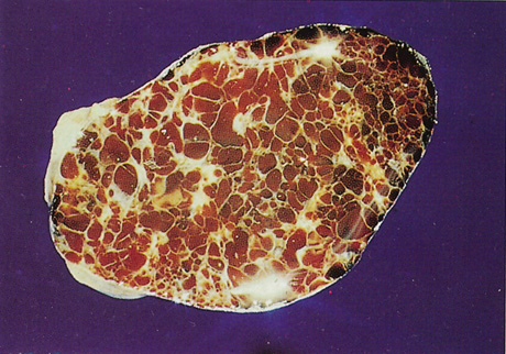
Fig. 7.
Cavernous hemangioma. Gross cut specimen showing large blood-filled spaces separated by fibrous septa and surrounded by a fine capsule. (Courtesy of Dr. Seymour Brownstein, University of Ottawa)