|
|
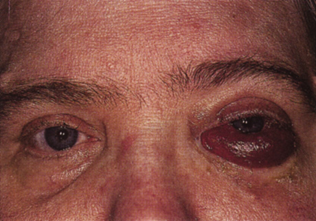
| Fig. 15. Direct arteriovenous (carotid cavernous) fistula. This 48-year-old woman presented with a 7-month history of periorbital discomfort, proptosis, diplopia, chemosis, and increased lacrimation treated as idiopathic orbital inflammatory syndrome with systemic immunosuppressants. No bruit was present, and intraocular pressure was normal. Computed tomography scan showed diffuse thickening of the left rectus muscles and an enlarged superior ophthalmic vein. Magnetic resonance angiography was interpreted as normal. She was referred for consultation service when she developed gradual visual loss to 20/400. Cerebral angiography confirmed the presence of a left direct arteriovenous fistula between the internal carotid artery and cavernous sinus. |