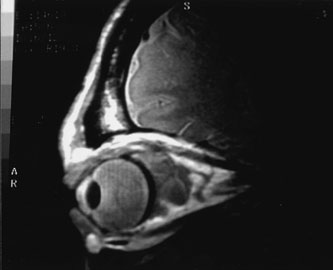
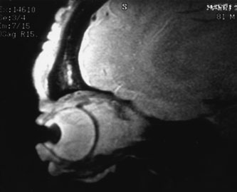
Fig. 10.
A
and
B.
No flow malformation
of the orbit. Sagittal magnetic resonance imaging showing dilated cystic spaces hypointense to muscle on T1-weighted images
(A)
and hyperintense to muscle on T2-weighted images
(B)
.