|
|
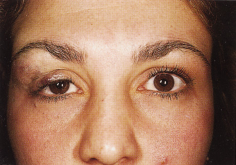
| Fig. 13. Venous flow malformation. This woman, who had a superficial component to her lesion, demonstrates the characteristically lumpy, violaceous mass causing swelling and disfigurement of the upper eyelid. (Courtesy of Dr. John V. Linberg, University of West Virginia, Morgantown, WV) |