|
|
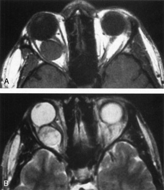
| Fig. 6. Cavernous hemangioma. Axial magnetic resonance imaging demonstrates a well-defined, homogeneous intraconal mass that is isointense to muscle and gray matter on T1-weighted image (A), and hyperintense on T2-weighted image (B). Note the displacement of the optic nerve and indented posterior globe. |