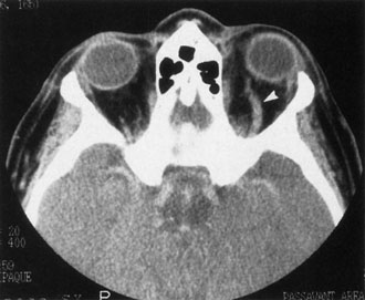
Fig. 18.
Direct arteriovenous fistula. Axial computed tomography scan of this patient showing the enlarged left superior ophthalmic vein (
white arrow
)
.
The rectus muscles of the left orbit were all diffusely enlarged.