|
|
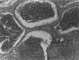
| Fig. 8. Cavernous hemangioma. Histopathologic appearance of the widely dilated vascular spaces filled with red blood cells. Abundant, loosely distributed smooth muscle is present in the vascular walls, and scattered inflammatory cells can be seen (HE, ´100). |