|
|
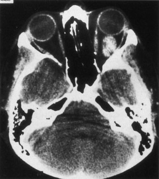
| Fig. 5. Cavernous hemangioma. Contrast-enhanced axial computed tomography scan shows a well-demarcated, oval intraconal mass in the lateral part of the middle third of the orbit. Note the enhancement within the lesion, which in this instance is inhomogeneous, but can also be homogeneous. |