Chapter 13
Retina
MARILYN C. KINCAID, MYRON YANOFF and BEN S. FINE
Main Menu   Table Of Contents

Search

CONGENITAL ANOMALIES
VASCULAR DISEASE
INFLAMMATION
DEGENERATIONS
HEREDITARY RETINAL DYSTROPHIES
SYSTEMIC DISEASES INVOLVING THE RETINA
TUMORS
RETINAL DETACHMENT
REFERENCES

CONGENITAL ANOMALIES

ALBINISM

Albinism can be divided into two groups: oculocutaneous, in which the skin and eye are hypopigmented, and ocular, in which only the eye is affected. Both ocular and oculocutaneous albinism are characterized by poor vision and nystagmus, although this is variable, even within a single pedigree.1 On clinical examination, no macular reflex is present. Histologically there is ganglion cell thickening in the macular area, but on serial sectioning no foveal pit is demonstrated. This is in contrast to albinoidism, in which there is variable but decreased pigmentation but a normal fovea, good vision, and no nystagmus.2

The most severe form of oculocutaneous albinism is tyrosinase-negative albinism, which is inherited as an autosomal recessive trait. Tyrosinase is an enzyme required for the conversion of tyrosine to dioxyphenylalanine (DOPA); lack of the enzyme means that no DOPA is available for further conversion to melanin. These persons have very fair skin, pale blond hair, and gray irises that show diffuse transillumination defects. Hairbulbs obtained from these persons have no melanosomes and do not convert tyrosine to melanin.2

Tyrosinase-positive albinism is somewhat less severe; these persons have minimal pigmentation, but the appearance is variable. Blacks are more commonly affected with this type of albinism than are whites. These patients have a positive hairbulb test.

A special type of autosomal recessive oculocutaneous albinism is Chédiak-Higashi syndrome. In addition to the albinism, the patient's neutrophils lack the ability to lyse ingested bacteria. These children are thus vulnerable to infection and die at an early age. The leukocytes contain characteristic inclusions, allowing the diagnosis to be made.

The cells of the uveal stroma and the pigmented epithelium contain giant spherical melanosomes 1 μm or more in diameter, some even as large as the cell nucleus. The disturbance appears to be one of packaging of melanosomes and of decreased number but not one of disordered melanogenesis.3

Hermansky-Pudlak syndrome is a disease that includes albinism and a hemorrhagic disorder. Thus, for both Chédiak-Higashi and Hermansky-Pudlak syndromes, the ophthalmologist may be in a position to make the initial diagnosis and appropriate referral.

Several types of ocular albinism exist, but in all the cutaneous pigmentation is relatively normal. All show abnormalities of the melanin of the neuroectoderm, while that of the neural crest-derived cells is relatively normal. Some patients previously classified as having “idiopathic nystagmus” have been found to have some type of ocular albinism. The classic type is Nettleship-Falls albinism, which is inherited as a sex-linked recessive. Histologically, both the epidermis and ocular pigment epithelium contain macromelanosomes. Female carriers can have characteristic pigmentary changes in the fundus and macromelanosomes, but they are asymptomatic.4

Forsius-Eriksson, another form of ocular albinism, is also inherited as a sex-linked recessive trait, but no macromelanosomes are seen histologically. Recently, based on electroretinographic (ERG) findings5 this disorder has been renamed Aland Island disease and reclassified as a form of incomplete congenital stationary night blindness. Still another type of ocular albinism resembles the Nettleship-Falls type except for an autosomal recessive mode of inheritance.

Albinoidism is a reduction in number of melanosomes of the neural crest. It can be a component of the Prader-Willi syndrome, which is associated with deletions in chromosome 15 (15q11-q13). In contrast, the melanin-bearing cells of the neuroepithelium are normal, and these patients thus have normal or near-normal vision and no nystagmus. Iris transillumination defects may occur.6

GROUPED PIGMENTATION (Fig. 1)

Fig. 1. Grouped pigmentation. A. Macroscopic appearance of pigmented plaques. Inset. Fundus shows a typical grouped pigmentation. B. Flat preparation of retinal pigment epithelium (RPE) showing a greater concentration of pigment granules in cells corresponding to grouped pigmentation. C. RPE in a normal area adjacent to the plaque. D. RPE in the area of the plaque shows a greater concentration of pigment granules. The overlying retina is detached artifactitiously. (A, macroscopic; inset, fundus; B, flat preparation, H&E, ×256; C, H&E, ×640; D, H&E, ×640)

The sensory retina is transparent. Pigmentation seen through it reflects underlying pigment from the retinal pigment epithelium or choroid. The hyperpigmentation of grouped pigmentation results from retinal pigment epithelial cells that are larger and contain more pigment granules than normal. The granules are large and spherical, and the cell density is higher than normal.7 Sometimes the grouped pigmentation resembles the footprints of an animal, hence the picturesque name “bear tracks.” The condition is benign and stationary. The area of retinal pigment epithelial hypertrophy may also be large and solitary, but the histology is identical to the grouped type. Lacunae of depigmentation may occur focally or diffusely.7

This phenomenon is generally harmless and of little clinical consequence. An exception is Gardeners syndrome, which is one form of hereditary gastrointestinal polyposis with nongastrointestinal manifestations, including benign soft tissue tumors, osteomas, and bilateral congenital retinal pigment epithelial hypertrophy.8 Another such syndrome is Turcot syndrome, which includes adenomatous polyps, hypertrophic pigment epithelium, and epithelial tumors of the central nervous syndrome. All may be part of a spectrum.9 The gastrointestinal polyps are adenomatous and thus have malignant potential. Because the retinal pigment epithelial hypertrophy is congenital, ophthalmic examination is potentially lifesaving in identifying persons at risk for these autosomally inherited conditions.10

COLOBOMA

During embryonic life, the hyaloid vessels enter the eye inferonasally through the fetal fissure, which extends from the optic nerve posteriorly to the iris anteriorly. Normally, this fissure closes so perfectly that no defect of retina, choroid, or sclera can be found clinically or histologically. With imperfect closure, defects of varying size result. These can vary from tiny, clinically insignificant defects to complete failure of the fissure to close, leading to microphthalmos with cyst.11

Clinically, the defects resemble bare sclera, but in fact they are lined by a thin glial lining incorporating retinal vasculature. The retina may also contain dysplastic rosettes, especially at the margins of the coloboma. The pigment epithelium and the choriocapillaris are absent in the colobomatons defect, and an absolute scotoma occurs. Colobomas are bilateral in 60% of patients and can be an isolated finding or be part of a syndrome such as trisomy 13.11

RETINAL DYSPLASIA (Fig. 2)

Fig. 2. Retinal dysplasia: four types of rosettes. A and B. Three-layer rosettes, which have the appearance of a mature retina secondarily thrown into folds. C. Two-layer rosette in which the innermost layer resembles a photoreceptor cell layer with an external limiting membrane and a relatively large lumen usually containing several undifferentiated cells. D and E. One-layer rosettes with a single layer of moderately well-differentiated neural cells, usually several cells in thickness, having an external limiting membrane-like structure surrounding the lumen. F. Primitive unilayer rosette in which a single layer of undifferentiated retinal cells surrounds the lumen with a tangle of fibrils centrally. (A, H&E, ×50; B, H&E, X200; C, H&E, X200; D, H&E, × 100; E, H&E, X 400; F, H&E, X 400)

Retinal dysplasia can be a solitary abnormality or can be associated with a variety of malformations; it can also be focal or diffuse. Instead of the normal linear arrangement of the nuclear layers, the cells form rosettes with varying degrees of differentiation. Clinically the involved areas appear translucent and grayish. The term dysplasia refers to this developmental anomaly and does not imply a premalignant condition; it is not the same as retinocytoma.12

Lahav and co-workers13 classified the dysplasias and described four types of dysplastic rosettes: (1) those with three nuclear layers, resembling full-thickness retina and simulating an artifactual cut; (2) two-layer rosettes; (3) one-layer rosettes with well-defined external limiting membranes; and (4) one-layer rosettes with a primitive nuclear layer or with a fibrillary tangle in the lumen. They found an overall incidence of 2.1% in their autopsy series.

LANGE'S FOLD (Fig. 3)

Fig. 3. Lange's fold (arrows, inset) consists of an artifactitious, inward fold of the retina at the ora serrata. (Main figure, H&E, × 101; inset, macroscopic)

Lange's fold is an inward fold of the peripheral retina at the ora serrata and is a artifact of fixation, present both grossly and microscopically. It is not present in the living eye.14 Although it can be seen as late as at 20 years of age, it is most common in infant eyes up to 2 years of age. Apparently, it results from decreased adhesion of the far peripheral sensory retina to the underlying retinal pigment epithelium; this adhesion normally develops with age.15

CONGENITAL NONATTACHMENT OF THE RETINA

Congenital nonattachment of the retina is a normal condition in the developing embryo up until the eighth week of gestation. If the detachment persists beyond this time, it is abnormal. Secondary superimposed abnormalities, such as retinal dysplasia, may occur. Histologically, total detachment of the retina, obliteration of the vitreous space, and retinal immaturity are seen.16

MYELINATED (MEDULLATED) NERVE FIBERS

Myelinated nerve fibers are typically congenital lesions. Usually, the lesion extends to the disc margin, but it can be anywhere in the retina. The lesion appears whitish with feathery margins and follows the orientation of the nerve fiber layer. Myelination is not continuous across the lamina cribrosa, so some discontinuity always exists, even in those myelinated fibers that do not extend to the disc.17 They can be minimal or extensive; patients who have extensive myelination may also have myopia, amblyopia, and strabismus. Oligodendroglial cells are believed to be the cells responsible for myelination; they normally populate the optic nerve but not the retina. The degree of visual field defect varies, but usually the defect is surprisingly minimal. Rarely, myelinated nerve fibers may be acquired.18 Demyelination of the myelinated fibers has been documented in multiple sclerosis and other demyelinating disease.

CONGENITAL NIGHT BLINDNESS

Congenital night blindness is a disorder of rod function or of neural transmission under scotopic conditions. Some types of congenital night blindness show a normal fundus appearance, but two types, Oguchi's disease and fundus albipunctatus, do not.19 Also, as noted above, Forsius-Eriksson albinism may be a form of incomplete congenital stationary night blindness with a variably albinotic fundus.5

Oguchi's disease is an unusual form of congenital night blindness. Under light-adapted conditions, the retina takes on an unusual gray-white sheen, called Mizuo's phenomenon. The phenomenon is reversible; in the dark-adapted state, the retina appears normal. Kuwabara and associates found an abnormal layer of amorphous material between the retinal pigment epithelium and the photoreceptors. Also, the photoreceptors lacked the normal lamellar arrangement of outer segment discs.19

FOVEOMACULAR ABNORMALITIES

It should be noted that the pathologist and the clinician use the terms fovea and macula differently. Anatomically, the foveola is about 350 μm in diameter and is the floor of the pit. It is thus slightly smaller than the foveal avascular zone, which is approximately 500 μm in diameter. The fovea anatomically is about 1.5 mm, or about the size of the optic disc. The anatomic macula is that area in which the ganglion cell layer is greater than a single cell layer in thickness. It is about 5 mm in diameter and thus corresponds approximately to the clinician's posterior pole.20

The fovea may be hypoplastic in such disorders as albinism, aniridia, microphthalmos, and hemeralopia. In these conditions there is incomplete development of the foveal pit, lack of xanthophyll pigment, and irregularities in the vasculature, without the normal foveal avascular zone.

So-called coloboma of the macula is usually secondary to toxoplasmosis. Some patients who have Leber's congenital amaurosis may have a foveal pigment abnormality resembling a coloboma.21

LEBER'S CONGENITAL AMAUROSIS

Leber's congenital amaurosis is characterized by subnormal vision or blindness at birth or shortly after, and it is usually inherited as an autosomal recessive disorder. Blindness for any reason in the neonatal period is characterized by searching nystagmus, but Leber's congenital amaurosis can be differentiated from most other causes of neonatal blindness by the markedly diminished or absent ERG response.22 Only a few other causes of neonatal blindness associated with ERG depression occur, most notably Zellweger's syndrome.23 The fundus appearance in Leber's congenital amaurosis varies from essentially normal to marked pigmentary migration in a spicule formation with optic atrophy and retinal vessel narrowing.22 A bull's-eye maculopathy has been reported in one patient.21 Associated systemic findings, including neuromuscular dysfunction, are not uncommon,22 and biochemical abnormalities have been described in some cases.23

Histologically, the sensory retina is immature, with ultrastructural abnormalities in the photoreceptor nuclei and inner segments and also in the retinal pigment epithelium and choriocapillaris.24

Back to Top
VASCULAR DISEASE
The retina is supplied by a dual circulation. The inner half of the sensory (neural) retina, extending outward to include about half of the inner nuclear layer, is supplied directly by the retinal vasculature. The outer half, and the retinal pigment epithelium, are supplied by diffusion from the choroid. The retinal vessels are end vessels, like those of the brain, and do not normally anastomose. Also, the retinal capillaries, like those of the brain, do not allow the passage of relatively small molecules, such as fluorescein.

The central retinal vessels remain histologic arteries and veins for only a short distance from the optic disc, thereafter becoming arterioles and venules, respectively. Nevertheless, the terms artery and vein are used clinically, and also histologically, to designate the respective vessels and their diseases.

RETINAL ISCHEMIA

The retina, like any other tissue, becomes ischemic from lack of blood supply. Choroidal vascular insufficiency is relatively uncommon posteriorly, although it can result from choroidal tumors such as melanoma, hemangioma, or metastatic tumor competing for the blood supply. Multiple emboli occurring in various disease processes can also occlude choroidal vessels.25

Peripherally, outer retinal ischemia is extremely common, recognized clinically as cobblestone or paving stone degeneration (Fig. 4). The prevalence of these lesions increases with age and is also higher in axial myopia. In these lesions, the retinal pigment epithelium and the outer half of the sensory retina are obliterated, so that the thinned inner nuclear layer is directly adherent to Bruch's membrane. Clinically, these lesions are seen as well-demarcated round pale areas because of the lack of retinal pigment epithelial cells and their pigment.26 Cobblestone lesions are most frequent inferiorly. Elshnig's spots, secondary to hypertension, are histologically identical but larger.27

Fig. 4. Paving stone degeneration. A. The retinal pigment epithelium (RPE) ends abruptly in the area of degeneration (between arrows). Insets 1 and 2. Typical circumferential lesions of paving stone degeneration. B. Higher magnification of (A). In the area of RPE disappearance, Bruch's membrane (arrows) is intact, but the overlying retina (especially the outer layers) and the underlying choroid show degenerative changes. (A, H&E, ×69; inset 1, macroscopic; inset 2, macroscopic; B, H&E, ×101)

In contrast, occlusion of the central retinal artery or its branches results in ischemic infarction of the inner retinal layers. Usually, these occlusions are embolic, most commonly from calcific, platelet, and cholesterol emboli.25 Emboli can result from a variety of other causes including amniotic fluid, bacteria, air, cloth particles from prosthetic heart valves,28 and talc emboli in intravenous drug abusers.29 Nonembolic causes include atherosclerosis (Fig. 5) and inflammatory processes that narrow and occlude the lumen from swelling of the vascular walls, such as cranial arteritis, Wegener's granulomatosis, and collagen vascular diseases.

Fig. 5. Atherosclerosis. A. Gross appearance Of the fundus of an eye removed postmortem. Arrow indicates plaque in the inferonasal arteriole, seen microscopically in (B). B. Frozen section stained with oil red-O shows lipid deposits (arrow) in atheromatous plaque. Note the fatty infiltration in the muscular wall at the edge of the artifactitious cleft (double arrow). (A, macroscopic; B, oil red-O, ×200)

Cholesterol emboli are commonly seen in patients who have ulcerated atheromatous plaques of the carotid arteries. Often, however, these emboll do not occlude the vessel because their shape does not readily conform to the shape of the vessel. These are seen clinically as Hollenhorst plaques and appear as bright shiny yellow emboli within the vessels (Fig. 6).30

Fig. 6. Emboli. A. Retinal vessel on the left (v) is occluded by an osmophilic fat globule. A smaller fat globule (arrow) is seen in the vessel on the right. The retinal arteriole fat emboli followed closed-chest cardiac massage. B. Cholesterol embolus in a retinal artery (arrow). Inset 1. Higher magnification of the embolus. Inset 2. Clinical appearance of a cholesterol embolus (arrow) in the same eye prior to enucleation. Multiple cholesterol emboli (Hollenhorst plaques) were present in the fundus of a 67-year-old man with carotid artery stenosis. (A, PD ×300; B, H&E ×40; inset 1, H&E × 101; inset 2, clinical)

In central artery occlusion, inner retinal edema occurs acutely, causing the normally transparent retina to become translucent and grayish with loss of underlying detail. Visual acuity is profoundly depressed or completely lost. Because no inner layers exist in the foveal pit, the fovea remains transparent. The normally perfused choroid and the RPE remain visible, giving rise to the clinically observed cherry-red spot. The retinal edema begins within a few hours after occlusion and becomes maximal in 24 hours.

Histologically, swelling of the inner retinal layers, extending outward to include a portion of the inner nuclear layer, contrasts to the intact outer layers.31

With time, the inner layers of the retina are lost and the retinal vessels become somewhat hyalinized. Because these occlusions are complete, the glial cells of the retina are also destroyed, so no reactive gliosis occurs. Histologically, the inner layers are homogenized, and the outer layers remain, up to and including a portion of the inner nuclear layer. In contrast, in glaucomatous atrophy, only the ganglion cell and nerve fiber layers are lost.

Complications of central artery occlusion, in addition to profound visual loss, include iris neovascularization and secondary angle-closure neovascular glaucoma in 2% of cases.32 Rarely, in some elderly persons, central retinal artery occlusion can be bilateral.

Branch arteriolar occlusion is similar to central arterial occlusion in both cause and appearance. The involved area is sharply demarcated from adjacent areas perfused by uninvolved arterioles. Hypertension with arteriolar sclerosis, diabetes mellitus, and malignant hypertension27 (Fig. 7) are other causes of branch arteriolar occlusion.

Fig. 7. Malignant hypertension in an 18-year-old man with hereditary interstitial nephritis. A. Retina is detached and disorganized with neovascularization into the vitreous. Inset. Macroscopic appearance. B. Intraretinal neovascularization (arrows). C. New vessels are leaving the retina (arrow) to enter the subvitreal space. D. Neovascularization is budding out of the retina (arrows) into the subvitreal space. (A, H&E, × 16; inset, macroscopic; B, H&E, ×69; C, H&E, ×101; D, H&E, ×101)

Capillary closure results in very localized ischemia and infarction of inner retinal layers. Clinically, these are cotton-wool spots; they are gray or white and contain feathery margins. They are common in a number of diseases, including diabetes mellitus, the acquired immunodeficiency syndrome (AIDS), collagen vascular diseases (especially lupus), leukemia, and other blood dyscrasias.33 They have been called soft exudates, in contrast to the hard exudates of capillary leakage, but histologically they are swollen axons in the nerve fiber layer. These fusiform swellings have been called cytoid bodies, because the eosinophilic central density superficially resembles a cell nucleus.34 Ultrastructurally, these fusiform densities are areas where the products of antegrade and retrograde axoplasmic flow have accumulated.34 As noted, the usual cause is edema but other causes of axoplasmic flow interruption give rise to the same clinical and histologic picture.

PANOCULAR VASCULAR INSUFFICIENCY

Ocular vascular insufficiency results from systemic hypotension or from local or regional circulatory compromise. In turn, this affects both the retinal and the choroidal circulations.

Atherosclerosis of the carotid artery is the most common cause of diffuse circulatory compromise,26 but there are others. Takayasu's disease is a rare disease of the major arteries, occurring most frequently in young Japanese women. The aortic lumen is gradually occluded by a granulomatous reaction to the arterial adventitia and media, with intimal hyperplasia. The corresponding syndrome, Takayasu's syndrome, refers to any process affecting the aortic arch with resulting circulatory compromise, including atherosclerosis and syphilis. Both eyes are affected. Clinical symptoms include amaurosis fugax and pain.36

Histologically, in all these diseases, wide-ranging effects occur. Cobblestone degeneration may extend posteriorly, indicating diffuse outer ischemic atrophy from choroidal insufficiency. Also, inner ischemic atrophy of the retina occurs from decreased retinal perfusion. Optic nerve head neovascularization has been reported. Anterior segment changes include iris neovascularization, iris necrosis, and cataract.26

TEMPORAL ARTERITIS

Temporal arteritis (cranial arteritis) is a granulomatous inflammation of arteries and arterioles that involves all layers of the vessel, narrowing and sometimes occluding them. It is most frequent in the elderly and is associated with an elevated erythrocyte sedimentation rate. Associated symptoms are those of ischemia and include scalp tenderness and jaw claudication. Despite the name, the disease can affect arteries elsewhere, including the coronary arteries, so cardiac angina and infarction can occur on this basis. Nonspecific symptoms are common and include weight loss and fever.

The most common name for this condition, temporal arteritis, derives from that easily palpated, superficial artery. When involved, the temporal artery is generally firm, tender, enlarged, and red; however, it can also be normal with a palpable pulse. Ophthalmic involvement, with partial or complete loss of vision, should lead to suspicion of the diagnosis; prompt action is needed to prevent further visual loss in the same or opposite eye. Other ophthalmic manifestations include ptosis and extraocular muscle palsies.

Histologically, a granulomatous reaction affects all layers of the artery, with fragmentation and patchy obliteration of the internal elastic lamina. At times, the inflammation is nongranulomatous or the epithelioid and giant cells are inconspicuous.37 “Skip areas” where the artery appears normal may also occur, so examination of multiple levels is imperative.38

Resolved temporal arteritis has a distinctive clinical picture. At times, a biopsy specimen is obtained weeks after corticosteroid treatment has been initiated, or the active inflammation resolves on its own. Such a specimen characteristically shows variation in thickness of the arterial media with fibrous scarring.37

RETINAL HEMORRHAGIC INFARCTION (Fig. 8)

Fig. 8. Retinal hemorrhagic infarction. A. Central retinal vein is occluded and recanalized. The artery is identified by an internal elastic lamina (arrow). B. Hemorrhagic infarction causing disorganization of the retina in a patient with polycythemia vera and central retinal vein occlusion. Inset. Fundus following central retinal vein occlusion in an otherwise healthy 18-year-old girl; no cause for the occlusion was found. C. Sheets of blood in the inner retinal layers produce the characteristic fundus appearance. Note the preservation of the photoreceptors. D. Rubeosis iridis is present in the same patient with polycythemia vera. E. Pigment (arrows) is hemosiderin in a long-standing occlusion. (A, elastica, ×136; B, H&E, ×69; inset, fundus; C, H&E, ×69; D, H&E, × 101; E, Prussian blue, × 176)

In contrast to arterial occlusion, retinal vein occlusion is usually thrombotic and results in varying amounts of intraretinal hemorrhage. Central retinal vein occlusion can be divided into two types. The first type is nonischemic and is sometimes called venous stasis retinopathy39 (an unfortunate term, since ocular ischemia from diffuse hypoperfusion, discussed earlier, is also sometimes called venous stasis retinopathy). Nonischemic central vein occlusion is characterized by a variable number of flame-shaped and dot and blot hemorrhages but with few or no cotton-wool spots. As documented by fluorescein angiography, capillary perfusion is nearly normal, and thus the sequelae of ischemia, including iris neovascularization and neovascular glaucoma, are very rare in the nonischemic type. The visual prognosis is good.39

In younger patients, more frequently men, this condition is usually caused by venous phlebitis and runs a benign course. Clinically, it appears as disc swelling associates with venous dilatation and tortuosity and scattered intraretinal hemorrhages. Visual acuity is retained or recovered. The late sequelae include perivenous sheathing and dilated veins at the optic nerve head. In older persons, probably arteriosclerosis is the usual cause.

Ischemic central vein occlusion is also called hemorrhagic vein occlusion because of extensive retinal hemorrhage (the “blood and thunder” fundus). Numerous cotton-wool spots may be seen, but even if they are obscured by the hemorrhage, fluorescein angiography helps to document the extensive capillary closure and ischemia. Iris neovascularization is frequently associated and characteristically occurs between 6 weeks and 6 months (so-called 90-day glaucoma).39 With either type of vein occlusion, hemorrhage into the macula can result in permanent visual loss.

For obscure reasons, primary open-angle glaucoma is associated with central vein occlusion in 8% to 20% of patients, either before or after the occlusion.40

Histologically, central retinal vein occlusion appears acutely as hemorrhagic necrosis, with massive hemorrhage involving all layers of the retina (Fig. 9). The hemorrhage may also extend under the internal limiting membrane, separating it from the underlying nerve fiber layer. Cytoid bodies and optic nerve head edema may be present. Whether complete or “impending,” a thrombus occurs in the central vein,40although as Hayreh subsequently noted,39 only one of Green's cases was nonischemic histologically. All the rest had neovascularization of iris, optic disc, or both, sometimes with inner retinal ischemic atrophy.

Fig. 9. Hemorrhage within the nerve fiber layer (arrow) tends to “track” along the nerve fibers, producing a flame-shaped hemorrhage observed clinically. An accumulation of blood in the potential space between the internal limiting membrane (ilm) and the nerve fiber layer produces submembranous intraretinal hemorrhage, which is generally restrained from entering the vitreous compartment by the strength of the overlying thick basement membrane. Pockets of blood accumulating within the bipolar cell layer or between Henle's fibers (photoreceptor axons) of the extramacular outer plexiform layer produce the dot and blot hemorrhages seen clinically. (H&E, × 115)

Later in the evolution of the occlusion, exudates may occur in the outer plexiform layer. Other late changes include disruption, gliosis, and hemosiderosis of the retina. The retinal blood vessel walls can be markedly thickened. The thrombus is organized and may be recanalized.40

The histology of branch vein occlusion is similar.41 Clinically, it has been observed that nearly all branch vein occlusions occur where the artery crosses over the vein, rather than under the vein, as viewed ophthalmoscopically.42

HYPERTENSIVE AND ARTERIOSCLEROTIC RETINOPATHY

Although arteriolar sclerosis and hypertension usually occur together, it is easier to understand the changes if they are graded separately. Both hypertensive retinopathy and arteriolar sclerotic changes are graded from I through IV.43

Grade I hypertensive retinopathy consists simply of generalized narrowing of the arterioles, so the blood column, which is all that is visible ophthalmoscopically, is narrowed. Grade II includes narrowing and focal spasms of the arterioles. In grade Ill, which includes the above changes, there are also intraretinal hemorrhages, cotton-wool spots, and hard exudates. The hemorrhages include flame-shaped hemorrhages of the nerve fiber layer and dot and blot hemorrhages of the inner nuclear and outer plexiform layers. Hard exudates consist of proteins and lipids that have leaked from the capillaries. The exudates preferentially collect in the macular area, because here the fibers of the outer plexiform layer are obliquely oriented. The exudates may thus form a macular star clinically. In grade IV, all the preceding changes are seen, along with optic disc edema.

The choroidal vasculature can also be involved in malignant hypertension. Retinal pigment proliferation in a patchy pattern can be seen. Elschnig's spots are areas of choriocapillaris occlusion and are thus similar to cobblestone degeneration except for their larger size and more posterior location. Siegrist's lines consist of increased pigmentation along a sclerotic choroidal vessel but are uncommonly seen.44

Arteriolar sclerotic changes are also graded from I to IV. Grade I is subintimal hyalin deposition and thickening of the vascular wall, so it is no longer completely transparent. Thus, an increased light reflex is seen clinically. Grade II adds arteriolovenular crossing defects. The arterioles and venules normally share a common adventitial sheath where they cross. With thickening and increased rigidity of the arteriolar wall, the venular wall is compressed. Clinically, this appears as a gap in the course of the venule where the arteriole crosses it. Grade III consists of the above changes and also increased thickening of the arteriole, so that the blood column is narrowed. The increased light reflex gives a copper-like appearance to the vessel, called “copper wire” change. In Grade IV, even more thickening of the arteriole occurs, along with extensive narrowing of the blood column so that it no longer appears red. The vessel thus appears white, or like a “silver wire.” The vessel may actually remain patent; patency can be documented with fluorescein angiography.27,43

PATTERNS OF RETINOPATHY

No retinal vascular change, whether hemorrhage, exudate, or microinfarction, is specific for a given ocular or systemic disease. Rather, it is the overall pattern, location, and combination of the findings that is characteristic.

Hemorrhagic Retinopathy (Fig. 10)

Fig. 10. Hemorrhagic retinopathy. A. Hemorrhage separating the vitreous body (v) from the internal limiting membrane of the retina (ilm) is a “true” subhyaloid hemorrhage. B. Massive accumulation of blood in the potential cleft (arrow) between the internal limiting membrane and the nerve fiber layer is a submembranous intraretinal hemorrhage. Such hemorrhages may appear with “fluid level” clinically, where they are somewhat inaccurately called subhyaloid hemorrhages. C. Large submembranous (intraretinal) hemorrhage may extend throughout the other layers of the retina as seen here and even rupture through the external limiting membrane to gain access to the subretinal space. D. Hemorrhages may be restrained from entering the vitreous compartment by the internal limiting membrane of the retina (as on the left), but this restraint diminishes rapidly whenever a thick basement membrane becomes thin (as over the optic nerve head on the right). (A, H&E, × 130; B, H&E, ×70; C, H&E, × 115; D, H&E, ×80)

Any of a number of blood dyscrasias and retinal diseases can cause a retinopathy in which hemorrhages predominate. In leukemia, for example, the decreased platelet count and anemia together are believed to be responsible for the hemorrhages.45 Increased blood viscosity, as with polycythemia and macroglobulinemia, also causes a fundus picture primarily of hemorrhages.46 Disseminated intravascular coagulation is another cause of a predominantly hemorrhagic fundus picture. Initially, for unknown reasons, the blood is hypercoagulable, with multiple thrombi forming in small vessels throughout the body. As the coagulation factors are consumed, the blood becomes hypocoagulable, resulting in diffuse hemorrhages. Histologically, fibrin can be identified in choroidal and retinal vessels.47

Exudative Retinopathy

Exudates occur when the normally competent, “tight,” capillaries leak proteins and lipids into the surrounding retina. These are seen clinically as yellow, shiny material. Sometimes these occur in a circular, or circinate, pattern surrounding a leakage site such as a microaneurysm. If the leaking microaneurysm is occluded by photocoagulation, the exudates may gradually resorb.48 Histologically, these are amorphous collections of eosinophilic, periodic acid-Schiff-positive material found predominantly in the outer plexiform layer.

DIABETES MELLITUS

Diabetes mellitus and its ophthalmic manifestations are discussed in the chapter “Diabetes Melli tus.”

COATS' DISEASE, LEBER'S MILIARY ANEURYSMS, RETINOPATHY OF PREMATURITY

These diseases are part of the differential diagnosis of retinoblastoma and are thus included in the chapter “Retinoblastoma.”

RETINAL MACROANEURYSMS (Fig. 11)

Fig. 11. Retinal arteriolar macroaneurysms are shown in a trypsin-digested preparation in (A)(arrows) and in cross-section in (B) (arrow). (A, PAS. ×75; B, H&E, ×69)

Retinal macroaneurysms, as the name implies, are outpouchings of the vessel walls of the large vessels, the arterioles.49 Like the microaneurysms of capillaries, macroaneurysms leak high-molecular-weight substances into the surrounding retina.50 They can also bleed, sometimes in a dumbbell configuration, with blood both beneath the retina and also just under the internal limiting membrane.51

Visual loss results from either exudate or hemorrhage into the macular area.

Macroaneurysms are frequently associated with hypertension or other systemic vascular disease.50

Histologically, the vessels are thickened by blood, fibrin, and macrophages.49

SICKLE CELL DISEASE

Sickle cell retinopathy is most frequent in sickle cell-hemoglobin C disease but also occurs in homozygous SS, sickle cell-thalassemia, and even sickle trait (hemoglobin SA). The retinopathy appears to be caused by hypoxia. The erythrocytes are more rigid and less able to pass through small capillaries. Hypoxia causes further sickling, in a self-perpetuating cycle.42

Typically, sickle cell retinopathy is a peripheral retinal disease. Nonproliferative changes include salmon patches, which are intraretinal hemorrhages; iridescent spots, areas of resolving hemorrhage with iron-containing macrophages beneath the internal limiting membrane; and black sunbursts, areas with proliferated retinal pigment epithelium in a bone spicule pattern.53

Proliferative disease occurs because of increased ischemia in peripheral retina. Arteriolar occlusion and shunt formation occur, both of which can be documented by fluorescein angiography. Neovascular fronds arise from the shunts and may lead to vitreous hemorrhage and secondary retinal detachment.52 Angioid streaks are associated with the sickle cell hemoglobinopathies, and these can give rise to neovascularization in the posterior pole.54

EALES' DISEASE

Eales' disease has no known cause. It occurs most frequently in young men, and clinical findings include peripheral capillary closure, vascular leakage, arteriovenous shunts, and segmental microaneurysms. Retinal nonperfusion and neovascularization are late changes. Some investigators have found an increased association with hypersensitivity to tuberculin protein (a false-positive tuberculin skin test) and an increased incidence of hearing or vestibular abnormalities. An association with multiple sclerosis has been reported but more recently discounted.55

HEMANGIOMA

Retinal hemangiomas can be capillary or cavernous. Retinal capillary hemangiomas are part of the yon Hippel-Lindau syndrome, one of the phakomatoses. Clinically, a single feeder arteriole and draining venule are identified, involved with a rounded vascular tumor.56

Conversely, a retinal cavernous hemangioma is often an isolated finding. In these hemangiomas, blood circulation is sluggish and fluid-clot levels can be identified by fluorescein angiography. Clinically, cavernous hemangiomas appear as dark, rounded masses within the retina.57 Some investigators have reported that central nervous system hemangiomas can be associated with the retinal lesions and suggest that patients and family members with retinal cavernous hemangiomas be referred for central nervous system evaluation and treatment.58

A third type of retinal hemangioma is the racemose hemangioma, a component of the Wyburn-Mason syndrome. No intervening capillary bed exists between the arteriole and venule; thus both large vessels become dilated, high-velocity shunts. Surprisingly, they seldom bleed and usually do not leak. Histologically, the involved vessels are relatively thin walled and focally replace the entire retinal thickness. A propensity to form arterial macroaneurysms has been reported in one patient.59

DISSEMINATED INTRAVASCULAR COAGULATION

Disseminated intravascular coagulation is a condition caused by release of tissue thromboplastin into the circulation, allowing massive clotting to occur throughout the circulatory system. The precise cause is unknown, but conditions such as gram-negative sepsis and respiratory distress syndrome have been associated. Because the platelets and clotting factors are consumed, the blood then progresses to a hypocoagulable state, hemorrhage, tissue hypoxia, and further release of thromboplastin. Histologically, fibrin-containing clots are present in the choriocapillaris, especially in the posterior pole, but can also be present in retinal vessels and in vessels of the anterior segment. Subsequent retinal and vitreous hemorrhages may occur.47

Back to Top
INFLAMMATION
A number of conditions can cause a retinal inflammatory infiltrate, including keratitis, uveitis, and scleritis. Sometimes this occurs in a perivenular pattern. Cystoid macular edema after cataract surgery can be associated with phlebitis.60

BACTERIAL INFECTION

Septic retinopathy is usually caused by bacterial emboli from subacute bacterial endocarditis, although any bacteremia can lead to septic retinopathy. Clinically, the emboli appear as white-centered hemorrhages, often called Roth's spots. Histologically, the emboli contain white blood cells and bacteria. Noninfectious causes of white-centered hemorrhages include leukemia and an elevated white blood cell count and fibrin thrombi from any cause.61

FUNGAL INFECTION

Endogenous fungal infections may cause fungal retinitis.62 Endogenous endophthalmitis is fungal more often than bacterial, and the most common causative fungus is Candida.63

VIRAL INFECTION

Several viruses can cause a clinically characteristic retinitis. There are two types of herpes simplex, type 1 and type 2. Classically, type 1 causes infections in nongenital sites, “above the belt,” while type 2 causes genital infection. Type 1 is the most frequent cause of sporadic, fatal encephalitis, but herpetic retinitis is much less common.64

Herpetic retinitis consists of irregular yellow-white patches of retinal necrosis with overlying hazy vitreous. With resolution of the acute inflammation, a patchy hypopigmentation and hyperpigmentation occurs. Histologically, full-thickness retinal and retinal pigment epithelial necrosis is seen along with characteristic nuclear inclusions.65 Typically the choroid contains a dense chronic inflammatory infiltrate, in contrast to cytomegalovirus infection in which the choroid usually shows minimal involvement.64

Congenital herpes simplex retinitis, in contrast, is usually caused by type 2 virus and is usually acquired during passage through the birth canal, although some newborns contract the infection in utero. Depending on the time of infection, the retinitis may appear active or quiescent with retinal scarring.66

Herpes zoster causes a retinitis and choroiditis either with direct tissue involvement by the virus or secondary to vasculitis and ischemia. After primary infection as varicella, or chickenpox, herpes zoster becomes dormant in a sensory ganglion and, after a usually unknown stimulus, is secondarily reactivated. Ophthalmic herpes zoster involves the territory of the first branch of the trigeminal nerve, including the eye. The inflammation can be very destructive with a granulomatous choroiditis.67

Herpes zoster can also be acquired in utero, often during the second trimester, leading to chorioretinal scarring with pigmentation. The macular scarring can resemble toxoplasmosis. Optic nerve hypoplasia has been associated with infection during the 16 th week of gestation.68

Cytomegalovirus has historically been associated with immunosuppression following renal transplant or associated with leukemia or chemotherapy. AIDS is now the most frequent predisposing condition, and cytomegalovirus retinitis is the most frequent cause of visual loss in the AIDS population.69 Cytomegalovirus retinitis is characterized by retinal destruction with hemorrhages and exudates in a “brush fire” pattern. The fundus appearance is so characteristic that the ophthalmologist is often called on to make the clinical diagnosis of systemic cytomegalovirus infection. Treatment with gancyclovir is the mainstay of therapy, as well as control of the underlying immunosuppressive process, although gancyclovir does not completely obliterate the Virus.70

Histologically, cytomegalovirus retinitis is characterized by full-thickness necrosis and destruction of the retina, often with rather abrupt transition to normal retina. Infected cells contain large, reddish, nuclear inclusions that can be seen at all retinal layers and also in the pigment epithelium. Ultrastructurally, these cells contain innumerable viral particles.

ACUTE RETINAL NECROSIS

Bilateral acute retinal necrosis syndrome was once considered a rare disease of unknown cause that occurred in immunocompetent persons. It has recently become more common, mostly because of its association with AIDS, and it frequently is unilateral (ARN). Most cases now are recognized as being caused by herpes zoster,71 sometimes with associated immunosuppression, but herpes simplex and cytomegalovirus can also cause this syndrome. The virus is most easily demonstrated during the acute phase of the disease and may be undetectable after necrosis or glial repair.72

Clinically, the retina shows vasculitis and patchy retinal whitening, indicating full-thickness necrosis. Uveitis and vitritis also occur. As the retinal necrosis becomes confluent, visual loss occurs. Late in the course of the disease, retinal holes may form at the junction of necrotic and intact retina. Retinal detachment is common. Central nervous system involvement with cerebrospinal fluid pleocytosis can sometimes be present. More recently, it has been recognized that mild forms of the syndrome can occur.73

ACQUIRED IMMUNODEFICIENCY SYNDROME

AIDS, first recognized in 1981, is caused by the human immunodeficiency virus (HIV), a retrovirus that destroys cellular immunity, leaving the patient vulnerable to multiple secondary infections and neoplasms. HIV itself causes a microangiopathy with selective loss of pericytes, believed due to the deposition of circulating immune complexes. The microangiopathy is manifest as cot-ton-wool spots in the retina74 and, less frequently, as hemorrhages, microaneurysms, and telangiectasias.75 In two patients with AIDS, the cotton-wool spots had calcified.76

The most common secondary infection in AIDS is cytomegalovirus, and in some patients cytomegaloviral retinitis is the first AIDS-defining opportunistic infection. Unlike HIV retinopathy, which usually is asymptomatic, cytomegaloviral retinitis is an important cause of visual loss, justifying treatment with gancyclovir.77 Other opportunistic infections include herpes zoster,78 atypical mycobacteria, cryptococcus,74 toxoplasmosis,79,80 and histoplasmosis.81 AIDS has been associated with particularly aggressive acute retinal necrosis caused by herpes zoster.78

Syphilis can also be acquired along with AIDS; and in that setting, syphilis tends to be more aggressive, with a short latency. In one study, nearly all the patients who had AIDS and syphilis also developed neurosyphilis, and sometimes even presented with symptoms of neurosyphilis. In the eye, syphilitic retinitis appears as a periphlebitis with dense vitritis and intraretinal hemorrhage.82

Recently, Pneumocystis carinii choroiditis, associated with disseminated infection, has been recognized. P. carinii, although previously believed to be a protozoan, is probably a fungus, and causes pneumonia in more than 80% of patients who have AIDS. Aerosolized pentamidine has been used to prevent pneumonia, but this treatment does not eradicate the organism in other body sites. The choroidopathy is an indication of systemic dissemination of the organism. The choroidal lesions are generally asymptomatic and appear as irregular yellowish lesions in deep retina, which gradually enlarge. The vitreous remains clear and the retina is usually uninvolved. The lesions correspond to relative visual field defects. Intravenous pentamidine allows resolution over a few weeks to months.83

Histologically, the involved areas of choroid contain eosinophilic acellular infiltrates. Silver stain discloses cystic or crescentic organisms.84

RETINITIS OF UNKNOWN CAUSE

Acute posterior multifocal placoid pigment epitheliopathy is an acute, self-limited disorder of unknown cause. Some researchers believe it is a primary inflammation of the retinal pigment epithelium, but others believe it is an underlying occlusion of the choriocapillaris. The disease occurs in young persons who usually have an underlying systemic disease. The choroidal lesions are yellow-white and typically show early blockage and late staining on fluorescein angiography. The lesions and the decreased acuity, if present, resolve, but permanent changes occur in the pigment epithelium. No histopathologic findings have been reported.85

Patients who have acute macular neuroretinopathy usually present with mild loss of visual acuity and associated scotomas. The lesions are reddish brown and irregular and are in or adjacent to the fovea. The lesions are most common in young women and resemble the changes seen in commotio retinae or in macular infarction in sickle cell anemia. In some recent cases, an associated acute systemic hypertension occurred.86 The disorder is uncommon, and no histology has been described.

Back to Top
DEGENERATIONS

MICROCYSTOID DEGENERATION

Microcystoid degeneration occurs in two forms, typical and reticular. Typical microcystoid degeneration is virtually universal in all eyes, except those of young children, and increases in extent with age. It is generally more extensive temporally and tends to be symmetrical between the two eyes. It begins at the ora serrata and extends circumferentially and posteriorly. Clinically and grossly, typical peripheral microcystoid degeneration appears as a grayish translucent area that contains myriad tiny interconnecting channels (Figs. 12 AND 13).87

Fig. 12. Typical microcystoid degeneration of the retina and adjacent cysts of the pars plana contain acid mucopolysaccharides (arrows) when stained with the Hale colloidal iron method. (Ora, region of the ora serrata) (AMP, × 50)

Fig. 13. Typical microcystoid degeneration. A. Spaces or channels lie initially within outer plexiform layer (i.e., between compressed bundles of photoreceptor axons and the remains of Müller glial cells). B. Higher magnification shows that the middle limiting membrane (a) limits the inner boundaries, the glial-neuronal columns (b) limit the lateral boundaries, and the photoreceptor cell bodies (outer nuclear layer) and the external limiting membrane (c) limit the outer boundaries of the microcystoid channels. C. Diphosphopyridine nucleotide (DPNH) diaphorase (nitro-blue tetrazolium [BT] method). The glial-neuronal columns show a dense precipitate of formazan, signifying the presence of the cytochrome oxidase system. (A, H&E, ×135; B, H&E, ×300; C, nitro-BT, × 125)

The cysts (technically a misnomer, since a cyst is defined histologically as an epithelial lined space) are at the level of the outer plexiform and inner nuclear layers of the retina. With time, the spaces may spread vertically to involve retinal layers above and below. The cysts appear empty with hematoxylin and eosin, but special stains show that they contain acid mucopolysaccharide (hyaluronic acid) sensitive to hyaluronidase. The cysts may coalesce to form schisis, or splitting; a schisis cavity is arbitrarily defined as being greater than 1.5 mm, or 1 disc diameter, in length.87

Reticular cystoid degeneration is much less common and seems to occur posterior to typical Cystoid degeneration. Because these cystic spaces are more superficial, the vasculature, including the capillaries, is prominent. The borders tend to follow the larger vessels, in contrast to typical cystoid degeneration, which shows a rounded border. The inner portion of the cysts is less transparent, so the clinical appearance is of beaten metal (Fig. 14).

Fig. 14. Reticular cystoid degeneration. A. Limited to the innermost layers of the retina. B and C. Reticular cystoid (arrows) and typical microcystoid degeneration (m) in the same regions of the retina. (A, H&E, × 165; B, H&E, × 165; C, H&E, ×40)

This type is found in only about 13% of eyes and is bilateral in 46% of those. Unlike typical Cystoid degeneration, no clear relation is seen with aging. Histologically, the cystoid spaces are at the level of the nerve fiber layer, the same location as in juvenile retinoschisis. Thus, the inner aspect of reticular Cystoid degeneration includes only nerve fiber layer remnants, retinal vessels, and internal limiting membrane.87

RETINOSCHISIS (Fig. 15)

Fig. 15. Typical degenerative senile (adult) type of retinoschisis. Rupture of the middle limiting membrane and the restraining glial-neuronal columns cleaves the retina into inner and outer layers--that is, retinoschisis. (H&E, × 80)

Retinoschisis is a splitting of the retinal layers. Arbitrarily, the Cystoid degeneration is called schisis when the spaces measure more than 1.5 mm linearly. Typical degenerative schisis forms from typical Cystoid degeneration and involves the same retinal layers. Generally it is clinically benign and does not tend to spread posteriorly. An absolute scotoma exists in the involved area, in Contrast to retinal detachment, which shows a relative scotoma.

Reticular schisis forms from reticular degeneration. With time, the inner layer consists of only the internal limiting membrane and retinal vessels. This inner layer may form a large, bullous separation. This type of schisis is significant both because of the propensity to extend posteriorly and because both inner and outer layers may develop holes.87 Juvenile X-linked retinoschisis, which typically involves the inferotemporal quadrant and also the macula, involves the same layers.88

COBBLESTONE DEGENERATION

Cobblestone or paving stone degeneration is so frequent in adult eyes that it is considered to be a degeneration, although, as discussed above, it is actually outer retinal ischemic atrophy.26

PERIPHERAL ALBINOTIC SPOTS

Peripheral retinal albinotic spots are areas with normal retinal pigment epithelial cells but loss of pigmentation. Fluorescein angiography documents that the pigment epithelial cells are intact, because transmission of dye occurs, but there is noleakage. The transition from normal to the involved area is abrupt, but without pigment epithelial hyperplasia.89

MYOPIA (Fig. 16)

Fig. 16. Myopic retinopathy. The retina posteriorly near the optic disc is stretched and thin. The retinal pigment epithelium and outer retinal layers are degenerated (arrow). (H&E, ×40)

Low or moderate myopia is generally associated with normal fundus findings. Pathologic myopia, in which increased axial length and a history of progression occurs, is associated with secondary macular and peripheral changes. The retina appears thinned because of the enlarged eye. Also a localized posterior scleral thinning, or staphyloma formation can occur, which increases the axial length still farther.90 Bruch's membrane shows discontinuities, called lacquer cracks, which may lead to subretinal neovascularization.91

Fuchs' dot represents a tiny pigmented disciform scar in the central macula, best evaluated with fluorescein angiography to detect occult subretinal neovascularization. As with all such vascular proliferations, associated serous and hemorrhagic retinal detachments may occur.92 In a few cases, the retinal pigment epithelial cells proliferate around the vessels, forming a tight seal, so that no leakage is clinically evident.19 Usually, however, leakage exists, and visual prognosis in these patients is poor.93

CENTRAL SEROUS CHOROIDOPATHY (Figs. 17 AND 18)

Fig. 17. Schematic diagram correlates the histologic changes on the left (A, B and C) with (a) the fundus changes, (b) the very early fluorescein stage, and (c) the late fluorescein stage. A. Simple small detachment of the retinal pigment epithelium (RPE). (NR, neural retina) B. Simple large detachment of the RPE. C. Small detachment of the RPE with overlying serous detachment of the neural retina (i.e., idiopathic central serous choroidopathy).

Fig. 18. Eosinophilic fluid is present in the subretinal pigment epithelial (RPE) space (PS) and in the subsensory retinal space (RS). Fluid presumably goes from the sub-RPE space into the subsensory retinal space through a break in the RPE (arrow).

Central serous choroidopathy (or central serous retinopathy, pigment epitheliopathy, or chorioretinopathy) is an idiopathic disorder usually occurring in young men. One of more leaks in the retinal pigment epithelium allow fluid from the choriocapillaris to collect in the potential space beneath the sensory retina. The result is a blister-like sensory retinal detachment with clinical metamorphopsia. On fluorescein angiography, the leakage point classically appears as a “smokestack” as the fluorescein enters the subretinal space through the leakage site. The borders of the detachment are fuzzy. In contrast, any accompanying retinal pigment epithelial detachment has sharp margins. The condition can recur, and the cause is not understood.94

A number of conditions can cause secondary retinal pigment epithelial leakage with retinal pigment epithelial and sensory retinal detachments, including scleritis, inflammation or tumors of the choroid (e.g., nevus and hemangioma),95 and subretinal neovascularization.96

AGE-RELATED MACULAR DEGENERATION

General Aging Changes

Age-related macular degeneration is one of the leading causes of legal blindness among older adults. Despite its name, age-related macular degeneration and associated visual loss are by no means inevitable; many older persons retain excellent vision. However, even in these persons, some age-associated changes typically occur in the photoreceptors, retinal pigment epithelium, and Bruch's membrane. Age-related macular degeneration involves the retinal pigment epithelium-photoreceptor complex and may be in part an accentuation of normal aging changes.

Over a person's lifetime, considerable stress is placed on the ability of the retinal pigment epithelium to digest the volume of material accumulated from the constantly shed photoreceptor outer segments. As it does in all tissue, lipofuscin accumulates in the retinal pigment epithelium with increasing age.97Lipofuscin, an autofluorescent pigment formed from peroxidized lipids, is an indigestible end result of cytoplasmic catabolism. The lipid-rich outer segments are the major source of lipofuscin. With increasing age, the amount of intracellular lipofuscin increases and the melanosomes reciprocally decrease in number.97,98 After age 90, virtually no intact melanosomes remain, with the remaining melanin being either combined with lipofuscin to form melanolipofuscin or found within lysosomes.99

With age, Bruch's membrane gradually increases in thickness, especially posteriorly, although the degree of thickening varies greatly from person to person.99 Bruch's membrane also becomes somewhat more disorganized, so that the individual layers are more difficult to discern.100 The retinal pigment epithelium seems to be the source of the changes. Transport of metabolic end products from the retinal pigment epithelium to the choriocapillaris may be slowed by changes in the outer layers of Bruch's membrane, leading to drusen formation.101 The macula tends to have more severe changes than the periphery; this may relate in part to the effect of focused light.99

Histologically, increased basophilia of Bruch's membrane is seen, indicating both calcification and also a progressive increase in lipid deposition (Fig. 19).101 With increased age, wide-banded (“100 nm”) collagen is also deposited in Bruch's membrane, not only between the retinal pigment epithelium and its basement membrane but also in the two collagenous zones of Bruch's membrane and between the outer collagenous zone and the basement membrane of endothelium of the choriocapillaris. Sarks102 found that the wide-banded collagen was most abundant between the infoldings of the retinal pigment epithelium plasma membrane and its basement membrane and called it a “basal laminar deposit.” Green and co-workers103 found this material most frequently within the inner aspect of Bruch's membrane and called it “diffuse drusen.” Wide-banded collagen deposition may be considered a nonspecific aging change.

Fig. 19. Senile macular degeneration. A. Basophilia of the region of Bruch's membrane (arrows) is a common senile change. Note the partial obliteration of the choriocapillaris. Such aging changes may be precursors of some senile degenerations in the macular region. B and C. Fragile basophilic Bruch's membrane, which shows numerous cracks. B. Positive result (arrows) with a special stain to demonstrate the presence of calcium. D. Electron micrograph shows dark areas representing calcification in the region of Bruch's membrane. (A, H&E, ×395; B, von Kossa, ×252; C, H&E, ×252; D, ×20,000)

With age, foveal cones may become less numerous, but this is not inevitable. Photoreceptor cell nuclei are sometimes seen histologically at the level of the inner segments, evidently migrating outward from the outer nuclear layer.99

Drusen

Drusen (German, “bumps” or “bodies”) are the initial clinical findings in age-related macular degeneration. Typical hard (hyaline) drusen contain complex lipids, which are undigestible end-products of outer segment digestion by the retinal pigment epithelium. By themselves, they do not cause visual loss, and many persons who have drusen never show other changes of macular degeneration.102 In an autopsy series of patients 36 to 94 years of age, no increase occurred in the number of drusen with advancing age.

The retinal pigment epithelium is intact but thinned overlying the drusen, so they may appear as window defects on fluorescein angiography, showing early hyperfluorescence and staining but no leakage. Not all drusen can be seen clinically. Very small ones have been identified in postmortem eyes by scanning electron microscopy that could not be seen clinically or grossly.106

Histologically, hard drusen are dense, rounded, homogeneous bodies beneath the basement membrane of the retinal pigment epithelium. They have a uniform consistency, are periodic acid-Schiff positive, and contain lipid. Ultrastructurally, they contain finely granular material, vesicles, and sometimes wide-banded collagen. They may represent focal metabolic abnormalities of the retinal pigment epithelium.103

Several theories exist to explain the formation of hard drusen. Some histologic evidence supports the theory of Burns and Feeney-Burns that they are formed from shedding of portions of the retinal pigment epithelial cell into Bruch's membrane, a process they called apoptosis.107

Some hard drusen may be formed by lipidization and degeneration of a retinal pigment epithelial cell, which is then surrounded and covered by adjacent proliferating retinal pigment epithelial cells to form a druse.108

In contrast to hard drusen, soft drusen have less well defined boundaries clinically and are histologically identical to a small retinal pigment epithelial detachment.108 The involved area is somewhat larger than that of a hard druse, and the material appears somewhat granular and less uniform. Eyes with soft or confluent drusen are more likely to develop visual loss.100 Ultrastructurally, soft drusen consist of vesicles, membranous debris, and wide-spread collagen.103

Sarks102 found drusen with features intermediate between these two types and postulated that they represented transitional forms between hard and soft drusen.

Thickening of Bruch's membrane is manifest clinically as diffuse drusen. Histologically, this change appears as thickening and nodularity of the inner aspect of Bruch's membrane, and the thickening can be massive.109 The retinal pigment epithelium is generally intact overlying the thickened Bruch's membrane but may show focal hyperplasia. Ultrastructurally, the involved areas contain abundant wide-banded (“100 nm”) collagen,102 and vesicular and linear profiles. Calcification can occur. These deposits are theorized to consist of plasma membrane and basement membrane products elaborated by the retinal pigment epithelium and to represent an extreme form of aging changes in Bruch's membrane. The thickening appears to weaken Bruch's membrane, predisposing it to splitting, and hence to the sequelae of age-related macular degeneration, including retinal pigment epithelial detachment, neovascularization, and scarring.109

Hard and soft drusen can be superimposed on a thickened Bruch's membrane, although extensive thickening is generally associated with soft drusen. Both extensive thickening and soft drusen imply a diffuse disorder of retinal pigment epithelial metabolism,103 perhaps a reduced ability of the retinal pigment epithelium to shed its end-stage metabolic products.110

With time, the retinal pigment epithelial cells overlying drusen atrophy and the drusen become fibrotic. In turn, they may calcify.102 These calcified drusen probably correspond to what are seen clinically as small, hard, glistening deposits.108

Window Defects

Window defects take their name from the abnormality seen on fluorescein angiography. These defects transmit fluorescein during choroidal filling and fade as the choroidal fluorescence fades. They do not change size or shape during the angiogram, nor do they stain late. These characteristics imply an intact retinal pigment epithelium with intercelluar junctions in place but with thinning and loss of pigment. Overlying photoreceptor cells can be lost as well.111

Window defects exist in many conditions and can also be an isolated angiographic finding. In age-related macular degeneration, they may correspond to the thinned pigment epithelium over the apex of hard drusen96 and can be a precursor to areolar atrophy.111

Areolar Atrophy

Areolar atrophy is the so-called “dry” or “atrophic” type of macular degeneration. Clinically, the involved areas are pale, are well demarcated, and allow an enhanced view of the underlying larger choroidal vessels. As the retinal pigment epithelium is lost, these areas transmit fluorescein brightly. Histologically, these areas show loss of the photoreceptors and the retinal pigment epithelium, with adhesion of outer plexiform layer against Bruch's membrane. Thus, areolar atrophy represents loss of the retinal pigment epithelium-photoreceptor complex. Preexisting drusen can also disappear in the areas of areolar atrophy.96

Most investigators agree that this type of atrophy is distinct from outer ischemic atrophy. The missing layers in outer ischemic atrophy are those normally supplied by the choroidal circulation. In contrast, in areolar atrophy, the inner nuclear layer and the outer plexiform layer remain intact, and the latter is adjacent to Bruch's membrane.103 Choriocapillaris degeneration occurs to a variable degree in areolar atrophy,112 and often the choriocapillaris is not lost.108 The normal hexagonal lobular pattern of the choroidal circulation tends to be replaced by a tubular vascular pattern.100

At one time it was assumed that an eye with areolar atrophy was not at risk for the neovascular complications of age-related macular degeneration, but it is now known that the “wet” or “exudative” complications can occur along with areolar atrophy. In one postmortem study, one third of all patients who had choroidal neovascularization also had areolar atrophy.96

Serous Retinal Pigment Epithelial Detachment (Figs. 20 AND 21)

Fig. 20. Schematic drawing of the evolutionary stages of senile disciform macular degeneration (disciform detachment of the neuroepithelium). (R, retina; RPE, retinal pigment epithelium; BR, Bruch's membrane; CC, choriocapillaris; CH, choroid; S, sclera)

Fig. 21. Senile disciform macular degeneration. A. Left eye shows a hemorrhage (h) in the subretinal pigment epithelial (RPE) space and eosinophilic coagulum in the subsensory retinal space (SR) in the macular area. The RPE (arrow) has undergone postmortem autolytic changes and is present artifactitiously as short segments in the SR (see higher magnification in inset). B, Other eye from the same patient shows RPE proliferation, disciform fibrous scar, and chronic nongranulomatous choroiditis in the macular area. (A, H&E, × 16; inset, H&E, ×40; B, PAS, ×40)

If soft drusen enlarge and coalesce, they form what clinically appears to be a serous detachment of the retinal pigment epithelium but which is actually a separation between the split layers of Bruch's membrane. This detachment represents a further step in the progressive weakening of Bruch's membrane, and such an eye is at high risk for choroidal neovascularization.103

The detached inner aspect of Bruch's membrane is also weakened by the degenerative process. In some cases, it ruptures, appearing clinically and histologically as a rip or tear in the retinal pigment epithelium, clinically seen as a linear rolled edge of retinal pigment epithelium adjacent to a pale area of retina. On fluorescein angiography, the rolled edge of retinal pigment epithelium is hypofluorescent and the denuded area is hyperfluorescent.113 Histologically, the tear involves both the retinal pigment epithelium and the inner aspect of Bruch's membrane and the margins are wrinkled and everted.103

Subretinal Neovascularization

Splitting of Bruch's membrane and soft drusen formation are the first steps in disciform degeneration, even though these degenerative changes may not necessarily be followed by neovascularization. When neovascularization does occur, new vessels extend from the choroid into the area between the two halves of the split Bruch's membrane. The stimulus is unknown but does not appear to be ischemia. These new vessels may initially be occult and difficult to identify, if the overlying retinal pigment epithelium is intact or hypertrophic. Clinical clues include subretinal blood and exudation, which, if extensive, lead to hemorrhagic or exudative retinal pigment epithelial detachment.103 Vessels growing inward from the choroid initially have the characteristics of capillaries. With time, the vessels become larger, taking on arteriolar and venular characteristics, with intervening capillaries.108

It has been shown in an experimental model of photic injury that capillary endothelium itself can digest Bruch's membrane, allowing inward vascular growth. Thus, the vessels can create their own defect in Bruch's membrane.114

Disciform Degeneration

Because the new vessels are fragile, they readily leak and bleed, leading to exudative or hemorrhagic detachment of the retinal pigment epithelium and inner aspect of Bruch's membrane. In time, the detachment becomes organized and fibrotic, forming what is clinically recognized as a fibrovascular disciform scar. Sometimes, hemosiderin can be identified in these scars, indicating a prior hemorrhage.103

The overlying retinal pigment epithelium may also proliferate, leading to a second component of the scar. This portion forms between the inner portion of Bruch's membrane and the sensory retina. Histologically, the retinal pigment epithelium may remain recognizable as pigmented cells retaining their polarity, or it may undergo fibrous metaplasia to form an avascular fibrous scar. Clinically, these two patterns correspond to dark hypofluorescent areas and pale hyperfluorescent areas, respectively. Usually, a combination of both exist, explaining the variegated pattern of disciform scars seen clinically.103

If there is a tear or defect in the retinal pigment epithelium and its accompanying inner layer of Bruch's membrane, the two separate areas of scarring become continuous. Such a defect would allow vascularization of the previously avascular inner portion of the disciform scar.103 Also, retinal vessels can anastomose to the choroidal vessels in the scar.115

The sensory retina overlying the disciform scar generally shows loss of the photoreceptors. Other degenerative changes include cystoid degeneration and lamellar and full-thickness macular holes.116

Sometimes extensive exudation occurs from the vessels of the disciform scar, leading to lipid and cholesterol deposition within and beneath the retina. This condition is called senile Coats' response as an analogy to Coats' disease, although the pathogenesis is different.96

A devastating, and fortunately very rare, complication of disciform scarring is massive hemorrhage.117 In this condition, loss of vision occurs suddenly, often with pain. The retina is completely detached by blood, and this hemorrhagic detachment can push the lens and iris forward, causing elevated intraocular pressure. In one series of six eyes from five patients, useful vision could not be restored in any eye despite surgical attempts to drain the blood in three of the eyes.117 Anticoagulants such as warfarin have been implicated as risk factors in several cases.118,119 In one case, an arteriole from the choroid was histologically identified as the bleeding source.108 The hemorrhage may break through the retina into the vitreous compartment, presenting clinically as a vitreous hemorrhage.

PRESUMED OCULAR HISTOPLASMOSIS SYNDROME (Fig. 22)

Fig. 22. Disciform macular detachment secondary to multifocal choroiditis, presumed to be histoplasmic choroiditis. A. Granulomatous inflammation involving the choroid primarily but breaking through Bruch's membrane-to involve the retina. B. Typical yeast forms of Histoplasmosa capsulatum are evident in the specially stained preparation. Free-lying retinal pigment epithelial melanin granules are also shown. (A, H&E, × 100; B, Grocott's methenamine silver, × 1000)

Presumed ocular histoplasmosis syndrome is another frequent cause of subretinal neovascularization and is a major cause of legal blindness in young and middle-aged adults.120 Certain areas of the United States are endemic for the fungus Histoplasma capsulatum. Affected persons often have a positive histoplasmin skin test and sometimes fibrotic or calcific foci in the lungs, indicative of remote infection. The ocular findings are typical and include peripapillary atrophy, macular subretinal neovascularization and disciform scarring, and peripheral chorioretinal lesions with discrete, “punched out” borders. Extramacular neovascularization can be seen as well. Histologically, choroidal inflammation occurs, and breaks in Bruch's membrane with neovascularization may be present.120

Usually, the organism is not identifiable. In some cases, however, foci of choroidal inflammation may be granulomatous, with nonviable organisms.121

More rarely, Histoplasma organisms may cause an acute endophthalmitis. One patient developed fungal endophthalmitis following intraocular surgery; the diagnosis was established on vitreous biopsy. The eye was ultimately enucleated, and organisms were found in the vitreous wick and overlying the retina but not in the choroid.122

CYSTOID MACULAR EDEMA

Cystoid macular edema is the result of many different diseases and conditions leading to diffuse permeability of retinal capillaries. Clinically, in fully developed cystoid edema, tiny cysts arranged like the petals of a flower are present around the foveola. Fluorescein angiography demonstrates the diffuse capillary leakage with pooling in the cystoid spaces. However, the pathology is incompletely understood, since so few cases come to enucleation. Clinical causes are as diverse as cataract extraction, vascular diseases such as diabetes and central vein occlusion, intraocular tumor, trauma, and accelerated hypertension.123

The classic understanding has been that cystoid spaces form in Henle's fiber layer. In the foveal region, these fibers of the outer plexiform layer are slanted and allow fluid to collect. However, Tso has shown that the cystoid spaces can form in the outer nuclear, inner nuclear, inner plexiform, and even the ganglion cell layer. The specific layers involved depend on the disease.123 The conclusions must be considered preliminary, however, since relatively few eyes with each disease are available for study and some eyes had more than one disease process.

Also controversial is whether the cystoid spaces are intracellular or extracellular. In a study of three eyes, one each with melanoma and diabetic retinopathy and one eye with both diseases, the fluid was intracellular, within damaged Müller cells. Extracellular fluid was a late change.124,125 Another study of one eye enucleated for melanoma, however, demonstrated extracellular fluid, with intact cells.126

MACULAR HOLES

Macular holes are often an idiopathic finding, and their pathogenesis is controversial. They can be lamellar or full thickness. Clinically, they are discrete, small, round areas of retinal discontinuity in the fovea. A fibroglial membrane and small drusen are often present, but the most frequent accompaniment is cystoid edema. Patients who have full-thickness holes typically have poor vision, but patients who have lamellar holes may have surprisingly good visual acuity.127

The most frequent cause of macular holes, both full thickness and lamellar, is trauma, including surgical trauma. Macular cysts are most frequently associated with diabetes. Age-related macular degeneration can be associated. Holes and cysts can also be idiopathic.127

Histologically, epiretinal membranes and cystoid edema are the usual accompaniments and tangenital traction may play a role. In enucleated and postmortem eyes, the membranes were glial and occasionally an operculum was observed.116 However, membranes from patients undergoing vitrectomy for impending holes were acellular, probably cortical vitreous.128

TOXIC RETINAL DEGENERATIONS

Some drugs, most notably chloroquine but also others such as clofazimine, cause a bull's-eye retinopathy that appears to be dose dependent. Along with the fundus appearance, other signs of toxicity include an abnormal ERG and changes in color vision. 129 Chloroquine itself has been shown histologically to cause inclusions of two types in the ganglion cells and inner nuclear layer, even though these are not manifest clinically, as well as late retinal pigment epithelial changes (Fig. 23).130

Fig. 23. Chloroquine retinal degeneration. inset A. A retinal ganglion cell from the ganglion cell layer is shown in light micrograph. The cytoplasm of the ganglion cell, as seen by electron microscopy in the main figure contains myriad clusters of curvilinear structures (CT) and membranous cytoplasmic bodies (MCB). The bodies are better seen in inset B; the suggestion of the continuity of the curvilinear structure with the membranous body is seen at the free arrows. (NUC, nucleus of the ganglion cell.) (Main figure, × 10,200; inset A, 1.5 μm section, toluidine blue, ×380; inset B, ×25,800)

Severe visual loss with a fundus pattern similar to gyrate atrophy has been reported with thioridazine toxicity. Histologically, the patient had photoreceptor outer segment atrophy and a secondary loss of retinal pigment epithelium and choriocapillaris.131

Tamoxifen is an antiestrogen drug used for advanced breast carcinoma. In some patients, it has produced a retinopathy characterized by white refractile opacities in superficial retinal layers. Histologically, small spherical lesions containing glycosaminoglycans have been found in nerve fiber and inner plexiform layers. Ultrastructurally, these lesions consisted of branching 6-nm filaments, and were interpreted as possibly representing axonal degenerative products.132

POSTIRRADIATION RETINOPATHY

The eye may be injured by radiant energy directed at tumors within or adjacent to the globe itself, or simply in the vicinity. With modern methods of shielding, damage can be reduced, but often it is impossible to protect the eye entirely. Radiation energy is deleterious to the capillary endothelium, leading to loss of the normal intraretinal blood-retinal barrier and resulting in hemorrhages and exudates.133 Secondary complications include retinal ischemia with neovascularization and vitreous hemorrhage.134,135

LIGHT ENERGY RETINOPATHY

Light in the visible and ultraviolet range can be focused by the lens onto the retina. If the radiant energy is intense enough, the absorbed energy can damage the retina. Probably the most frequent cause is sun gazing with inadequate protection, often by schizophrenic patients. Other causes include watching a solar eclipse with inadequate protection or sun gazing while under the influence of hallucinogenic drugs. Damage occurs at the level of the photoreceptors, with retinal pigment epithelial alteration. The resulting macular damage may resolve with recovery of good vision.136

The operating microscope has been shown to cause retinal light toxicity, although some controversy exists about its clinical significance. Since the operating microscope is not quite coaxial, the lesions noted clinically are not quite foveal. Thus, central vision may be preserved but a paracentral scotoma is present, with pigment epithelial changes.137 Similar lesions have been created by endoillumination during vitrectomy.138

It is not clear whether the damage is thermal or photochemical, although it appears to be primarily photochemical.139 Lowering the temperature is somewhat protective, however, as indicated by a study comparing the light damage induced during vitrectomy with room temperature versus body temperature infusion fluid.140

Shorter wavelengths, in the ultraviolet and blue range, seem especially damaging, so aphakic patients would be at particular risk.137 However, near infrared light, as emitted by operating microscopes, is also toxic.139

TRAUMATIC RETINOPATHY (Figs. 24 AND 25)

Fig. 24. Traumatic retinopathy. A. Retinal pigment epithelium (RPE) is interrupted (between arrows) at the site of the chorioretinal scar. Inset 1. Macroscopic appearance of the same pigment retinopathy, shown at higher magnification in inset 2. B. Pigment has migrated into the retina around the blood vessel (arrow). Note the almost complete atrophy of the retina and RPE proliferation. (A, H&E, ×40; insets 1 and 2, macroscopic; B, H&E, ×101)

Fig. 25. A and B. Migration of the melanin in macrophages and retinal pigment epithelium (RPE) into the retina, mainly around the blood vessels (arrows). Inset. Characteristic moth-eaten appearance of the involved retina and pigment around the blood vessels.

Traumatic retinopathy is one of the conditions simulating retinitis pigmentosa, but it can be localized or diffuse. The traumatic insult stimulates hyperplasia of the retinal pigment epithelium, which in turn migrates into the sensory retina. Previous reports had implied that the condition was progressive, but an eye studied 10 years after traumatic insult demonstrated no evidence of progression.141

Choroidal ruptures are actually tears of the choroid, Bruch's membrane, and retinal pigment epithelium and can result from direct trauma or contusion. The indirect ruptures are located at the posterior pole and are arranged concentrically around the optic disc. Clinically, they appear as well-circumscribed crescentic pale areas, but there can be varying amounts of pigment epithelial proliferation.142

In their study, Aguilar and Green found that early ruptures were associated with hemorrhages and that fibroblastic proliferation started as soon as 4 days after trauma. Sometimes the proliferation could be massive and extend into the vitreous. The retina showed outer atrophy to full-thickness discontinuity with scarring, depending on the traumatic insult.142

Recently, a characteristic lesion has been identified in infants who have been abused, called “shaken baby” syndrome. Along with varying amounts of retinal hemorrhage, this consists of a peculiar paramacular retinal fold caused by posterior vitreous traction. Clinically, the lesions appear as elevated whitened ridges surrounding the macula. Associated nonocular findings include neurologic damage from intracranial hemorrhage.143 The theory is that the infant's head is relatively large and the neck muscles are weak, allowing considerable flexion-extension injury to the central nervous system. Others have postulated that shaking alone is insufficient to cause this retinal finding and that direct head trauma is also necessary.144 However, one infant in a recent series had no clinical or pathologic evidence of direct head trauma but only shaking.145

Back to Top
HEREDITARY RETINAL DYSTROPHIES

VITREORETINAL DYSTROPHIES

Vitreoretinal dystrophies comprise a group of hereditary diseases with ocular, and sometimes systemic, findings.146

Juvenile Retinoschisis

Juvenile retinoschisis, which typically involves the inferotemporal quadrant and also the macula, involves the same layers as reticular retinoschisis in adults, described earlier. Central vision is reduced, and an absolute field defect occurs in the area of the schisis. The macula superficially appears similar to Cystoid macular edema, but no leakage occurs on fluorescein angiography.88

Goldmann-Favre Maculopathy

Goldmann-Favre maculopathy is a rare disorder inherited as an autosomal recessive trait. Visual acuity is markedly reduced, and geographic lesions occur at the level of the pigment epithelium along the vascular arcades, coupled with macular schisis. Later, optic atrophy is seen along with further visual reduction. No histopathology is available.147

Familial Exudative Vitreoretinopathy

Familial exudative vitreoretinopathy is an autosomal dominant dystrophy that superficially resembles Coats' disease or retinopathy of prematurity. The retina typically has preretinal acellular fibrous membranes peripherally, with retinal traction and macular dragging.148 Later intraretinal exudation occurs, leading to total detachment. Probably it is primarily a disease of small retinal vessels that are abnormally permeable, although not telangiectatic, while the vitreous changes are secondary.149

Stickler's Syndrome

The arthro-ophthalmopathy of Stickler is an autosomal dominant condition involving the eye and sometimes the orofacial region, the skeletal system, and joints. High myopia, strabismus, presenile cataract, and open-angle glaucoma are seen, but the striking finding is vitreous degeneration and syneresis, a so-called optically empty vitreous cavity. Fundus changes can be progressive and include perivascular pigmentation overlying atrophic pigment epithelium, lattice degeneration, and retinal breaks. Over 50% of patients develop retinal detachment, and these can be difficult to repair. Histologically, advanced cases show complete retinal detachment and preretinal membranes that extend through the retinal holes into the retroretinal space.150

Wagner's Disease

Differentiating Wagner's disease from Stickler's syndrome can be difficult, since many cases now believed to be Stickler's syndrome were originally described as Wagner's disease. It seems that Wagner's disease, another autosomal dominant vitreoretinal dystrophy, is rare, and characterized by low to moderate myopia, degenerative fundus changes simulating retinitis pigmentosa, and gliotic membrane formation, but without retinal detachment.146

Lattice Degeneration (Fig. 26)

Fig. 26. Lattice degeneration of the retina. A. Heavily pigmented ovoid lesion in the retinal periphery parallels the ora serrata. B. Retinal digest of the lesion in (A). Note the increased density of the vitreous attachments, particularly anteriorly (arrow). C. Section of the anterior part of the lesion. The vitreous base (o) is contracted anteriorly. The glial cells (g) proliferating within the inner retinal layers have grown along the retracted vitreal surface. Arrows indicate the side of the delicate original internal limiting membrane of the peripheral retina (poorly visualized by light microscopy). Note the loss of the photoreceptors along the external limiting membrane (x). D. Electron micrograph shows the glial cells, their characteristic dense attachments, and their villous projections. Inset. Light micrograph of glia proliferating along the “opened” inner surface of the lesion. Note the formation of a surface “membrane” beyond which project delicate villi (arrows). E, inset. Electron micrograph illustrates the terminal barlike arrangement of the external glial (i.e., Müller) cells. Glial microvilli (mv) project into the subretinal space. Light micrograph of the outer retinal surface at approximately midlesion. There is loss of the photoreceptors, and the external limiting membrane (x) of the retina is interrupted by ingrowing proliferating pigment epithelial cells (arrows). (A, macroscopic; B, trypsin digest; C, 1.5 μm section, toluidine blue, ×300: D, × 12,378; inset, 1.5 μm section, toluidine blue, × 1260; E, ×532; inset, 1.5 μm section, toluidine blue, ×525)

Lattice degeneration is a very common peripheral vitreoretinal degeneration, usually located circumferentially at or anterior to the equator.151 It is characterized by inner retinal thinning, a pocket of overlying fluid vitreous, sclerotic retinal vessels traversing the involved area, and secondary pigment epithelial hyperplasia and migration. The retina can thin, even to the point of hole formations and scanning electron microscopy demonstrates a smooth membrane consisting of everted photoreceptor remnants.152 Radial perivascular lattice shares the same clinical and histologic features but extends along a vessel.

CHOROIDAL DYSTROPHIES

Gyrate atrophy is caused by a defect in the enzyme ornithine aminotransferase, an enzyme found in the mitochondrial matrix. There are two subtypes, one of which is responsive to vitamin B6, the cofactor of the enzyme, and has a less severe progression. The ocular findings are characteristic, consisting of confluent, sharply demarcated areas of depigmentation in the midperiphery.

Histologically, in one case,154 the retina was relatively normal in the posterior pole but showed abrupt photoreceptor and pigment epithelial loss in the atrophic area. The choriocapillaris extended into the atrophic region for a short distance. Focally, the pigment epithelium was hyperplastic. The mitochondria in other ocular tissues were dilated with disruption of the cristae. Preretinal membranes were also present. Based on this case, the authors concluded that the primary abnormality resided in the photoreceptors.

Other choroidal dystrophies are covered in the chapter, “Uvea.”

STARGARDT'S DISEASE AND FUNDUS FLAVIMACULATUS

For a long time, it was believed that Stargardt's disease and fundus flavimaculatus were separate entities. However, persons with and without macular involvement have been found in the same pedigree. Usually it is inherited as an autosomal recessive, but it can be autosomal dominant.155

Clinically, the retina shows poorly defined yellow-white linear or fishtail opacities in deep retina. Fluorescein angiography shows hypofluorescence of new lesions, but later, as the lesions are resorbed, they may be hyperfluorescent. If no macular involvement exists, visual acuity tends to remain good. With macular involvement, a gradual, progressive visual loss occurs, secondary to atrophic degeneration, with a bull's eye appearance to the macula.156

Most of the cases studied histologically have shown the primary abnormality to reside in the pigment epithelium. Accumulation of a form of lipofuscin results in markedly enlarged cells, increasing in heterogeneity more posteriorly.156 Similar findings were noted in a patient who had the autosomal dominant form.155 This accumulation began within the first few months of life.157 However, in a case without maculopathy, no lipofuscin was found in the pigment epithelium, although the cells did contain lipid membranes.158

VITELLIFORM DYSTROPHY

Vitelliform dystrophy (Best's disease) is inherited as an autosomal dominant trait. In its classic form, a striking yellow deposit is seen at the macula or elsewhere in the posterior pole. It is smooth and round and has been likened to the yolk of an egg. At this stage, visual acuity is surprisingly good and it does not decrease until the yolk “scrambles,” becoming irregular in appearance. The hallmark of the disorder is a depressed electro-oculogram (EOG) and a normal electroretinogram (ERG); these findings can be used to diagnose persons who do not yet show a fundus abnormality.

Controversy exists about the primary defect, whether it lies in the photoreceptors or in the retinal pigment epithelium. Lipofuscin accumulates within the apices of the pigment epithelial cells. In the macular region, an acellular fibrillar material has been found beneath the pigment epithelium.160,161 These findings may, therefore, represent disordered shedding of photoreceptor outer segments160 or diffusely impaired metabolism of the pigment epithelial cells.161,162 Secondary subretinal neovascularization may occur.

An adult form of vitelliform dystrophy, a pigment epithelial dystrophy, causes mild visual acuity loss in middle age, a normal or only slightly abnormal EOG, and pale macular lesions. Histologically, findings are varied. In one report, the subfoveal pigment epithelium was atrophic, bordered by hypertrophic cells. Between the atrophic cells and Bruch's membrane were fibrous plaques.163 In another case, the subfoveal pigment epithelium had proliferated to form a nodule and the cells contained abundant lipofuscin granules and focal calcification.164 It is possible that the cases represent different stages of the same disease.

FENESTRATED SHEEN MACULAR DYSTROPHY

Fenestrated sheen macular dystrophy is a rare condition described in only four families to date. Clinically, small, irregular, reddish lesions occur in deep retina, without alteration in retinal thickness. By the fourth decade, they become more annular in appearance. Visual acuity remains good. In some, pigment epithelial mottling is seen in later life, and some patients have reduced ERG amplitudes.

RETINITIS PIGMENTOSA (Figs. 24 AND 25)

Retinitis pigmentosa can be an isolated condition, inherited as an autosomal recessive, autosomal dominant, or X-linked recessive trait. The changes of retinitis pigmentosa can also be seen in a number of ocular, central nervous system, and systemic conditions.166 The bone spicule appearance additionally can be a normal degenerative change in the peripheral fundus.167(Table 1)


TABLE ONE. Causes of Secondary Retinitis Pigmentosa


CauseMajor FindingsInheritance
Alstrom's syndromeObesity, diabetes mellitus, nystagmusPossible autosomal recessive
Bacterial or protozoal retinitis (e.g., congenital or acquired syphilis or toxoplasmosis)According to specific agent 
Bardet-Biedl syndromeMental retardation, obesity, hypogenitalism, polydactylyAutosomal recessive
Bassen-Kornzweig syndromeAcanthocytosis, heredodegenerative neuromuscular disease, abetalipoproteinemiaAutosomal recessive
Cockayne's syndromeProgressive infantile deafness, dwarfism, progeria, oligophrenia, changes in Bowman's membraneAutosomal recessive
CystinosisPolyuria, growth retardation, rickets, progressive renal failureAutosomal recessive
Drug-induced retinopathy (e.g., vitamin A, chloroquine, or chlorpromazine intoxication)According to specific agent 
Flynn-Aird syndromeCataracts, ataxia, dementia, epilepsy, cutaneous changesAutosomal dominant
Friedreich's ataxiaPosterior column disease, nystagmus, ataxiaAutosomal recessive
Goldmann-Favre diseaseVitreous degeneration, preretinal strands, juvenile retinoschisisAutosomal recessive
Hallervorden-Spatz diseaseExtrapyramidal signs related to degenerative changes in the basal ganglia, which are rust brown at autopsyAutosomal recessive
Hallgren's syndromeCongenital deafness, vestibulocerebellar ataxia, mental deficiency, psychoses, nystagmus, cataractAutosomal recessive
Hereditary olivopontocerebellar degenerationAtaxia of all extremities, slurred speech, writhing athetosisAutosomal dominant
Imidazole aminoaciduriaSeizures, mental deterioration, excess carnosine and anserine excretionsAutosomal recessive
Juvenile familial nephrophthisisCystic disease of the renal medullaAutosomal recessive
Kartagener's syndromeDextrocardia, bronchiectasis, sinusitisAutosomal recessive
Kearns-Sayre syndromeProgressive external ophthalmoplegia, heartblocks 
Laurence-Moon syndromeMental retardation, hypogenitalism, spastic paraplegiaAutosomal recessive
Leber's congenital amaurosis of retinal originNystagmus, zonular cataracts, keratoconus and keratoglobus, mental retardationUsually autosomal recessive but rarely autosomal dominant
Lignac-Fanconi syndromeRenal dwarfism, osteoporosis, chronic nephritisAutosomal recessive
Mucopolysaccharidoses  
Myotonic dystrophyMyotonia, frontal baldness, endocrinopathy, cataractsAutosomal dominant
Neuronal cerolipofuscinosisLate infantile and juvenile forms of amaurotic idiocy or Batten-Spielmeyer-Vogt syndromeAutosomal recessive
Organization of retinal hemorrhages  
Pelizaeus-Merzbacher diseaseDiffuse cerebral sclerosis, extrapyramidal signs, mental deteriorationSex-linked recessive
Refsum's syndromeChronic polyneuritis, cardiac abnormalities, phytanic acid stored in tissuesAutosomal recessive
Trauma  
Turner's syndromeInfertility, short stature, shield chest, low hairline, 45,X0 
Usher's syndrome Viral retinitis (e.g.,congenital rubella)Familial congenital deafness According to specific agentAutosomal recessive


Clinically, in primary retinitis pigmentosa the manifestations are least severe in the autosomal dominant form and most severe in the X-linked form. In all patients, an early symptom is decreased night vision. Gradually, the visual fields become more constricted. The fundus shows the characteristic spicule arrangement of pigment, waxy pallor of the optic disc, and marked attenuation of the arterioles.166 Female carriers of the X-linked form can have normal fundi or patchy involvement of the fundus with some visual reduction.168

Dark adaptation becomes progressively more abnormal, and visual field testing characteristically shows ring scotomas. The ERG is abnormal in both the light-adapted and dark-adapted state.169 Fluorescein angiography shows the pigment epithelial changes as mottling, with focal window defects.170

Histologically, the earliest changes occur in the equatorial zone, extending from there both peripherally and centrally. Nuclei of the photoreceptors migrate outward, with subsequent degeneration of photoreceptors. Pigment epithelial cells migrate into the retina, mostly around vessels, but also in small clusters. The remaining retina adheres to Bruch's membrane. Cystoid macular edema may occur, or hole formation, and frequently epiretinal membranes are present. Other macular abnormalities include disciform changes. Gliosis overlies the optic disc.171 The photoreceptor loss is greatest in the areas of poorest vision clinically.169 The outer layers of retina also show gliosis.172 As noted clinically, carriers of X-linked retinitis pigmentosa can show many of the same histologic features.168

The overlying vitreous contains cells, which are shown on aspiration to include pigment epithelial cells, uveal melanocytes, macrophages, and retinal astrocytes, along with free pigment.173

PATTERN DYSTROPHIES OF THE RETINAL PIGMENT EPITHELIUM

A number of congenital abnormalities of the retinal pigment epithelium have been described and characterized by autosomal dominant inheritance, good visual acuity, color vision, and dark adaptation but ERG abnormalities. Depending on the arrangement of the pigment, they have been given picturesque names such as butterfly-shaped dystrophy and macroreticular dystrophy.

No histology is available. The dystrophies are probably variants of a single disease process, since families with different presentations within the same pedigree have been reported.174,175

CRYSTALLINE RETINOPATHY

Crystalline retinopathy (Bietti's dystrophy) is a congenital condition involving the cornea and the retina, although onset of symptoms is typically in the third decade. The inheritance pattern is not established; but because siblings and children born of consanguineous parents have been reported, it is most likely autosomal recessive. It has been reported in patients all over the world.176

The retinal changes cause a loss of vision that is variable but may be profound. The patient complains of loss of night vision and visual field loss; thus this condition is part of the differential diagnosis of retinitis pigmentosa. The retinal pigment epithelium and, to a variable degree, the choriocapillaris are atrophic. Small shiny crystals at all levels in the retina at the posterior pole are the most striking feature.

The electroretinogram has been reported as being normal with white light but abnormal with blue light.177 Others have reported an increasingly abnormal ERG with disease progression.176 The EOG ratio may be normal or subnormal, with a tendency to decrease in value over time. Visual fields are initially normal177 but later become constricted.176 Color vision can be abnormal with a suggestion of tritan deficiency.176 Because the retinal disease may be localized or diffuse, Wilson and co-workers suggested that Bietti's crystalline dystrophy may represent a group of conditions.

The corneal changes are subtle but consist of sparkling, refractile crystals seen most densely at the corneal periphery, becoming more rarefied centrally. Ultrastructurally, the crystals are electron-lucent clefts sometimes surrounded by electron-dense material, suggestive of lipid. Wilson and co-workers found similar crystals and deposits in circulating lymphocytes, suggesting a metabolic abnormality.176 However, serum levels of lysosomal enzymes are normal, and thus this condition is probably not a lysosomal storage disorder.177

Calcium oxalate crystals can accumulate in the retina either from the inborn metabolic error of primary hyperoxaluria,178 secondary to methoxyflurance abuse or other poisoning,179 or from renal failure and malnutrition.180 They appear as whitish, variably refractile particles in deep retina, associated with small, round areas of pigment hyperplasia. With time, additional pigment proliferation occurs in a geographic pattern with decreased visual acuity. In one patient, diffuse retinal vascular nonperfusion was seen.180

Histologically, birefringent calcium oxalate crystals are seen within sensory retina, particularly within vessel walls.180

ANGIOID STREAKS (Fig. 27)

Fig. 27. Angioid streaks. A break in Bruch's membrane is present between the arrows in (A) (and at higher magnification in [BJ). The patient had Paget's disease of the bone and the angioid streaks. A, inset. Angioid streaks around the optic disc. B, inset. Massive involvement of the posterior of the eye with angioid streaks. (A, PAS, ×136; inset, fundus; B, PAS, ×252; inset, fundus)

Angioid streaks are idiopathic in about 50% of cases but are also associated with several diseases of connective tissue. The most frequent association is with pseudoxanthoma elasticum, an autosomal dominant disorder of elastic tissue.181 Other reported associations include sickle cell anemia,52 other hemolytic anemias,54 and Paget's disease.181

Angioid streaks, as the name implies, resemble blood vessels. They appear as irregular radiating reddish-brown cracklike lesions extending outward from the optic disc. Because of their shape and color, they superficially resemble blood vessels. The overlying pigment epithelium may be disrupted or depigmented, so the streaks transmit fluorescence. Subretinal neovascularization, and ultimately a disciform scar may also occur.

Histologically, the streaks correspond to discontinuities in Bruch's membrane, which is thickened and calcified at the level of the elastic layer.181 In the past, iron deposition was theorized to account for the increased brittleness of Bruch's membrane, but iron is often not demonstrable and calcification appears to cause the brittleness.54

MUCOPOLYSACCHARIDOSES

Mucopolysaccharidoses are discussed in the chapter “Cornea.”

MUCOLIPIDOSES (TABLE 2)


TABLE TWO. Mucolipidoses


DiseaseEponymEnzyme DefectTissue StorageOcular SignsInheritance
Gm1-gangliosidosis, type IGeneralized gangliosidosis; Norman-Landing's diseaseß-GalactosidaseKeratan sulfate(cornea); Gm1- ganglioside in retinal ganglion cells and elsewhereCorneal clouding; macular cherry-red spot -Autosomal recessive
Gm1-gangliosidosis, type IILate onset Gm1-gangliosidosisß-GalactosidaseKeratan sulfate(viscera); Gm1-ganglioside (brain only)Not importantAutosomal recessive
Fucosidosis α-L-FucosidaseFucose-containing glycolipidsBull's eye maculopathyAutosomal recessive
Mannosidosis α-A and B MannosidasesMannose-containing glycolipidsCorneal and reticular opacities; pale optic discAutosomal recessive
Juvenile sulfatidosis, Austin type Arylsulfatases A, B, and CSulfated mucopoly- saccharides (Alder- Reilly granules in leukocytes and Buhot cells in bone marrow)Pale optic disc; retinal hypopigmentationAutosomal recessive
Mucolipidosis ILipomucopoly- saccharidosisNot knownAcid mucopolysaccharides and glyco lipids -Corneal opacities; macular cherry-red spotAutosomal recessive
Mucolipidosis III-cell diseaseß-galactosidaseAcid mucopolysaccharides and glycolipids (derived from lysosomes?); pecu- liar fibroblast inclusionsCorneal opacities; macular cherry-red spotAutosomal recessive
Mucolipidosis IIIPseudo-Hurler polydystrophyNot knownAcid mucopolysaccharides and glycolipidsCorneal cloudingAutosomal recessive
Mucolipidosis IVBermanNot knownAcid mucopolysaccharides and glyco lipidsCorneal cloudingAutosomal recessive
Mucolipidosis V Not knownAcid mucopolysaccharides and glycolipidsCorneal clouding; retinal degenerationNot known
LipogranulomatosisFarber's diseaseCeramidaseCeramide and hematosideMacular cherry-red spotAutosomal recessive
Syndrome of sea blue histiocyteChronic Niemann-Pick disease (Silverstein's syndrome)Not knownDi- and trihexosylceramide, sphingomyelin, and psychosinMacular cherry-red spotAutosomal recessive


Mucolipidoses are diseases that combine clinical features of the mucopolysaccharidoses and the sphingolipidoses, but without mucopolysacchariduria. The enzyme defect is known for some of these. In a number of these diseases, storage of an abnormal material in the retinal ganglion cells causes retinal thickening and a cherry-red spot.182

SPHINGOLIPIDOSES (TABLE 3; Fig. 28)


TABLE THREE. Sphingolipidoses


DiseaseEponymEnzyme DefectTissue StorageOcular SignsInheritance
Gm2-gangliosidosis, type ITay-Sachs diseaseHexosaminidase AGm2-ganglioside and ceramide trihexosideMacular cherry-red spotAutosomal recessive
Gm2-gangliosidosis, type IISandhoff's diseaseHexosaminidase A and BGm2-gangliosideMacular cherry-red spotAutosomal recessive
Gm2-gangliosidosis, type IIILate onset Gm2 gangliosidosis; late infantile or juvenile amaurotic idiocyPartial deficiency hexosaminidase AGm2-gangliosideNot importantAutosomal recessive
Gm2-gangliosidosis, type IVType AB Gm2-gangliosideMacular cherry-red 
Gm3-gangliosidosis UDP- GalNAc:Gm3-ganglioside in brain and liverNoneNot known
  Gm 3   
  N-acetylgalactos-aminyltransferase   
Neuronal cerolipofuscinosisLate infantile or juvenilePeroxidase deficiencyLipofuscinMacular abnormalities (not cherry red spot), optic atrophy, secondary retinitis pigmentosaAutosomal recessive
 Batten-Spielmeyer-Vogt syndrome ( Sjögren, Mayou, Jansky-Bielschowsky, Kufs)    
Essential lipid histiocytosisInfantile Niemann-Pick disease type ASphingomyelinaseSphingomyelin and cholesterolMacular cherry-red spotAutosomal recessive*
Lactosyl ceramidosis Lactosyl ceramide; ß-GalactosidaseLactosyl ceramideNoneAutosomal recessive†
Primary splenomegalyGaucher's disease(infantile neuropathic form)ß-Glucosidase (glucocerebrosidase)Ceramide glucoside (glucocerebroside)PinqueculaAutosomal recessive‡
Angiokeratoma corporis diffusum universaleFabry's diseaseα-GalactosidaseCeramide trihexosideCorneal lesions; tortuous retinal blood vessels containing lipid depositsSex-linked recessive
Globoid leukodystrophyKrabbe's disease; infantile diffuse cerebral sclerosisGalactocerebroside ß-galactosidaseCeramide galactoside (galactocere- broside)Optic atrophy; nystagmusAutosomal recessive
Infantile metachromatic leukodystrophySulfatide lipidosisArylsulfatase ASulfated glycolipids; metachromatic granules in retinal ganglion cells(mainly the large ones)Grayness of macula; macular cherry-red spot; optic atrophyAutosomal recessive

* Types B, C, D, and E are also present. B shows sphingomyelinase deficiency and is a chronic form with no CNS involvement and no ocular involvement. C, D, and E show no sphingomyelinase deficiency; only C (also called subacute, juvenile, or chronic neurovisceral) shows ocular (macular) involvement.
† Ceramide lactoside shows a partial deficiency of sphingomyelinase and is probably a variant of Niemann-Pick disease.
‡ An adult type (nonneuropathic form) may also have an autosomal dominant inheritance.


Fig. 28. Tay-Sachs disease. A. Macular area shows the swollen cytoplasm of the ganglion cells. Inset. Typical cherry-red spot fundus appearance. B. Peripheral retinal of the same patient shows less involvement, with the cytoplasm of one ganglion cell (arrow) especially swollen. C. Macular area of another patient shows marked involvement of the ganglion cells, which contain PAS-positive material. D. Peripheral retinal ganglion cells are also involved. E. By electron microscopy of the eye shown in C and D, ganglion cells containing whorled laminated bodies that accumulate to fill the cells can be seen. The accumulated ganglioside produces opacification of the retina wherever there are ganglion cells (most prominent in foveomacular area). F. Another area of ganglion cells shows a more dense lamination that may be seen in the accumulating substance. (A, PAS, × 176; Inset, fundus; B, PAS, × 176; C, H&E, ×252; D, PAS, ×44; E, ×22,000; F, ×20,000)

The sphingolipidoses are inborn errors of metabolism in which one of the sphingolipids, or ceramides, accumulates in tissues. All of the enzyme defects are known and can be assayed. Many of these diseases are characterized by retinal thickening and a cherry-red spot or by formation of a macular halo.183

Back to Top
SYSTEMIC DISEASES INVOLVING THE RETINA
Many systemic diseases have retinal manifestations, on the basis of vascular change, neural abnormality, or storage of abnormal material. These are discussed elsewhere in this chapter or in other chapters in this text.

DEMYELINATING DISEASES

A number of diseases cause demyelination in the central nervous system, of which the best known is multiple sclerosis. Although the nerve fibers are normally not myelinated within the eye, they normally are myelinated in the optic nerve after crossing the lamina cribrosa. Thus, diseases that demyelinate, including multiple sclerosis184 and inborn metabolic errors such as Krabbe's disease,185 cause optic atrophy and blindness.

Back to Top
TUMORS

MASSIVE RETINAL GLIOSIS

Clinically, massive retinal gliosis appears as a well-vascularized, salmon pink nodule, usually involving the peripheral retina, although they can arise anywhere. They can be solitary or multiple and can resemble other retinal tumors.186 Histologically, they replace and thicken the retina and contain thickened retinal vessels. Lipid may be seen within the underlying pigment epithelium. The gliotic nodules have been shown to contain glial fibrillary acidic protein172 and carbonic anhydrase isoenzyme C, an enzyme specific for Müller cells. Because proliferating Müller cells also elaborate glial fibrillary acidic protein, the cells of massive retinal gliosis have tentatively been identified as Müller cells.186

PHAKOMATOSIS

See the chapter “Congenital Anomalies.”

RETINAL PIGMENT EPITHELIAL TUMORS

See the chapter “Malignant Melanoma.”

RETINOBLASTOMA AND PSEUDOGLIOMA

See the chapter “Retinoblastoma and Pseudoglioma.”

METASTATIC TUMORS

Metastatic tumors of the retina are very uncommon. The reason for this is unclear because the retina is well vascularized. Most intraocular metastatic tumors occur in the choroid and then may secondarily involve the retina. Clinically, retinal metastatic tumors appear as solitary or multiple whitish nodules clearly involving retina and overlying vitreous. They can simulate fungal endophthalmitis. Histologically, tumor replaces a portion of retina and extends into overlying vitreous.187

Leukemia can cause a hemorrhagic retinopathy, as described above, but also can form discrete tumors of leukemic cells.188 Intraocular lymphoma (so-called reticulum cell sarcoma) involves the retina and vitreous, especially when the central nervous system is also involved. Clinically, there can be retinal pigment epithelial changes resembling fundus flavimaculatus.189

Back to Top
RETINAL DETACHMENT
(FIG. 29)

Fig.29. True retinal detachment. A. Recent retinal detachment shows degeneration of the photoreceptors and fluid in the subretinal space. B. Chronic retinal detachment shows marked degeneration of the outer retinal layers and fluid in the subretinal space. (A, H&E, ×176; B, H&E, ×101)

In reality, retinal detachment is a retinal separation. In embryonic life, the neural (sensory) retina, derived from the inner neuroectodermal layer, is normally separate from the retinal pigment epithelium, the outer neuroectodermal layer. Retinal detachment simply recapitulates that state. (A true retinal detachment would occur beneath the basement membrane of the retinal pigment epithelium, separating it from the rest of Bruch's membrane.) The attachment between the two neuroectodermal layers (sensory retina and pigment epithelium) is physiologic and biochemical, rather than anatomic,15 so detachments are common artifacts in histologic preparations as well.

Retinal detachment may be caused by fluid leakage from the underlying tissues into the subsensory retinal space, as in primary and metastatic tumors of the choroid, inflammatory diseases of choroid or sclera, and idiopathic central serous chorioretinopathy. This leakage results in bullous detachment of the sensory retina, which shifts according to the position of the patient.95

The other major cause of retinal detachment is vitreous traction, which can lead to detachment with or without formation of a hole in the retina. Tears from vitreous traction can lead to detachment, called “rhegmatogenous” from the Greek word for “tear” or “rent.” Rhegmatogenous detachments can result from abnormalities of the vitreous, retina, or both.

A defect in the retina by itself does not usually cause detachment; traction must also occur at the margin of the defect (Fig. 30). Thus, for example, holes in the center of lattice degeneration do not lead to retinal detachment, since the overlying vitreous is fluid.190

Fig. 30. Horseshoe tear of the retina. A. Rounded edges (arrows) show that the tear is a true retinal tear (artifact shows sharp edges). Note the vitreous (v) is adherent to the anterior lip of the tear, causing traction. Inset. Macroscopic appearance. B. Another case similar to (A). Again, the vitreous (v) is adherent to the anterior lip of the tear, causing traction. Inset. Macroscopic appearance. (A, main figure, PAS, ×40; inset, macroscopic. B, main figure, H&E, × 16; inset, macroscopic.)

One sign of vitreous traction is retinal pit formation. These are partial-thickness retinal defects that appear as tiny holes alongside retinal vessels. Histologically they represent focal inner retinal loss. Sometimes the portion of retina is identifiable in overlying vitreous (Fig. 31).191

Fig. 31. Retinal pits. A. Macroscopic appearance of retinal pits arranged along a thickened and sheathed vessel. The optic disc is to the left. B. Several retinal pits are visible in the vicinity of sclerotic blood vessels (v). C. Internal limiting membrane ends abruptly (arrow) as it approaches the sclerotic vessel (v) and the associated retinal defect or pit (P). D. Higher magnification of (C) to show the termination of the internal limiting membrane (arrow) and the proliferation of the glial membrane onto its inner surface (preretinal glial membrane). (A, macroscopic; B, H&E, ×36; C, Wilderls reticulin stain, ×90; D, Wilder's reticulin stain, ×325)

CONDITIONS PREDISPOSING TO DETACHMENT

A number of conditions predispose to retinal detachment. High myopia is believed to cause increased stretch because of the enlarged eye. Retinal detachment is also more frequent after cataract extraction. Heredity may play a role.

Lattice degeneration causes retinal thinning, but additionally a firm vitreous adhesion occurs on either side of the lattice. Traction at the vitreous adhesion makes a detachment more likely.190

PATHOLOGIC CHANGES FOLLOWING RETINAL DETACHMENT

Pathologic changes following retinal detachment include regression of photoreceptors, intraretinal cyst formation, calcium oxalate crystal deposition, and gliosis. The outer retinal layers may atrophy because of loss of nutrient supply from the choriocapillaris. An unusual type of drusen is generated by the proliferating retinal pigment epithelium. Large intraretinal cysts can develop in long-standing retinal detachments.192

Preretinal193 and retroretinal membranes194 are formed by glial and pigment epithelial tissue; because of myofibroblastic differentiation, the membranes shrink, causing fixed folds in the detached retina.

Scleral buckling surgery is the usual means for correcting a retinal detachment. The purpose of the surgery is to relieve vitreous traction with the buckle and create retinal adhesion by means of a chorioretinal scar. Histologically, pigment epithelial hyperplasia and migration is seen in the area of the scar, with variable amounts of retinal gliosis. Preretinal membranes are frequent, sometimes with macular pucker. Other complications of the surgery include Cystoid macular edema, ciliochoroidal effusion, and choroidal neovascularization.195

After surgery, the visual results can be disappointing, even when anatomic success is achieved, particularly when the macula is detached. Barr found that histologically outer retinal atrophy occurred in some eyes and other eyes showed macular edema, both of which would explain decreased vision. However, the poor vision in some eyes had no obvious cause.196

Back to Top
REFERENCES

1. Castronuovo S, Simon JW, Kandel GL et al: Variable expression of albinism within a single kindred. Am J Ophthalmol 111:419, 1991

2. King RA, Lewis RA, Townsend D et al: Brown oculocutaneous albinism: Clinical, ophthalmological, and biochemical characterization. Ophthalmology 92; 1496, 1985

3. Valenzuela R, Morningstar WA: The ocular pigmentary disturbance of human Chédiak-Higashi syndrome: A comparative light- and electron-microscopic study and review of the literature. Am J Clin Pathol 75:591, 1981

4. Wong L, O'Donnell FE Jr, Green WR: Giant pigment granules in the retinal pigment epithelium of a fetus with X-linked ocular albinism. Ophthalmic Paediatr Genet 2:47, 1983

5. Weleber RG, Pillers DM, Powell BR et al: Åland Island eye disease (Forsius-Eriksson syndrome) associated with contiguous deletion Syndrome at Xp21: Similarity to incomplete congenital stationary night blindness. Arch Ophthalmol 107:1170, 1989

6. Hittner H, King R, Riccardi V et al: Oculocutaneous albinoidism as a manifestation of reduced neural crest derivatives in the Prader-Willi syndrome. Am J Ophthalmol 94:328, 1982

7. Lloyd WC III, Eagle RC Jr, Shields JA et al: Congenital hypertrophy of the retinal pigment epithelium: Electron microscopic and morphometric observations. Ophthalmology 97: 1052, 1990

8. Traboulsi El, Maumenee IH, Krush AJ et al: Pigmented ocular fundus lesions in the inherited gastrointestinal polyposis syndromes and in hereditary nonpolyposis colorectal cancer. Ophthalmology 95:964, 1988

9. Munden PM, Sobol WM, Weingeist TA: Ocular findings in Turcot syndrome (glioma-polyposis). Ophthalmology 98:111, 1991

10. Traboulsi El, Maumenee IH, Krush AJ et al: Congenital hypertrophy of the retinal pigment epithelium predicts colorectal polyposis in Gardner's syndrome. Arch Ophthalmol 108:525, 1990

11. Pagon RA: Ocular coloboma. Surv Ophthalmol 25:223, 1981

12. Fulton AB, Craft JL, Howard RO, Albert DM: Human retinal dysplasia. Am J Ophthalmol 85:690, 1978

13. Lahav M, Albert DM, Wyand S: Clinical and histopathologic classification of retinal dysplasia. Am J Ophthalmol 75:648, 1973

14. Kalina RE: A histopathologic postmortem and clinical study of peripheral retinal folds in infant eyes. Am J Ophthalmol 71:446, 1971

15. Gartner S, Henkind P: Lange's folds: A meaningful ocular artifact. Ophthalmology 88: 1307, 1981

16. Levine RA, Gray DL, Gould N et al: Warburg syndrome. Ophthalmology 90: 1600, 1983

17. Straatsma BR, Foos RY, Heckenlively JR, Taylor GN: Myelinated retinal nerve fibers. Am J Ophthalmol 91:25, 1981

18. Baarsma GS: Acquired medullated nerve fibres. Br J Ophthalmol 64:651, 1980

19. Vaghefi HA, Green WR, Kelley JS et al: Correlation of clinicopathologic findings in a patient: Congenital night blindness, branch retinal vein occlusion, cilioretinal artery, drusen of the optic nerve head, and intraretinal pigmented lesion. Arch Ophthalmol 96:2097, 1978

20. Orth DH, Fine BS, Fagman W, Quirk TC: Clarification of foveomacular nomenclature and grid for quantization of macular disorders. Trans Am Acad Ophthalmol Otolaryngol 83:OP506, 1977

21. Smith D, Oestreicher J, Musarella MA: Clinical spectrum of Leber's congenital amaurosis in the second to fourth decades of life. Ophthalmology 97:1156, 1990

22. Noble KG, Cart RE: Leber's congenital amaurosis: A retrospective study of 33 cases and a histopathological study of one case. Arch Ophthalmol 96:818, 1978

23. Hayasaka S, Hara S, Mizuno K et al: Leber's congenital amaurosis associated with hyperthreoninemia. Am J Ophthalmol 101:475, 1986

24. Mizuno K, Takei Y, Sears ML et al: Leber's congenital amaurosis. Am J Ophthalmol 83:32, 1977

25. Patrinely JR, Green WR, Randolph ME: Retinal phlebitis with chorioretinal emboli. Am J Ophthalmol 94:49, 1982

26. Kahn M, Green WR, Knox DL, Miller NR: Ocular features of carotid occlusive disease. Retina 6:239, 1986

27. Tso MOM, Jampol LM: Pathophysiology of hypertensive retinopathy. Ophthalmology 89:1132, 1982

28. Arruga J, Sanders MD: Ophthalmologic findings in 70 patients with evidence of retinal embolism. Ophthalmology 89:1336, 1982

29. Kresca LJ, Goldberg MF, Jampol LM: Talc emboli and retinal neovascularization in a drug abuser. Am J Ophthalmol 87:334, 1979

30. Brownstein S, Font RL, Alper MG: Atheromatous plaques of the retinal blood vessels: Histologic confirmation of ophthalmoscopically visible lesions. Arch Ophthalmol 90:49, 1973

31. Zimmerman LE: Embolism of central retinal artery. Arch Ophthalmol 73:822, 1965

32. Gartner S, Henkind P: Neovascularization of the iris (rubeosis iridis). Surv Ophthalmol 22:291, 1978

33. Enzenauer RJ, Stock JG, Enzenauer RW et al: Retinal vasculopathy associated with systemic light chain deposition disease. Retina 10:115, 1990

34. Wolter JR: Axonal enlargements in the nerve-fiber layer of the human retina. Am J Ophthalmol 65: 1, 1968

35. Ashton N, Harry J: The pathology of cotton wool spots and cytoid bodies in hypertensive retinopathy and other diseases. Trans Ophthalmol Soc UK 83:91, 1963

36. Kahn M, Knox DL, Green WR: Clinicopathologic studies of a case of aortic arch syndrome. Retina 6:228, 1986

37. McDonnell PJ, Moore GW, Miller NR et al: Temporal arteritis: A clinicopathologic study. Ophthalmology 93:518, 1986

38. Albert DM, Ruchman MC, Keltner JL: Skip areas in temporal arteritis. Arch Ophthalmol 94:2072, 1976

39. Hayreh SS: Classification of central retinal vein occlusion. Ophthalmology 90:458, 1983

40. Green WR, Chan CC, Hutchins GM, Terry JM: Central retinal vein occlusion: A prospective histopathologic study of 29 eyes in 28 cases. Retina 1:27, 1981

41. Frangieh GT, Green WR, Barraquer-Somers E, Finkelstein D: Histopathologic study of nine branch retinal vein occlusions. Arch Ophthalmol 100:1132, 1982

42. Weinberg D, Dodwell DG, Fern SA: Anatomy of arteriovenous crossings in branch retinal vein occlusion. Am J Ophthalmol 109:298, 1990

43. Scheie HG: Evaluation of ophthalmoscopic changes of hypertension and arteriolar sclerosis. Arch Ophthalmol 49:117, 1953

44. Kishi S, Tso MOM, Hayreh SS: Fundus lesions in malignant hypertension: I. A pathologic study of experimental hypertensive choroidopathy. Arch Ophthalmol 103:1189, 1985

45. Rosenthal AR: Ocular manifestations of leukemia: A review. Ophthalmology 90:899, 1983

46. Khouri GG, Murphy RP, Kuhajda FP, Green WR: Clinicopathologic features in two cases of multiple myeloma. Retina 6: 169, 1986

47. Ortiz JM, Yanoff M, Cameron JD, Schaffer D: Disseminated intravascular coagulation in infancy and in the neonate: Ocular findings. Arch Ophthalmol 100:1413, 1982

48. Reeser F, Fleischman J, Williams GA, Goldman A: Efficacy of argon laser photocoagulation in the treatment of circinate diabetic retinopathy. Am J Ophthalmol 92:762, 1981

49. Rabb MF, Gagliano DA, Teske MP: Retinal arterial macroaneurysms. Surv Ophthalmol 33:73, 1988

50. Fichte C, Streeten BW, Friedman AH: A histopathologic study of retinal arterial aneurysms. Am J Ophthalmol 85:509, 1978

51. Perry HD, Zimmerman LE, Benson WE: Hemorrhage from isolated aneurysm of a retinal artery. Arch Ophthalmol 95:281, 1977

52. Nagpal KC, Goldberg MF, Rabb MF: Ocular manifestations of sickle hemoglobinopathies. Surv Ophthalmol 21:391, 1977

53. Romayananda N, Goldberg MF, Green WR: Histopathology of sickle cell retinopathy. Trans Am Acad Ophthalmol Otolaryngol 77:OP652, 1973

54. Jampol LM, Acheson R, Eagle RC Jr et al: Calcification of Bruch's membrane in angioid streaks with homozygous sickle cell disease. Arch Ophthalmol 105:93, 1987

55. Renie WA, Murphy RP, Anderson KC et al: The evaluation of patients with Eales' disease. Retina 3:243, 1983

56. Whitson JT, Welch RB, Green WR: Von Hippel-Lindau disease: Case report of a patient with spontaneous regression of a retinal angioma. Retina 6:253, 1986

57. Messmer E, Laqua H, Wessing A et al: Nine cases of cavernous hemangioma of the retina. Am J Ophthalmol 95:383, 1983

58. Pancurak J, Goldberg MF, Frenkel M, Crowell RM: Cavernous hemangioma of the retina: Genetic and central nervous system involvement. Retina 5:215, 1985

59. Tilanus MD, Hoyng C, Deutman AF et al: Congenital arteriovenous communications and the development of two types of leaking retinal macroaneurysms. Am J Ophthalmol 112:31, 1991

60. Kincaid MC, Green WR, Iliff WJ: Granulomatous reaction to Choyce-style intraocular lens. Ophthalmic Surg 13:292, 1982

61. Duane TD, Osher RH, Green WR: White centered hemorrhages: Their significance. Ophthalmology 87:66, 1980

62. Weiss JN, Hutchins RK, Balogh K: Simultaneous Aspergillus endophthalmitis and cytomegalovirus retinitis after kidney transplantation. Retina 8: 193, 1988

63. Brod RD, Flynn HW Jr, Clarkson JG et al: Endogenous Candida endophthalmitis: Management without intravenous amphotericin B. Ophthalmology 97:666, 1990

64. Pepose JS, Kreiger AE, Tomiyasu U et al: Immunocytologic localization of herpes simplex type 1 viral antigens in herpetic retinitis and encephalitis in an adult. Ophthalmology 92: 160, 1985

65. Partamian LG, Morse PH, Klein HZ: Herpes simplex type 1 retinitis in an adult with systemic herpes zoster. Am J Ophthalmol 92:215, 1981

66. Reynolds JD, Griebel M, Mallory S, Steele R: Congenital herpes simplex retinitis. Am J Ophthalmol 102:33, 1986

67. Hedges TR III, Albert DM: The progression of the ocular abnormalities of herpes zoster: Histopathologic observations of nine cases. Ophthalmology 89: 165, 1982

68. Lambert SR, Taylor D, Kriss A et al: Ocular manifestations of the congenital varicella syndrome. Arch Ophthalmol 107:52, 1989

69. Bloom JN, Palestine AG: The diagnosis of cytomegalovirus retinitis. Ann Intern Med 109:963, 1988

70. Pepose JS, Newman C, Bach MC et al: Pathologic features of cytomegalovirus retinopathy after treatment with the antiviral agent gancyclovir. Ophthalmology 94:414, 1987

71. Culbertson WW, Blumenkranz MS, Pepose JS et al: Varicella zoster virus is a cause of the acute retinal necrosis syndrome. Ophthalmology 93:559, 1986

72. Freeman WR, Thomas EL, Rao NA et al: Demonstration of herpes group virus in acute retinal necrosis syndrome. Am J Ophthalmol 102:701, 1986

73. Duker JS, Nielsen JC, Eagle RC Jr et al: Rapidly progressive acute retinal necrosis secondary to herpes simplex virus, type 1. Ophthalmology 97: 1638, 1990

74. Pepose JS, Holland GN, Nestor MS et al: Acquired immune deficiency syndrome: Pathogenic mechanisms of ocular disease. Ophthalmology 92:472, 1985

75. Jabs DA, Green WR, Fox R et al: Ocular manifestations of acquired immune deficiency syndrome. Ophthalmology 96:1092, 1989

76. Tanenbaum M, Russell S, Richmond P, Gass JDM: Calcified cytoid bodies in acquired immunodeficiency syndrome. Retina 7:84, 1987

77. Jabs DA, Enger C, Bartlett JG: Cytomegalovirus retinitis and acquired immunodeficiency syndrome. Arch Ophthalmol 107:75, 1989

78. Forster DJ, Dugel PU, Frangieh GT et al: Rapidly progressive outer retinal necrosis in the acquired immunodeficiency syndrome. Am J Ophthalmol 110:341, 1990

79. Heinemann R-H, Gold JMW, Maisel J: Bilateral Toxoplasma retinochoroiditis in a patient with acquired immune deficiency syndrome. Retina 6:224, 1986

80. Parke DW II, Font RL: Diffuse toxoplasmic retinochoroiditis in a patient with AIDS. Arch Ophthalmol 104:571, 1986

81. Macher A, Rodrigues MM, Kaplan W et al: Disseminated bilateral chorioretinitis due to Histoplasma capsulatum in a patient with the acquired immunodeficiency syndrome. Ophthalmology 92:1159, 1985

82. Levy JH, Liss RA, Maguire AM: Neurosyphilis and ocular syphilis in patients with concurrent human immunodeficiency virus infection. Retina 9: 175, 1989

83. Shami MJ, Freeman W, Friedberg D et al: A multicenter study of Pneumocystis choroidopathy. Am J Ophthalmol 112:15, 1991

84. Rao NA, Zimmerman PL, Boyer D et al: A clinical, histopathologic, and electron microscopic study of Pneumocystis carinii choroiditis. Am J Ophthalmol 107:218, 1989

85. Laatikainen LT, Immonen I JR: Acute posterior multifocal placoid pigment epitheliopathy in connection with acute nephritis. Retina 8: 122, 1988

86. O'Brien DM, Farmer SG, Kalina RE, Leon JA: Acute macular neuroretinopathy following intravenous sympathomimetics. Retina 9:281, 1989

87. Foos RY: Senile retinoschisis: Relationship to cystoid degeneration. Trans Am Acad Ophthalmol Otolaryngol 74:33, 1970

88. Laatikainen L, Tarkkanen A, Saksela T: Hereditary X-linked retinoschisis and bilateral congenital retinal detachment. Retina 7:24, 1987

89. Schlernitzauer DA, Green WR: Peripheral retinal albinotic spots. Am J Ophthalmol 72:729, 1971

90. Curtin B J: The posterior staphyloma of pathologic myopia. Trans Am Ophthalmol Soc 75:67, 1977

91. Hotchkiss ML, Fine SL: Pathologic myopia and choroidal neovascularization. Am J Ophthalmol 91:177, 1981

92. Levy JH, Pollock HM, Curtin BJ: The Fuchs' spot: An ophthalmoscopic and fluorescein angiographic study. Ann Ophthalmol 9: 1433, 1977

93. Hampton GR, Kohen D, Bird AC: Visual prognosis of disciform degeneration in myopia. Ophthalmology 90:923, 1983

94. Yannuzzi LA, Shakin JL, Fisher YL, Altomonte MA: Peripheral retinal detachments and retinal pigment epithelial atrophic tracts secondary to central serous pigment epitheliopathy. Ophthalmology 91:1554, 1984

95. Kincaid MC, Green WR, Kelley JS: Acute ocular leukemia. Am J Ophthalmol 87:698, 1979

96. Green WR, Key SN III: Senile macular degeneration: A histopathologic study. Trans Am Ophthalmol Soc 75: 180, 1977

97. Weiter J J, Delori FC, Wing GL, Fitch KA: Retinal pigment epithelial lipofuscin and melanin and choroidal melanin in human eyes. Invest Ophthalmol Vis Sci 27:145, 1986

98. Feeney-Burns L: The pigments of the retinal pigment epithelium. Curr Topics Eye Res 2:119, 1980

99. Feeney-Burns L, Burns RP, Gao C-L: Age-related macular changes in humans over 90 years old. Am J Ophthalmol 109:265, 1990

100. Eagle RC Jr: Mechanisms of maculopathy. Ophthalmology 91:613, 1984

101. Feeney-Burns L, Ellersieck MR: Age-related changes in the ultrastructure of Bruch's membrane. Am J Ophthalmol 100:686, 1985

102. Sarks SH: Drusen and their relationship to senile macular degeneration. Aust J Ophthalmol 8:117, 1980

103. Green WR, McDonnell PJ, Yeo JH: Pathologic features of senile macular degeneration. Ophthalmology 92:615, 1985

104. van der Schaft TL, de Bruijn WC, Mooy CM et al: Is basal laminar deposit unique for age-related macular degeneration? Arch Ophthalmol 109:420, 1991

105. Coffey AJH, Brownstein S: The prevalence of macular drusen in postmortem eyes. Am J Ophthalmol 102:164, 1986

106. Ulshafer RJ, Allen CB, Nicolaissen B Jr, Rubin ML: Scanning electron microscopy of human drusen. Invest Ophthalmol Vis Sci 28:683, 1987

107. Burns R, Feeney-Burns L: Clinico-morphologic correlations of drusen of Bruch's membrane. Trans Am Ophthalmol Soc 78:206, 1980

108. Green WR: Clinicopathologic studies of senile macular degeneration. in Nicholson DH (ed): Ocular Pathology Update. New York, Masson, 1980

109. Kenyon KR, Maumenee AE, Ryan SJ et al: Diffuse drusen and associated complications. Am J Ophthalmol 100:119, 1985

110. El Baba F, Green WR, Fleischmann J et al: Clinicopathologic correlation of lipidization and detachment of the retinal pigment epithelium. Am J Ophthalmol 101:576, 1986

111. Keno DD, Green WR: Retinal pigment epithelial window defect, Arch Ophthalmol 96:854, 1978

112. Henkind P, Gartner S: The relationship between retinal pigment epithelium and the choriocapillaris. Trans Ophthalmol Soc UK 103:444, 1983

113. Yeo JH, Marcus S, Murphy RP: Retinal pigment epithelial tears: Patterns and prognosis, Ophthalmology 95;8, 1988

114. Heriot WJ, Henkind P, Bellhorn RW: Choroidal neovascularization can digest Bruch's membrane: A prior break is not essential. Ophthalmology 91:1603, 1984

115. Green WR, Gass JDM: Senile disciform degeneration of the macula: Retinal arterialization of the fibrous plaque demonstrated clinically and histopathologically. Arch Ophthalmol 86: 487, 197 1

116. Guyer DR, Green WR, de Bustros S, Fine SL: Histopathologic features of idiopathic macular holes and cysts. Ophthalmology 97: 1045, 1990

117. Wood WJ, Smith TR: Senile disciform macular degeneration complicated by massive hemorrhagic retinal detachment and angle closure glaucoma. Retina 3:296, 1983

118. Wolter JR, McWilliams JR: Rupture of disciform macular degeneration: Causing massive retroretinal hemorrhage. Am J Ophthalmol 59: 1044, 1965

119. El Baba F, Jarrett WH II, Harbin TS Jr et al: Massive hemorrhage complicating age-related macular degeneration: Clinicopathologic correlation and role of anticoagulants. Ophthalmology 93: 1581, 1986

120. Sheffer A, Green WR, Fine SL, Kincaid M: Presumed ocular histoplasmosis syndrome. Arch Ophthalmol 98:335, 1980

121. Khalil MK: Histopathology of presumed ocular histoplasmosis. Am J Ophthalmol 94:369, 1982

122. Pulido JS, Folberg R, Carter KD, Coonan P: Histoplasma capsulatum endophthalmitis after cataract extraction. Ophthalmology 97:217, 1990

123. Tso MOM: Pathology of Cystoid macular edema. Ophthalmology 89:902, 1982

124. Fine BS, Brucker AJ: Macular edema and cystoid macular edema. Am J Ophthalmol 92:466, 1981

125. Yanoff M, Fine BS, Brucker AJ, Eagle RC Jr: Pathology of human cystoid macular edema. Surv Ophthalmol 28(suppl):505, 1984

126. Gass JDM, Anderson DR, Davis EB: A clinical, fluorescein angiographic, and electron microscopic correlation of cystoid macular edema. Am J Ophthalmol 100:82, 1985

127. Frangieh GT, Green WR, Engel HM: A histopathologic study of macular cysts and holes. Retina 1:311, 1981

128. Smiddy WE, Michels RG, de Bustros S et al: Histopathology of tissue removed during vitrectomy for impending idiopathic 
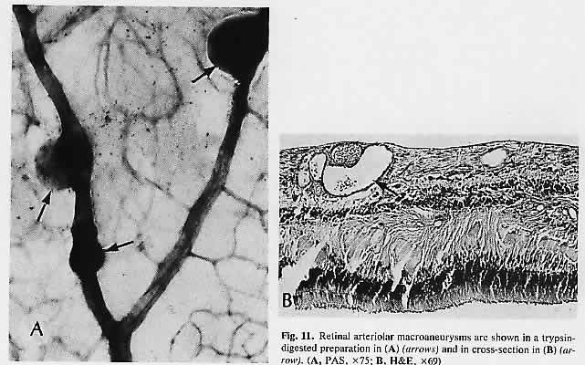
macular holes. Am J Ophthalmol 108:360, 1989

129. Craythorn JM, Swartz M, Creel DJ: Clofazimine-induced bull's eye retinopathy. Retina 6:50, 1986

130. Ramsey MS, Fine BS: Chloroquine toxicity in the human eye: Histopathologic observations by electron microscopy. Am J Ophthalmol 73:229, 1972

131. Miller FS III, Bunt-Milam AH, Kalina RE: Clinical-ultrastructural study of thioridazine retinopathy. Ophthalmology 89:1478, 1982

132. Kaiser-Kupfer MI, Kupfer C, Rodrigues MM: Tamoxifen retinopathy: A clinicopathologic report. Ophthalmology 88:89, 1981

133. Brown GC, Shields JA, Sanborn G et al: Radiation retinopathy. Ophthalmology 89: 1494, 1982

134. Midena E, Segato T, Piermarocchi S et al: Retinopathy following radiation therapy of paranasal sinus and nasopharyngeal carcinoma. Retina 7: 142, 1987

135. Boozalis GT, Schachat AP, Green WR: Subretinal neovascularization from the retina in radiation retinopathy. Retina 7:156, 1987

136. Tso MOM, Wallow IHL, Powell JO, Zimmerman LE: Recovery of the rod and cone cells after photic injury. Trans Am Acad Ophthalmol Otolaryngol 76: 1247, 1972

137. McDonald HR, Irvine AR: Light-induced maculopathy from the operating microscope in extracapsular cataract extraction and intraocular lens implantation. Ophthalmology 90:945, 1983

138. Kuhn F, Morris R, Massey M: Photic retinal injury from endoillumination during vitrectomy. Am J Ophthalmol 111:42, 1991

139. Michels M, Sternberg P Jr: Operating microscope-induced retinal phototoxicity: Pathophysiology, clinical manifestations and prevention. Surv Ophthalmol 34:237, 1990

140. Rinkoff J, Machemer R, Hida T, Chandler D: Temperature-dependent light damage to the retina. Am J Ophthalmol 102:452, 1986

141. Bastek JV, Fops RY, Heckenlively J: Traumatic pigmentary retinopathy. Am J Ophthalmol 92:621, 1981

142. Aguilar JP, Green WR: Choroidal rupture: A histopathologic study of 47 cases. Retina 4:269, 1984

143. Gaynon MW, Koh K, Marmor MF, Frankel LR: Retinal folds in the shaken baby syndrome. Am J Ophthalmol 106:423, 1988

144. Elner SG, Elner VM, Arnall M, Albert DM: Ocular and associated systemic findings in suspected child abuse: A necropsy study. Arch Ophthalmol 108:1094, 1990

145. Massicotte SJ, Folberg R, Torczynski E et al: Vitreoretinal traction and perimacular retinal folds in the eyes of deliberately traumatized children. Ophthalmology 98:1124, 1991

146. Maumenee IH: Vitreoretinal degeneration as a sign of generalized connective tissue diseases. Am J Ophthalmol 88:432, 1979

147. Nasr YG, Cherfan GM, Michels RG, Wilkinson CP: Goldmann-Favre Maculopathy. Retina 10:178, 1990

148. Brockhurst RJ, Albert DM, Zakov ZN: Pathologic findings in familial exudative vitreoretinopathy. Arch Ophthalmol 99:2143, 1981

149. Boldrey EE, Egbert P, Gass JDM, Friberg T: The histopathology of familial exudative vitreoretinopathy: A report of two cases. Arch Ophthalmol 103:238, 1985

150. Blair NP, Albert DM, Liberfarb RM, Hirose T: Hereditary progressive arthro-ophthalmopathy of Stickler. Am J Ophthalmol 88:876, 1979

151. Straatsma BR, Zeegen PE, Fops RY et al: Lattice degeneration of the retina: XXX Edward Jackson memorial lecture. Am J Ophthalmol 77:619, 1974

152. Robinson MR, Streeten BW: The surface morphology of retinal breaks and lattice retinal degeneration: A scanning electron microscopic study. Ophthalmology 93:237, 1986

153. Parelhoff ES, Wood WJ, Green WR, Kenyon KR: Radial perivascular lattice degeneration of the retina. Ann Ophthalmol 12:25, 1980

154. Wilson DJ, Weleber RC, Green WR: Ocular clinicopathologic study of gyrate atrophy. Am J Ophthalmol 111:24, 1991

155. Lopez PF, Maumenee IH, de la Cruz Z, Green WR: Autosomal-dominant fundus flavimaculatus: Clinicopathologic correlation. Ophthalmology 97:798, 1990

156. Eagle RC Jr, Lucier AC, Bernardino VB Jr, Yanoff M: Retinal pigment epithelial abnormalities in fundus flavimaculatus: A light and electron microscopic study. Ophthalmology 87: 1189, 1980

157. Steinmetz RL, Garner A, Maguire JI, Bird AC: Histopathology of incipient fundus flavimaculatus, Ophthalmology 98:953, 1991

158. McDonnell PJ, Kivlin JD, Maumenee IH, Green WR: Fundus flavimaculatus without maculopathy: A clinicopathologic study. Ophthalmology 93:116, 1986

159. Mohler CW, Fine SL: Long-term evaluation of patients with Best's vitelliform dystrophy. Ophthalmology 88:688, 1981

160. Frangieh GT, Green WR, Fine SL: A histopathologic study of Best's macular dystrophy. Arch Ophthalmol 100:1115, 1982

161. O'Gorman S, Flaherty WA, Fishman GA, Berson EL: Histopathologic findings in Best's vitelliform macular dystrophy. Arch Ophthalmol 106: 1261, 1988

162. Weingeist TA, Kobrin JL, Watzke RC: Histopathology of Best's macular dystrophy. Arch Ophthalmol 100:1108, 1982

163. Patrinely JR, Lewis RA, Font RL: Foveomacular vitelliform dystrophy, adult type: A clinicopathologic study including electron microscopic observations. Ophthalmology 92:1712, 1985

164. Jaffe G J, Schatz H: Histopathologic features of adult-on-set foveomacular pigment epithelial dystrophy. Arch Ophthalmol 106:958, 1988

165. Sneed SR, Sieving PA: Fenestrated sheen macular dystrophy. Am J Ophthalmol 112:1, 1991

166. Pagon RA: Retinitis pigmentosa. Surv Ophthalmol 33:137, 1988

167. Bastek JV, Siegel EB, Straatsma BR, Fops RY: Chorioretinal juncture: Pigmentary patterns of the peripheral fundus. Ophthalmology 89: 1455, 1982

168. Szamier RB, Betson EL: Retinal histopathology of a carrier of X-chromosome-linked retinitis pigmentosa. Ophthalmology 92:271, 1985

169. Bunt-Milam AH, Kalina RE, Pagon RA: Clinical-ultra-structural study of a retinal dystrophy. Invest Ophthalmol Vis Sci 24:458, 1983

170. Meyer KT, Heckenlively JR, Spitznas M, Fops RY: Dominant retinitis pigmentosa: A clinicopathologic correlation. Ophthalmology 89:1414, 1982

171. Gartner S, Henkind P: Pathology of retinitis pigmentosa. Ophthalmology 89: 1425, 1982

172. Rodrigues MM, Bardenstein D, Wiggert B et al: Retinitis pigmentosa with segmental massive retinal gliosis: An immunohistochemical, biochemical, and ultrastructural study. Ophthalmology 94: 180, 1987

173. Albert DM, Pruett RC, Craft JL: Transmission electron microscopic observations of vitreous abnormalities in retinitis pigmentosa. Am J Ophthalmol 101:665, 1986

174. Hsieh RC, Fine BS, Lyons JS: Patterned dystrophies of the retinal pigment epithelium. Arch Ophthalmol 95:429, 1977

175. de Jong PTVM, Delleman JW: Pigment epithelial pattern dystrophy: Four different manifestations in a family, Arch Ophthalmol 100:1416, 1982

176. Wilson DJ, Weleber RG, Klein ML et al: Bietti's crystalline dystrophy: A clinicopathologic correlative study. Arch Ophthalmol 107:213, 1989

177. Harrison RJ, Acheson RR, Dean-Hart JC: Bietti's tapetoretinal degeneration with marginal corneal dystrophy (crystalline retinopathy): Case report. Br J Ophthalmol 71:220, 1987

178. Small KW, Letson R, Scheinman J: Ocular findings in primary hyperoxaluria. Arch Ophthalmol 108:89, 1990

179. Novak MA, Roth AS, Levine MR: Calcium oxalate retinopathy associated with methoxyflurane abuse. Retina 8:230, 1988

180. Wells CG, Johnson RJ, Qingli Let al: Retinal oxalosis: A clinicopathologic report. Arch Ophthalmol 107: 1638, 1989

181. Dreyer R, Green WR: The pathology of angioid streaks: A study of twenty-one cases. Trans Pa Acad Ophthalmol Otolaryngol 31: 158, 1978

182. Cibis GW, Harris DJ, Chapman AL, Tripathi RC: Mucolipidosis I. Arch Ophthalmol 101:933, 1983

183. Matthews JD, Weiter JJ, Kolodny EH: Macular halos associated with Niemann-Pick type B disease. Ophthalmology 93:933, 1986

184. Arnold AC, Pepose JS, Hepler RS, Foos RY: Retinal periphlebitis and retinitis in multiple sclerosis: k Pathologic characteristics. Ophthalmology 91:255, 1984

185. Baker RH, Trautman JC, Younge BR et al: Late juvenile-onset Krabbe's disease. Ophthalmology 97:1176, 1990

186. Nork TM, Ghobrial MW, Peyman GA, Tso MOM: Massive retinal gliosis: A reactive proliferation of Müller cells. Arch Ophthalmol 104: 1383, 1986

187. Leys AM, Van Eyck LM, Nuttin BJ et al: Metastatic carcinoma to the retina. Arch Ophthalmol 108:1448, 1990

188. Kincaid MC, Green WR: Ocular and orbital involvement in leukemia. Surv Ophthalmol 27:211, 1983

189. Gass JDM, Weleber RG, Johnson DR: Non-Hodgkin's lymphoma causing fundus picture simulating fundus flavimaculatus. Retina 7:209, 1987

190. Foos RY, Simons KB: Vitreous in lattice degeneration of retina. Ophthalmology 91:452, 1984

191. Meyer E, Kurz GH: Retinal pits: A study of pathologic findings in two cases. Arch Ophthalmol 70:640, 1963

192. Smiddy WE, Green WR: Retinal dialysis: Pathology and pathogenesis. Retina 2:94, 1982

193. Kampik A, Kenyon KR, Michels RG et al: Epiretinal and vitreous membranes: Comparative study of 56 cases. Arch Ophthalmol 99: 1445, 1981

194. Schwartz D, de la Cruz Z, Green WR, Michels RG: Proliferative vitreoretinopathy: Ultrastructural study of 20 retroretinal membranes removed by vitreous surgery. Retina 8:275, 1988

195. Wilson DJ, Green WR: Histopathologic study of the effect of retinal detachment surgery on 49 eyes obtained post mortem. Am J Ophthalmol 103:167, 1987

196. Barr CC: The histopathology of successful retinal reattachment. Retina 10: 189, 1990

Back to Top