|
|
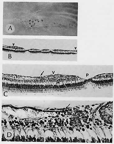
| Fig. 31. Retinal pits. A. Macroscopic appearance of retinal pits arranged along a thickened and sheathed vessel. The optic disc is to the left. B. Several retinal pits are visible in the vicinity of sclerotic blood vessels (v). C. Internal limiting membrane ends abruptly (arrow) as it approaches the sclerotic vessel (v) and the associated retinal defect or pit (P). D. Higher magnification of (C) to show the termination of the internal limiting membrane (arrow) and the proliferation of the glial membrane onto its inner surface (preretinal glial membrane). (A, macroscopic; B, H&E, ×36; C, Wilderls reticulin stain, ×90; D, Wilder's reticulin stain, ×325) |