|
|
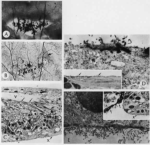
| Fig. 26. Lattice degeneration of the retina. A. Heavily pigmented ovoid lesion in the retinal periphery parallels the ora serrata. B. Retinal digest of the lesion in (A). Note the increased density of the vitreous attachments, particularly anteriorly (arrow). C. Section of the anterior part of the lesion. The vitreous base (o) is contracted anteriorly. The glial cells (g) proliferating within the inner retinal layers have grown along the retracted vitreal surface. Arrows indicate the side of the delicate original internal limiting membrane of the peripheral retina (poorly visualized by light microscopy). Note the loss of the photoreceptors along the external limiting membrane (x). D. Electron micrograph shows the glial cells, their characteristic dense attachments, and their villous projections. Inset. Light micrograph of glia proliferating along the “opened” inner surface of the lesion. Note the formation of a surface “membrane” beyond which project delicate villi (arrows). E, inset. Electron micrograph illustrates the terminal barlike arrangement of the external glial (i.e., Müller) cells. Glial microvilli (mv) project into the subretinal space. Light micrograph of the outer retinal surface at approximately midlesion. There is loss of the photoreceptors, and the external limiting membrane (x) of the retina is interrupted by ingrowing proliferating pigment epithelial cells (arrows). (A, macroscopic; B, trypsin digest; C, 1.5 μm section, toluidine blue, ×300: D, × 12,378; inset, 1.5 μm section, toluidine blue, × 1260; E, ×532; inset, 1.5 μm section, toluidine blue, ×525) |