|
|
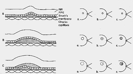
| Fig. 17. Schematic diagram correlates the histologic changes on the left (A, B and C) with (a) the fundus changes, (b) the very early fluorescein stage, and (c) the late fluorescein stage. A. Simple small detachment of the retinal pigment epithelium (RPE). (NR, neural retina) B. Simple large detachment of the RPE. C. Small detachment of the RPE with overlying serous detachment of the neural retina (i.e., idiopathic central serous choroidopathy). |