|
|
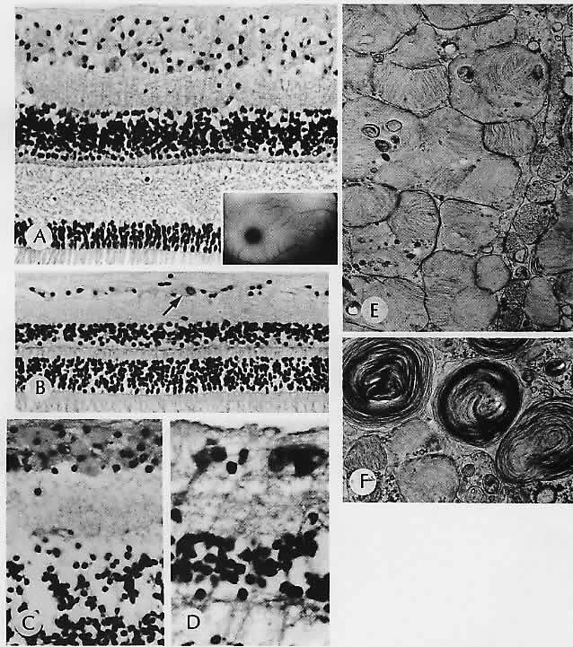
| Fig. 28. Tay-Sachs disease. A. Macular area shows the swollen cytoplasm of the ganglion cells. Inset. Typical cherry-red spot fundus appearance. B. Peripheral retinal of the same patient shows less involvement, with the cytoplasm of one ganglion cell (arrow) especially swollen. C. Macular area of another patient shows marked involvement of the ganglion cells, which contain PAS-positive material. D. Peripheral retinal ganglion cells are also involved. E. By electron microscopy of the eye shown in C and D, ganglion cells containing whorled laminated bodies that accumulate to fill the cells can be seen. The accumulated ganglioside produces opacification of the retina wherever there are ganglion cells (most prominent in foveomacular area). F. Another area of ganglion cells shows a more dense lamination that may be seen in the accumulating substance. (A, PAS, × 176; Inset, fundus; B, PAS, × 176; C, H&E, ×252; D, PAS, ×44; E, ×22,000; F, ×20,000) |