|
|
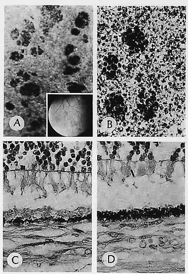
| Fig. 1. Grouped pigmentation. A. Macroscopic appearance of pigmented plaques. Inset. Fundus shows a typical grouped pigmentation. B. Flat preparation of retinal pigment epithelium (RPE) showing a greater concentration of pigment granules in cells corresponding to grouped pigmentation. C. RPE in a normal area adjacent to the plaque. D. RPE in the area of the plaque shows a greater concentration of pigment granules. The overlying retina is detached artifactitiously. (A, macroscopic; inset, fundus; B, flat preparation, H&E, ×256; C, H&E, ×640; D, H&E, ×640) |