|
|
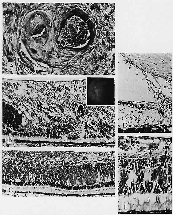
| Fig. 8. Retinal hemorrhagic infarction. A. Central retinal vein is occluded and recanalized. The artery is identified by an internal elastic lamina (arrow). B. Hemorrhagic infarction causing disorganization of the retina in a patient with polycythemia vera and central retinal vein occlusion. Inset. Fundus following central retinal vein occlusion in an otherwise healthy 18-year-old girl; no cause for the occlusion was found. C. Sheets of blood in the inner retinal layers produce the characteristic fundus appearance. Note the preservation of the photoreceptors. D. Rubeosis iridis is present in the same patient with polycythemia vera. E. Pigment (arrows) is hemosiderin in a long-standing occlusion. (A, elastica, ×136; B, H&E, ×69; inset, fundus; C, H&E, ×69; D, H&E, × 101; E, Prussian blue, × 176) |