Chapter 15
Lens
NORMAN J. KLEIMAN and BASIL V. WORGUL
Main Menu   Table Of Contents

Search

EMBRYOLOGY OF THE LENS
THE ADULT LENS
CONGENITAL ANOMALIES OF THE LENS
CONGENITAL ANOMALIES INVOLVING TRANSPARENCY
CONGENITAL LENTICULAR CATARACT
MOLECULAR GENETICS OF CATARACT
CONCLUSION
REFERENCES

The present understanding of lens anatomy can be traced to the very beginnings of microscopy. The lens has the distinction of being among the first tissues to be examined microscopically; van Leeuwenhoek himself described the fiber organization of lenses from cow,1,2 ox,1,2 hare, rabbit, cod, bird,2 and, most significant, human3,4 eyes. Despite those pioneering observations, however, subsequent studies concentrated, for the most part, on cataracts and cataract surgery. Nevertheless, by the second half of the nineteenth century, numerous lens studies had been reported,5 which led to renewed interest in lens microanatomy.* These early investigations have set the stage for present-day studies of lens biology and pathology.
* In an 1899 treatise on the lens, Stricker5 listed 3433 references spanning the years 1532 to 1898; of these, 2790 were published after 1850.

The lens is not simply a “bag of protein,” as one popular reductionist metaphor opined, but is instead a highly organized cytosystem exquisitely designed to carry out its sole function (i.e., to refract incoming light upon the retina). This singular role necessitated the evolution of unique anatomic characteristics, an appreciation of which can be best acquired by first understanding the nuances of the embryogenesis of the lens.

Back to Top
EMBRYOLOGY OF THE LENS
Morphologically, the development of the human lens begins at the 4-mm embryonic stage (Table 1). A section through the area of the forebrain (Fig. 1) reveals that lateral outpocketings in the totally internalized neural ectoderm, called the optic vesicles, come in close apposition to the surface ectoderm. The union of these tissues initiates the formation of the lens placode (Fig. 2), an area of columnarization or “palisading”6 of the monolayered surface ectoderm in the region adjoining the neural ectoderm. This induction is the first of a series that governs the formation and growth of the lens.7,8 The precise nature of the inductive agent(s) is unknown, but it is believed that a multistep process occurs that does not require direct contact between cell types.9–13 Although the process is still not well understood, it is likely that a number of inducing agents and/or growth factors are involved.14–19


TABLE ONE. Important Stages in the Development of the Human Lens


Description of Developmental EventEmbryo LengthApproximate Age
Lens thickening appears4 mm2 weeks
Lens pit formed5 mm2 weeks+
Lens vesicle formed7 mm4 weeks
Lens vesicle separated10 mm4 weeks+
Primary lens fibers beginning12 mm5 weeks
Lens capsule, deep layer, forming13 mm5 weeks+
Secondary lens fibers beginning25 mm7 weeks
Y sutures recognizable35 mm8 weeks+
Vascular capsule fully developed40 mm9 weeks
Zonular lamella of capsule forming70 mm3 months
Retrogression of posterior vascular capsule begins110 mm4 months
Retrogression of capsulopupillary vessels110 mm4 months
Retrogression of pupillary membrane250 mm6 months
Retrogression of vascular capsule complete Birth
Adult nucleus and cortex form Throughout the remainder of life


Fig. 1. A section through the area of the forebrain of a 4-mm human embryo. Lateral outpocketings of the completely internalized neural ectoderm (NE) form the optic vesicles (OV). The surface ectoderm (SE) is still, at this stage, totally undifferentiated. Carnegie embryo 6097. (Smelser GK: Embryology and morphology of the lens. Invest Ophthalmol 4:398, 1965)

Fig. 2. A section through the newly forming lens placode (LP) of a 4.5-mm human embryo. The surface ectoderm has been induced to elongate by the neural ectoderm (shown here already beginning to differentiate into the presumptive retina). The lens pit (fovea lentis) is barely discernible as a slight indentation below center in the surface of the placode (arrow). Carnegie embryo 8119. (Smelser GK: Embryology and morphology of the lens. Invest Ophthalmol 4:398, 1965)

Although the lens placode appears stratified, it remains a single layer of columnar cells. The apparent multilayering is caused by the staggered position of nuclei in adjacent cells. The variable location of nuclei along the length of their respective cells may be due to a cell cycle-dependent migration of the nuclei of the lens placode cells. The work of Zwaan and his associates has shown that, following deoxyribonucleic acid (DNA) synthesis, the nuclei located basally in the lens placode migrate to the cell apices prior to mitosis.20 Following cell division, the nuclei of the resulting daughter cells return to the basal location.

A small depression called the fovea lentis (lens pit) appears below the center in the lens placode (see Fig. 2). This will be the focus of the invagination of the surface ectoderm that forms the lens vesicle. As the crowding of the lens placode cells increases, the lens pit begins to deepen (Fig. 3). Because the lens pit was initially off-center, invagination of the surface ectoderm occurs asymmetrically. Concurrently, the optic vesicle invaginates as it undergoes conversion into the double-layered optic cup. Flat cells appear on the outer surface of the lens plate, predominantly in the area of the lens pit (see Fig. 3). At this stage (5 mm), patches of similar cells may be found elsewhere over the embryo. These epitrichial cells are the beginning (periderm) of the simple squamous epithelium that is destined to cover the entire embryo. Although they first accumulate in the concavity of the lens pit, the epitrichial cells begin to disappear by the 7-mm embryonic stage (4 weeks). Pari passu with their elimination, the lens pit deepens considerably, and the opening to the surface ectoderm, the lens stalk, constricts to the point where the lens pit may now be referred to as the lens vesicle. Following complete closure, the vesicle begins to separate from the presumptive cornea at approximately the 9-mm stage (Fig. 4). Phagocytic macrophages, whose role is to remove dead cells and debris, have been detected in the interspace between the vesicle and ectoderm as well as in the lens lumen.21 At this time, the vesicle is spherical and approximately 0.2 mm in diameter. Its wall is about 40 μm thick.

Fig. 3. At the 5-mm stage, the lens placode is clearly beginning to invaginate asymmetrically, and epitrichial cells appear in the deepening lens cup. Carnegie embryo 7394. (Smelser GK: Embryology and morphology of the lens. Invest Ophthalmol 4:398, 1965)

Fig. 4. A section through the lens vesicle of a 10-mm embryo. Complete closure of the vesicle has occurred, and separation from the surface ectoderm is commencing. The posteriorly situated cells of the vesicle are already beginning to elongate into the primary fibers. Note the presence of several epitrichial cells trapped in the lumen of the vesicle. Carnegie embryo 6517. (Smelser GK: Embryology and morphology of the lens. Invest Ophthalmol 4:398, 1965)

Because the vesicle represents an invagination of surface ectoderm, the basal portions of its cells are oriented externally, whereas the apices of the cells face the lumen. Because of this relationship, components of the basal lamina (secreted by the lens cells throughout life) are deposited externally and encase the tissue in a membranous envelope, later to be called the lens capsule. Electron microscopy reveals that capsule formation begins at about the 10-mm stage,22 just after the vesicle and surface separate. Capsule formation is initiated by the appearance of a second basal lamina deposited in a discontinuous manner23 beneath the original basement membrane of the lens vesicle cells. (The readily discernible periodic acid-Schiff (PAS) stain positive structure familiar to the light microscopist will not appear until approximately the 13-mm stage.)

Concomitant with the beginnings of capsule formation is the appearance of the first vessels of the tunica vasculosa lentis (Fig. 5), a vascular net that will completely encompass the lens by the 40-mm stage. It begins as small capillaries emanating from the hyaloid artery and covering only the posterior portion of the lens (posterior vascular capsule). From the posterior vascular capsule, palisade vessels, visible at the 12-mm stage, proceed anteriorly around the equator where they anastomose with choroidal veins (capsulopupillary portion). From these arise the vessels that will cover the anterior lens surface, in conjunction with branches of the long ciliary arteries, to form the so-called pupillary membrane (anterior vascular capsule). Fully developed by the 40-mm stage, the vascular capsule will completely disappear before or soon after birth. Phagocytic macrophages have been implicated in its destruction.24 (Not infrequently, however, a hyaloid remnant or a small portion of the pupillary membrane persists in otherwise normal eyes.

Fig. 5. The tunica vasculosa lentis, showing its major pathways and associations with other vasculature of the eye in a human fetus 85 mm in length. Note the hyaloid artery (a) and long posterior ciliary artery (b).

Inside the lens, the 10-mm stage also heralds a number of differentiating activities. At the biochemical level, expression and synthesis of various lens structural proteins, the crystallins25 can be detected.26–30 In the mammalian lens, the crystallins are divided into three groups, termed α, ß, and Γ on the basis of their chromatographic elution profiles, acid sequence, and immunologic cross-reactivities. Typically, α crystallin transcripts are expressed first, followed closely by ß crystallin. In contrast, Γ crystallin expression and synthesis is closely linked to the later elongation phase of the terminally differentiating fiber cell.

Morphologically, the posteriorly situated cells of the vesicle (i.e., those nearest the presumptive retina) begin to elongate (see Fig. 4). As they do, they encroach upon the lumen. Because those cells that are located at the posterior pole elongate first, they advance ahead of the more equatorially placed cells. As a result, the cavity of the lens vesicle becomes increasingly more crescentic and smaller (Fig. 6). The elongation of the posteriorly placed lens vesicle cells represents another step in the string of inductions controlling lens formation and development. In a classic series of experiments, Coulombre and Coulombre observed that if the lens vesicle is rotated 180° during the elongation process, the induction pattern can be reversed: the elongation of the cells, which are now located anteriorly, terminates, whereas the original anterior cells, those now nearest the retina, lengthen.31 This interaction between retina and lens tissue persists into adulthood.32–35

Fig. 6. A section through the eye of a 15-mm human embryo showing the elongation of the primary fibers as they encroach upon the lumen. The crescentic lumen of the lens vesicle still contains some epitrichial cells. Note the anterior migration of the nuclei of the primary fibers, producing an anuclear zone in the posterior portion of the lens and a bow pattern at the lens equator. Carnegie embryo 6520. (Courtesy of Victoria Ozanics, Columbia University)

The posteriorly located vesicle cells elongate until, at approximately the 16-mm embryonic stage, the lumen is completely obliterated. The nuclei of these transformed epithelial cells (now referred to as the primary fibers of the lens) assume a position more anterior than posterior along the length of the fibers. At this stage, the cells of the anterior portion of the original lens vesicle remain morphologically unchanged. Hereafter, this monolayer of cells will be referred to as the lens epithelium, and upon it all future growth of the lens depends. Except for the progressive denucleation of the innermost cells of the primary fiber mass, and the reversion of the lens epithelium to squat cuboidal cells (Fig. 7), little change occurs in the lens as the embryo grows from the 16-mm to the 25-mm stage. At the 25-mm stage, the lens epithelium displays a tremendous burst of proliferative activity, and the epithelial cells in the area of the equator begin to differentiate into fiber cells (Fig. 8). It is believed that a variety of growth factors,15 including the insulinlike growth factors34 and fibroblast growth factor,16 as well as various cellular proto-oncogenes,36,37 play essential roles in this process. The change in wet weight of the embryonic human lens from 8 to 12 weeks' gestation is shown in Figure 9.

Fig. 7. A section through the eye of a human embryo at approximately the 18-mm stage. The lumen of the lens vesicle has been completely eliminated by the forward progression of the primary fibers. The anterior ends (the apices of the fibers) adjoin the apices of the epithelial cells. The epithelial cells appear more cuboidal than at previous stages, and the innermost cells of the primary fiber mass are beginning to denucleate. Carnegie embryo 5537. (Courtesy of Victoria Ozanics, Columbia University)

Fig. 8. When the embryo has reached approximately 25 mm in length, the lens epithelium undergoes extensive proliferative activity. The newly formed cells begin to internalize at the equator and elongate into secondary fibers. Fibrogenesis is marked by the reorientation of the cells in the anteroposterior direction. The basal portions of the cells elongate posteriorly, while the apices of the cells proceed anteriorly. Continued deposition of new cells at the equator causes previously deposited cells to be buried in the lens substance. A continual accretion of these cells at the equator is the basis of the continued growth of the lens throughout life.

Fig. 9. At about the 25-mm stage (7–8 weeks' gestation) a tremendous burst of proliferative activity begins in the human lens, signaled by the terminal division of equatorial epithelial cells into mature lens fiber cells and a concomitant increase in lens weight. This process continues throughout life, albeit at a much slower rate following maturity. (Courtesy of Dr. Patricia Burke, Columbia University)

Following terminal cell division in the preequatorial area, just anterior to the lens equator, lens epithelial cells move into the equatorial region, where they rotate 90° and elongate (see Fig. 8). As they lengthen, the anterior ends pass in front of, and the posterior ends behind, the primary fiber cell mass, thereby completely surrounding and internalizing it. As new fibers are added around the primary fibers (secondary fibergenesis), each new layer grows longer than its predecessor. A secondary fiber cell, however, is never long enough to reach both the anterior and posterior poles of the lens. Instead of converging at a single point, within each layer or growth shell, the cell termini abut in a manner producing visual patterns known as lens sutures.38

The first recognizable suture is Y-shaped (at about the 35-mm stage) and is present in layers laid down from early embryogenesis to birth (Fig. 10A). Lens suture patterns become increasingly more complex with each succeeding growth shell. For example, young adults typically have nine suture lines arrayed in a starburst pattern (Fig. 10B), whereas older individuals have a more complex and multibranched sutural arrangement (Fig. 10C).39–41

Fig. 10. A. Diagrammatic representation of the anterior (left) and posterior (right) suture patterns of a newborn human lens. Note the offsetting anterior and posterior suture patterns as a consequence of opposite curvature of each end of the fiber cell. B. Scalar diagrammatic representation of the anterior (left) and posterior (right) suture patterns of a young adult human lens. Note the starburst pattern with nine equally spaced radial spokes. C. Scalar diagrammatic representation of the anterior (left) and posterior (right) suture patterns of an aged human lens. The number of spokes has increased, and the anterior and posterior suture patterns have become asymmetric with respect to each other. (Courtesy of Dr. Jer Kuszak, Rush Presbyterian St. Lukes Medical Center)

As the lens increases in size, it becomes apparent that each layer of fiber cells will overlap equatorially but not necessarily in the anterior and posterior regions. This results in the equatorial diameter becoming significantly greater than the diameter along the anteroposterior axis (Fig. 11). Functionally, this manner of fiber cell deposition provides for the transformation of the lens from a spherical shape during embryogenesis to a more refractively favorable biconvex lentoid shape in the adult eye.

Fig. 11. The relationship between the anteroposterior diameter and the equatorial diameter as the function of secondary fiber deposition during intrauterine life. Before secondary fibrogenesis begins (at approximately 25 mm), the anteroposterior diameter can actually exceed the equatorial diameter. However, once secondary fiber formation is initiated, equatorial growth predominates. (Mann I: The Development of the Human Eye. London, British Medical Association 1964)

Back to Top
THE ADULT LENS
The internal anatomy of the adult human lens is shown in Figure 12. From birth, the lens undergoes little qualitative change, although quantitatively its volume, weight, and size continue to increase throughout life (Table 2). Male lenses are heavier than female lenses of patients the same age,42 although the rate of increase in weight is approximately the same. The lens becomes markedly ellipsoid during the early portion of postnatal life, but after the 20th year the rate of anteroposterior growth exceeds the rate of equatorial growth, thus causing the lens to become rounder with increasing age.43–46 The length of the anteroposterior axis increases at a rate of about 0.023 mm/year throughout life44,45,47,48 (Fig. 13), whereas the equatorial axis increases from about 6 to 9 mm during the first 15 years of life, changing little thereafter. A large variation is observed in the radius of curvature of the anterior and posterior surfaces of the adult lens. The average radius of curvature of the anterior surface is approximately 10 mm (8–14 mm), and that of the posterior surface is about 6 mm (4.2–7.5 mm).45,49,50 The radius of curvature becomes increasingly smaller with age (Fig. 14).44–46

Fig. 12. The internal anatomy of the human lens; the capsule (C) alone or the capsule and epithelium together (CE) are peeled back from the anterior surface. A wedge of the lens has been removed to emphasize internal anatomy. AC, adult cortex; AN, adult nucleus; AS, anterior suture; CZ, central zone; EN, embryonic nucleus; FN, fetal nucleus; GZ, germinative zone; LB, lens bow; LE, lens epithelium; MR, meridional rows; PS, posterior suture; TZ, transitional zone.


TABLE TWO. The Change in Wet Weight, Volume, and Front Diameter of the Lens with Age


AgeWeight (mg)Volume (mm)Front Diameter (mm)
1–3 months92.8906.31
10–11 months124.5120.47.46
1–10 years146.8142.58.06
10–20 years152.8148.38.47
20–30 years172162.98.67
30–40 years190.3177.38.97
40–50 years202.4188.19.09
50–60 years223.3205.49.44
60–70 years230.12139.49
70–80 years237.1218.39.64
80–90 years258.1238.79.62


Fig. 13. The increase in the anteroposterior diameter of the lens as a function of age, determined ultrasonically (Δ) and by use of the slit-image camera (O) (Compiled from multiple sources.45,47,110)

Fig. 14. The change in the curvature of the anterior and posterior surfaces of the lens with increasing age, as determined ultrasonically (Δ) and by use of the slit-image camera (·, O). (Compiled from multiple sources.45,47,110)

THE LENS EPITHELIUM

Like the skin (its embryonic sibling), the lens grows throughout life, albeit at a rate that decreases with age. From the start of secondary fiber formation at the 25-mm embryonic stage, all lens growth becomes dependent on the mitotic activity of the lens epithelium.

The epithelium, a polarized monolayer of cells occupying the anterior surface of the adult lens, is derived from the original lens vesicle cells that did not differentiate into primary fibers. Estimates of cell number range from 350,00051 to 100,000,00052 in the mature human lens. The epithelium does not extend to the posterior side of the lens because the cells originally located there have elongated into primary fibers. Because it adheres tenaciously to the overlying capsule, it is possible to remove the entire population of epithelial cells. Once isolated, the intact epithelium can be mounted on a slide and subjected to qualitative and quantitative histocytologic analyses. Techniques such as autoradiography53 and histochemistry of lens epithelial whole-mount preparations have revealed that epithelium is organized into several distinct subpopulations, distinguished primarily by their proliferative kinetics,54,55 although there are some morphologic differences among them.

The cells located centrally on such preparations (i.e., those that would occupy the anterior pole of the lens in sagittal section) are polygonal with round nuclei when viewed on a whole mount and squamous with elliptically shaped nuclei when viewed in section (Fig. 15). They are flat, with a considerable range of cell diameters, an average height of approximately 6 μm, and a width of 13 μm,56 and they are endowed with all of the organelles typical of epithelial cells. Cells located more posteriorly tend to be smaller, and, consequently, cell density increases toward the equator.51 In the adult male, the age-adjusted mean cell density is about 5000 cells/mm2, and it is about 5800 cells/mm2 in females.57 The average cell density declines with age. In epithelium from cataractous lenses, both values are lower.58,59

Fig. 15. Photomicrographs showing the various regions of the human lens epithelium in sagittal section and on a whole-mount preparation. The 1-μm sagittal sections are stained by the Feulgen reaction, thus revealing only the nuclei of the cells. When viewed sagittally (a), the central zone cells are quite flat, causing the great distance seen between adjacent nuclei on a whole mount (b). In the germinative zone (c), the cells appear more cuboidal, and the nuclei are positioned nearer to the apices of the cells. Nuclei of the superficial lens cortical fibers are seen subjacent to the cells of the germinative zone (arrows). Viewed on a whole mount, this region (d) is marked by more closely packed nuclei and the presence of mitotic cells. (The arrow points to a metaphase figure.) At the equator, the cells are more columnar, and the nuclei are still located apically (e). As the cells begin to reorient in the anteroposterior direction, the nuclei maintain a more anterior (apical) position along the length of the elongating cells, thereby producing the bow pattern. The equatorial line (E) is an area of densely packed nuclei at the base of the meridional rows (MR) and is best seen on a whole mount (f). The sagittal section (e) reveals that this is a region where the nuclei of ajacent cells of the meridional rows (MR) are superimposed so that the nuclei appear to be compacted.

Lens epithelial cells contain defined apical, lateral, and basal membranes; their apical surface interfaces with the apical membranes of newly formed fiber cells.60 Considerable endocytosis occurs, presumably reflecting transport of nutrients between epithelial and fiber cells.61 It is likely that no tight junctions62 and very few gap junctions are found in this region. In contrast, at the lateral membranes, characterized by complex interdigitations, are found desmosomes (macula adherens) and plentiful numbers of gap junctions.63

The central zone cells do not normally undergo mitosis. In fact, the mitotic index for cataractous epithelia from aged individuals has been reported to be as low as 0.0004%,64 although values as high as 0.008% for both normal and cataractous lenses have been reported.51 However, experimental animal studies have shown that the central zone cells can respond to myriad mitotic stimuli, which include injury,54,55,65–72 explantation,54,55,73 hormones,74–76 and experimental uveitis.77–80 Because they are relatively easy to manipulate, these normally noncycling cells constitute a model system of particular interest to investigators studying the processes of wound healing and the control of the cell cycle.54,55 The cells of the anterior pole have also gained the attention of gerontologists, a fact that is not surprising because it is believed that these cells remain centrally located without migrating out of the region. They are, as Muggleton-Harris states, “a true collection of aged cells.”80

Biochemically, lens epithelial cells are relatively unremarkable. A variety of cytoskeletal proteins and components, including actin, vimentin, myosin, microtubules, spectrin, and intermediate filaments, have been reported. An unusual feature is the existence of an extensive actin-myosin network forming a polygonal array.81–83

Unlike the cells of the central zone, the germinative zone cells, located peripherally on a whole-mount preparation, actively divide (see Fig. 15). In sagittal section (see Fig. 15), where they are located pre-equatorially, the cells are smaller and more cuboidal and contain more mitochondria than cells of the central zone.84 The cells are joined by a large number of lateral interdigitations (Fig. 16).

Fig. 16. A transmission electron micrograph of cells near the germinative zone of an adult human lens epithelium. The cells contain a number of organelles typical of epithelia. The lateral borders between the cells are highly convoluted and are marked by numerous desmosomes (arrows) and junctional complexes located near the apices. C, capsule; F, fiber; N, nucleus.

Following a terminal cell division, one or both of the daughter cells of the germinative zone are displaced into the adjacent transitional zone where differentiation is initiated. Mitosis is rarely found here, although it may occur in some pathologic conditions, such as intraocular inflammation.79

The cytoplasm of the cells in the transitional zone shows a large increase of ribosomes (polysomes) and an increase in the number of multivesicular bodies. Microtubules are also pronounced in these cells. Closer to the equator, the cells become more columnar and assume a pyramidal shape, the basal portion being wider than the apex.

LENS FIBERS

As the transitional zone cells continue to elongate, they queue up in columns known as the meridional rows, a precise register seen best on whole-mount preparations (see Fig. 15). In sagittal section, this corresponds to the region where the cells elongate and begin to internalize in the upper strata of the lens (see Fig. 15). They elongate by sending a basal process posteriorly beneath the capsule and the apex anteriorly beneath the epithelium. During the elongation process, the cell nucleus assumes a position more anterior than posterior in the cell. As the anterior termini of the cells extend toward the suture, the nuclei are positioned anterior to those of the more superficial cells. This progressive anterior shift of the cell nuclei, as the fibers internalize, collectively produces the pattern known as the lens bow (Fig. 17).

Fig. 17. A sagittal section through the equatorial area of a rat lens showing the characteristic bow pattern produced by the nuclei of newly elongating cells. Note the loss of stainability and eventual disappearance of the nuclei of the deep bow area (DB). This section, which is stained with toluidine blue, also illustrates the basophilia of the cytoplasm of the most superficial fibers (SB) caused by a preponderance of ribosomes. In this case, the capsule (C) stains metachromatically (pinkish purple).

The cells composing the superficial bow region are filled with free ribosomes, polysomes, and rough endoplasmic reticulum. Cilia projecting from the apices are not uncommon, and basal bodies of cilia are frequently seen in the cytoplasm. As the cells of the bow region elongate, the nuclei assume the long slender shape of the fibers (Figs. 18 AND 19). During this process, the nucleoli increase in size85–87; at the light microscopic level, the cytoplasm stains intensely with the thiazine dyes (see Fig. 17), a basophilia attributed to an exaggerated ribosomal content.88,89 Both the nucleolar changes and the ribosomal increase reflect the characteristically elevated protein synthesis of the cells of this region.90 The subcapsular basal cytoplasm contains a large number of mitochondria and multivesicular bodies. Several types of cytoskeletal elements (see Fig. 18) appear in abundance in these cells: microtubules (about 25 nm in diameter),91 microfilaments (approximately 5 nm in diameter),92,93 and intermediate fibrils (approximately 10 nm in diameter).93,94 These cytoskeletal structures, which for the most part align parallel to the long axis of the cell, appear to play an important role in differentiation and morphogenesis of the lens fiber.95,96

Fig. 18. A transmission electron micrograph of the bow area of the normal human lens. The nuclei assume the elongated shape of the lens fibers. Note the distinct nucleolus. In this area some interdigitations between cells can be seen, as well as numerous microtubules (arrows). (Kuwabara T: Microtubules in the lens. Arch Ophthalmol 79:189, 1968)

Fig. 19. Scanning electron micrograph of the area of the bow in the rat lens following critical point drying and mechanical fracturing. Low-power scanning electron micrograph (inset) shows the elongated nuclei of internalizing cells. When viewed as increased magnification, fluffy deposits are seen on the surface. Because fracturing occurs between the double-layered membranes of the nuclei, the fluffy material comprises the remains of the outer membrane around nucleopores (× 20,000; inset × 5000).

Electron-microscopic analyses of the area of the meridional rows (i.e., where the lens fibers begin to internalize) reveal that adjacent cells enter the region in a staggered fashion, producing potential furrows between alternating cells (Fig. 20). This cadre may be responsible for the precise alignment of the cells as they enter the region. It has been hypothesized97 that the tension of the overlying capsule, in conjunction with the laterally placed and subjacent fibers, dictates the manner in which newly elongating cells align in the meridional rows. If this is indeed the case, it must then follow that an effect on any one of these parameters might result in “misdirected” fibergenesis and altered cytoarchitecture. It is interesting that the loss of meridional row alignment (Fig. 21) is associated with the development of a number of cataracts of varying causes in experimental animal,79,97,98 as well as in human,99 lenses. In the animal studies, it was shown that meridional row disorganization temporally precedes cataractogenesis. It remains to be determined whether there is a causal link between the loss of meridional row cytoarchitecture and the development of a number of types of cataract.

Fig. 20. An electron micrograph of the posterior third of the meridional row area in a normal rat lens when viewed in cross section. The basal extremity of a newly elongating cell (E ) seemingly has no alternative other than to assume the position dictated by the overlying capsule (C) and the presence of previously deposited lateral (L) and subjactent (S) fibers. Note the staggered arrangement of the nuclei (N) of the meridional rows.

Fig. 21. A. Lens epithelial whole-mount preparation from a normal young adult lens showing the area of the meridional rows. B. the same area taken from a lens with a nuclear cataract. C. The meridional rows of a lens with a posterior subcapsular cataract showing the loss of organization and the posterior migration of cells. The dark strand is a zonule fiber. D. The meridional rows are completely disorganized at the equator in an epithelium taken from a mature steroid cataract. (Streeten BW, Eshaghian J: Human posterior subcapsular cataract. Arch Ophthalmol 96:1653, 1978)

The differentiation of epithelial cells into fibers can occur at a remarkable rate, as indicated by animal studies.100–104 With the use of tritiated thymidine autoradiography, it has been shown that in the rat an epithelial cell becomes a mature lens fiber as deep as 0.4 mm into the lens substance within 5 weeks of labeling. Extrapolation suggests that during that period, approximately five new fiber layers were added each day. In older rats (1 year old) the rate is consistent with an average cellular deposition of one layer of cells per day; thus, the rate of fibergenesis, like that of mitosis, decreases with age.

Although the correlation between mitosis and differentiation has not escaped the experimentalist, there is some question as to the nature of the association. For example, it is a general assumption that mitosis “pushes” fiber formation103 (i.e., that increased proliferation gives rise to increased fibergenesis).101 This view is generally accepted, although the opposing view, that fiber formation “pulls” mitosis (i.e., increased fibergenesis causes elevated mitosis), is also possible.105

As the cells progressively internalize, the number of cytoplasmic inclusions begins to decrease. Polysomes and rough endoplasmic reticulum disappear, and mitochondria, as well as remnants of the Golgi apparatus, migrate to the termini of the fibers. The cytoplasm takes on a homogeneous appearance, and eventually the nuclear envelope breaks down, the DNA is degraded106 (Fig. 22), and the nucleus disappears.107,108 The last morphologic vestige of nucleation is a disintegrating nucleolus. According to Kuwabara, concomitant with denucleation, the basal attachment of the fiber to the posterior capsule terminates, and the fiber is completely internalized.87 Eventually, all recognizable organelles disappear from the cytoplasm of the lens fiber.109

Fig. 22. The sequence of events associated with the denucleation of the lens fiber. A. An elongated nucleus in the cell of a superficial bow fiber. B. The nucleus of a cell located deeper in the lens cortex. The nucleolus at this time appears more dense. C. As the fiber becomes more internalized, the nucleus rounds up, while the nuclear and cytoplasmic matrices become less distinguishable. The nucleolus, now very dense, displays a spokelike configuration. D. The nuclear envelope vesiculates as the nucleus breaks down. E. Remnants of some ribonucleoprotein material are the last remains of the nucleus. (Kuwabara T, Imaizumi M: Denucleation process of the lens. Invest Ophthalmol 13:973, 1974)

Fibergenesis and fiber deposition are continuous; cells that were superficial and nucleated become increasingly internalized and anuclear. Consequently, the central area of the lens (i.e., the “nucleus”; not to be confused with the cell nucleus) becomes increasingly larger. In reality, there are several “nuclear” regions. The innermost, which is made up of the original primary fiber cell mass, is referred to as the embryonic nucleus. During fetal life, secondary fiber formation produces what will later be called the fetal nucleus, which will be demarcated by the Y suture. The size of the embryonic and fetal nuclei together remains constant throughout life (Fig. 23).110 The later fibers, those that begin to be deposited during the first few years of postnatal life, become part of the fiber mass called the adult nucleus. Because fibers are continually being added to the lens, the size of the adult nucleus is always increasing. The area surrounding the adult nucleus, the lens cortex, contains the most recently created fibers, including the nucleated fibers of the bow. At the equator, the nucleated zone of the cortex averages 300 to 500 μm,87 and mature fibers can measure over a centimeter long. A cross section at the equator of the lens shows the superficial cells to be rectangular to hexagonal (see Fig. 20), about 7 μm wide, and approximately 5 μm thick. The cells are packed in a highly organized and tight fashion.111 This close packing and orderly arrangement is believed to contribute to lens transparency.112 Recent findings that lens optical quality is greatest where fiber cells are uniformly arranged support this conclusion.39,41 One indication of the degree of packing is the limited intercellular space found in the lens, which is approximately 5% to 12%, depending on the species studied and the analytic method employed.113–115

Fig. 23. A comparison of the sagittal width of the various regions of the lens as a function of age. Note that the overall increase in the diameter of the whole lens is due primarily to an increase in the thickness of the cortex, whereas the nuclear region undergoes very little change. (Brown NAP: Lens change with age and cataract; slit-image photography. Ciba Foundation Symposium 19, The Human Lens in Relation to Cataract. New York, Associated Scientific, 1973)

The fibers become thinner and wider as they approach the sutures. At this point, they appear to make a sharp bend before terminating. A section through this area reveals a great deal of interdigitation and an almost total loss of organization compared with that seen in the more equatorial area. Ultrastructural analysis suggests that the ends of some fiber cells fuse near the sutures to maintain spacing and columnar organization and to control the orientation of the curvature at the cell tips.116 The interdigitations occur at the termini of cells of the same generation, but from different quadrants in the lens. The joining of the fiber cell anterior ends gives rise to the anterior suture lines, whereas the posterior suture lines are formed by the abutments of the posterior cell processes. The interaction between two apposing cells, in the form of a tongue-and-groove or ball-and-socket type of interdigitation117 (Fig. 24), appears to be less complex in the most superficial layers of the lens than in the deeper cortex. The changes in the cell surface as the lens fiber grows can be best appreciated by scanning electron microscopy, a technique that Harding and his associates118 have used to document elaborately these changes in the rabbit lens (Fig. 25).

Fig. 24. Surface of the lens fiber, showing various membrane specializations associated with the mature cell. Rows of ball-and-socket interdigitations are found in the region of the angles formed by the fiber hexagon, whereas tongue-and-groove attachments are associated with the flat surfaces of the cells (Dickson DH, Crock GW: Interlocking patterns on primate lens fibers. Invest Ophthalmol 11:809, 1972)

Fig. 25. The differences in lens fiber morphology from various regions of the rabbit lens. The uppermost schematic and the scanning electron micrographs are keyed to the labeled sagittal diagram of the lens. The numbers correspond to those shown on the eight scanning electron micrographs. The fibers in each case are oriented so that the narrow side of the hexagon is exposed. The scale bar in the first plate represents 1 μm. (Harding CV, Susan S, Murphy H: Scanning electron microscopy of the adult rabbit lens. Ophthalmol Res 9:443, 1976)

Mature fiber cells, located deep within the lens, appear morphologically quite different from younger cells, containing considerable membrane convolutions and microvilli. These increase with age. Detailed electron-microscopic studies indicate, even deep within the nucleus, that the integrity of the fiber cell is maintained (Fig. 26).119 Occasionally, a ball-and-socket interdigitation of greater density than the surrounding cytoplasm is noted (Fig. 27).27120 It has been suggested that these structures are related to ribonucleoprotein (RNP) containing “terminal bodies.”121 It has been hypothesized that the existence of these spherical structures, about 1 μm in diameter (Fig. 28), may help explain the apparent disappearance of RNP particles in the maturing fiber.

Fig. 26. Scanning electron photomicrograph of the fiber surfaces in the region of the embryonic nucleus of a normal human lens. Note the preponderance of the tongue-and-groove interdigitations present on these fibers. (Courtesy of Dr. Clifford V. Harding, Kresge Eye Institute)

Fig. 27. A transmission electron micrograph of ball-and-socket interdigitations, in longitudinal (left) and cross section (right). Of particular note is the increased density of the cytoplasm contained within these processes. (Cohen AI: The electron microscopy of the normal human lens. Invest Ophthalmol 4:443, 1965)

Fig. 28. Electron micrographs of terminal bodies from the region of the posterior suture in the rat lens cortex. A difference in composition and density between the large and small subunits is clearly seen. The highly organized lattice arrangement of the smaller sphere is shown here in both longitudinal (A) and cross sections (B). The scale markers represent 0.1 μm. (Worgul BV, Iwamoto T, Merriam GR Jr: RNA-containing cytoplasmic inclusions at the termini of maturing fibers in the rat lens. Ophthalmol Res 9:388, 1977)

In addition to the cytoplasmic interdigitations, other specializations of the fiber surface are observed. Desmosomes are found in newly formed and superficial fiber cells but are completely lacking in the deeper fibers.122 Freeze-fracture and electron-microscopic analyses123,124 have shown that as much as 65% of fiber cell membranes in the chick lens and 30% in the rat lens represent gap junctions (Fig. 29). Amino acid sequence analysis125 and cDNA cloning126 have demonstrated that the principal gap junction protein is unique to the lens. The human lens also is rich in gap junctions.127 The presence of such a high proportion of gap junctions (it is known, for example, that only 3% to 5% of the total membranes in the “gap junction-rich” liver are gap junctions) permits extensive cell-to-cell communication and transfer of cellular components in the absence of an energy requirement. The entire lens, in fact, can be thought of as electrically coupled.128–130 These properties permit the lens to maintain viability despite its avascularity, and they explain how deep lens fiber cells can survive even though they are some distance from the surface and from the lens epithelium, their primary nutritional pathway. Because of the active role the cell membrane and its specializations must play in lenticular integrity, there is no doubt that many disorders of the lens, including some types of cataracts, will ultimately be traced to membrane defects.

Fig. 29. Scanning electron micrograph of the surface of chick cortical lens fiber cells that are covered with an extensive array of closely arranged plaques. By freeze-fracture techniques these plaques have been shown to be gap junctions. (Kuszak J, Maisel H, Harding CV: Gap junctions of chick lens fiber cells. Exp Eye Res 27:495, 1978)

THE LENS CAPSULE

The lens capsule is a tremendously hypertrophied basement membrane elaborated by the lens epithelium anteriorly and by superficial fibers posteriorly.131 At the light-microscopic level, the capsule appears quite homogeneous and is PAS-positive. In the most superficial level in the region of the equator, it stains metachromatically with thiazine dyes.132 The thickness of the capsule, which varies with age, also varies according to location: it is thickest both pre-equatorially and postequatorially and is thinnest posteriorly (Table 3).


TABLE THREE. The Difference in Capsule Thickness in Various Areas of the Lens as a Function of Age


 Thickness of the Lens Capsule in mm
AgeAnterior PoleMaximum of the Anterior SurfaceEquatorMaximum of the Posterior SurfacePosterior Pole
14 days683182.5
2.5 years8127182
7 years8139172
9 years8158222
15 years91414233
19 years122317263
23 years111814213
26 years101810173
32 tears121616212.3
35 years142117234
36 years92116223.4
40 years162216183
41 years111818233
48 years112215283.4
53 years142516233
56 years182314163
71 years1421992.3


Although the capsule appears homogeneous at the light-microscopic level, the electron microscope reveals a structure consisting of up to 40 lamellae (Fig. 30). The capsule appears to increase in thickness by the deposition of discrete lamellae,133 each approximating the thickness of a typical unit basal lamina (40 nm). The superficial layer of the capsule appears fibrillar when viewed by scanning electron microscopy. In the anterior region, the fibrils do not show the high degree of orientation seen in the equatorial area. In the latter they are oriented parallel to one another and to the long axis of the zonular fiber (see Fig. 30).134

Fig. 30. A transmission electron micrograph of the capsule-fiber interface in the region just slightly posterior to the equator. The capsule (C) is shown to consist of lamellae, each of which approximates the thickness of a unit basal lamina (40 nm). The basal terminus of a fiber (F) with extensive desmosomal junctions to adjacent fibers (arrows) is also shown. The scale marker equals 0.5 μm. (Courtesy of Dr. Takeo Iwamoto, Columbia University)

The major component of the human lens capsule is collagen type IV,135 but other extracellular matrix constituents, including collagens type I and III,136 laminin,136,137 and fibronectin,137 have been identified. Studies in rats suggest that changes in the composition of the capsule during embryogenesis influence lens cell development.138

The capsule was once thought to have a molding effect on the lens.139 However, more recent experiments reveal that decapsulation produces no change in the equatorial diameter and increases the anteroposterior diameter only slightly.46,140 It is likely that the capsule plays a critical role in maintaining the proper cytoarchitecture of the lens during fibergenesis.

ZONULES OF ZINN

One of the most important functions of the capsule is that it serves as a point of attachment for the zonular fibers or zonules of Zinn, a system of fibers that emanates from the ciliary body and attaches to the equator of the lens anteriorly and posteriorly, thereby suspending the lens in the visual axis. It is believed that the so-called zonular lamella of the lens capsule consists of the point of interaction of the zonular fibers and the capsule proper. The fibers are dense bundles up to 60 μm in diameter made up of 0.35- to 1-μm fibers, each in turn composed of microfibrils 8 to 12 nm in diameter.141–143 Biochemically, they contain noncollagenous acidic glycoproteins144 and are immunologically related to microfibrils of elastic tissue.145 The bundles vary in thickness according to the area of the lens in which they insert. Those that insert anteriorly and posteriorly are the largest (up to 100 μm in diameter), whereas those inserting into the equatorial region are often smaller, ranging from 10 to 15 μm in diameter. As the zonules reach the capsule, they separate into smaller bundles; at the actual point of insertion on the capsular surface, loose fibrils can be seen blending into the fibrillar meshwork (Fig. 31). This fibrillar layer of the zonular lamella ranges from 0.5 to 1 μm in thickness.143 Scanning electron microscopic studies have revealed the major zonular fiber pathways and their insertions (Figs. 32 AND 33).146 There appear to be four groups of zonules, depending on their origin in the pars plicata or pars plana and their insertion on the lens.147

Fig. 31. A. Scanning electron micrograph of the insertion of the zonules into the lens capsule. The area denoted by the arrow in A is shown in a higher magnification in B. The scale bar equals 5 μm, illustrating the fibrillar nature of the capsule (C) and the zonules (Z). Note the high degree of orientation of the capsular fibrils. (Farnsworth PN, Mauriello JA, Burke-Gadomski P, et al: Surface ultrastructure of the human lens capsule and zonule attachments. Invest Ophthalmol 15:36, 1976)

Fig. 32. A scanning electron microscopic view of the anterior face of the lens with the ciliary body folded back. A direct pathway of the anterior zonules can be traced between the ciliary processes of the pars plicata (ppli) to the pars plana (ppla). The scale bar equals 500 μm. (Farnsworth PN, Burke P: Three-dimensional architecture of the suspensory apparatus of the lens of the rhesus monkey. Exp Eye Res 25:563, 1977)

Fig. 33. A major pathway to the anterior (A), equatorial (E), and posterior (P) zonules and their relationship to the lens, the ciliary process (CP), and the anterior hyaloid membrane (AHM). (Farnsworth PN, Burke P: Three-dimensional architecture of the suspensory apparatus of the lens of the rhesus monkey. Exp Eye Res 25:563, 1977)

Back to Top
CONGENITAL ANOMALIES OF THE LENS
It should now be apparent that the lens is, indeed, a highly organized cytosystem, as unique anatomically as it is functionally. Its singularity is further illustrated by the nature of the pathologies that can be expressed in the organ. It is interesting that, although the lens grows throughout life, perturbations in lens morphogenesis or growth during intrauterine life account for the largest percentage of lens defects in people under the age of 50.

From the foregoing discussion one can readily recognize that a defect in normal lens development may reflect changes associated with the lens itself, which would therefore be ectodermal in origin. Alternatively, an anomaly might be the result of an exogenous influence involving the vascular capsule, thus having a mesodermal basis. Although this embryologic distinction has been used previously for broad classification schemes,148,149 the lack of understanding regarding the causation of a number of congenital lens disorders should preclude the use of such systems. Thus, rather than attempting to categorize congenital anomalies on an embryonic basis, this chapter will deal with the abnormalities in relation to their physical expression (Fig. 34). The first major grouping, then, takes into consideration the presence or, to be more precise, the absence of a lens, for among the rarest but more obvious of congenital lens defects is aphakia.

Fig. 34. Congenital abnormalities of the lens.

CONGENITAL APHAKIA

Congenital aphakia may result from developmental failure of the lens “anlage” (primary congenital aphakia) or, more commonly, from a degeneration and absorption of the lens at some stage after the lens vesicle forms (secondary congenital aphakia).

First described by Baker in 1887,150 primary congenital aphakia or “true congenital aphakia”151 occurs when the induction of the surface ectoderm fails. This may be due to a defect in the inducer, the neural ectoderm, or the target tissue, the ectoderm itself. Because this involves an altered differentiation in early ocular ectoderm, it should not be surprising that such a defect would almost certainly be associated with other abnormalities of the anterior segment. For example, severe microphthalmia, microcornea, and nystagmus are typical sequelae to primary aphakia. In fact, the frequency of associated complications has led to some question as to whether primary congenital aphakia can occur in an otherwise normal, or nearly normal, eye at all.149,152

The more common secondary congenital aphakia is generally marked by the presence of lens remnants or by a totally aphakic eye with microphthalmus and other malformations. Occasionally, however, secondary congenital aphakia may be found in an otherwise normal eye. The cause is obscure but it is believed that an intrinsic defect in the surface ectoderm, which forms the lens vesicle, or some exogenous noxious influence, such as intrauterine inflammation,153 may cause degeneration of the primary fibers.

Assuming the presence of the lens, the remaining congenital anomalies are grouped into two general categories: in the first, the primary defect is a loss of transparency in an otherwise normal lens, whereas the other is principally characterized by an abnormal geometry or atypical location of the lens within the globe. These categories incorporate most of the aberrations that might befall the lens. The anomalies may have such a profound adverse effect that they not only may reduce the role of the lens as a functional asset but, in fact, might make it a liability to the visual system. Because they represent the rarer condition, it is perhaps best to discuss first those congenital anomalies that are caused by geometric aberrations.

GEOMETRIC ANOMALIES

A defect in the size or shape of the lens, even when there is no other anomaly, can be severe enough to alter its refractive properties. For example, in the extremely rare case of microphakia in an otherwise normal eye, a high refractive error occurs because of the small size and often spherical shape of the lens. Because microphakia is almost always associated with a spherophakic lens (microspherophakia), it undoubtedly reflects an interference with lens growth occurring during the fetal period (i.e., before normal secondary fibergenesis produced the lentoid shape). A recent case of microspherophakia was described154 in which the lens nucleus was shifted anteriorly and the cortical fibers were found to be 20% of their normal thickness. This suggests atypical rather than arrested fibergenesis. The condition is typically bilateral and may be familial. Although in rare instances it does occur as an isolated anomaly,155 it is more commonly associated with other defects of the lens, such as ectopia lentis,154,155 persistent remnants of the tunica vasculosa,156 and megalocornea.155 It is also often associated with mesodermal dystrophies such as Marfan and Weill-Marchesani syndromes. A small lens may also have an effect on the development of the rest of the eye,157 resulting in a commonly observed association with microphthalmus.

The nature of the cessation of growth is speculative. Because of its frequent association with abnormal and apparently hypoplastic zonules, it has been suggested that the lack of proper support caused the lens to develop abnormally.158 An alternative view suggests that the abnormal development might be traced to malnutrition caused by a dysfunction in the tunica vasculosa lentis.156 In the latter case, the zonular anomaly would be secondary to the lens changes, a situation that is supported by the aforementioned study,154 wherein the microspherophakic lens displayed abnormal nuclear cytoarchitecture (so that the primary lesion would have occurred well before the zonules could become associated with the lens).

Unlike the spherophakic condition, other congenital anomalies associated with aberrant lens shape are for the most part unilateral and nonfamilial. These may be conveniently divided into three main defects. One involves an anomalous curvature of either the anterior or posterior surface of the lens and can assume the form of a conical (lenticonus) or hemispheric (lentiglobus) bulge. A second type involves a depression or umbilication in the surface of the lens. Finally, a third anomaly in shape, a coloboma, is simply a notching or concavity at the margin of the lens. Although all are quite rare, posterior lentiglobus is the most common.

Lentiglobus

Posterior lentiglobus, typically a unilateral condition, usually involves only the outermost layers of the adult nucleus and the cortex and is often associated with opacities in the region of the bulge. Histologically, the capsule is found to be thin over the globus itself. Abnormal nuclei are frequently seen within the tissue mass and may belong to lens epithelial cells that have migrated into this region. A hyaloid remnant is often, but not always, seen adherent to the globus. A number of hypotheses have been used to explain the origin of lentiglobus. One suggests that it is caused by traction from the hyaloid remnant, although a remnant is not always associated with the lentiglobus. The relative noninvolvement of the fetal nucleus indicates that the defect develops late in embryonic life. At the turn of the century, Pergens suggested that the condition might actually represent a “phakoma.”159 Although the concept of any sort of “lentoma” is generally eschewed,160 this theory received a great deal of support, based on the findings that, histologically, the globus is associated with the presence of a “hyperplastic” epithelium beneath the posterior capsule.161,162 The most widely accepted theory, but one not vigorously supported by evidence, is that the globus is a localized lenticular hernia resulting from a weakness of the posterior capsule.152

The rarer anterior lenticonus (lentiglobus) is often bilateral, and (as in the case of the posterior variety) an opacity is usually associated with the defect. Upon histologic examination, it can be seen that the normal cytoarchitecture of the affected region has been lost. Vacuoles and irregular nuclei are seen in the cells.163 The source of the abnormality is still unknown, although it has been suggested that the problem might represent a more pervasive developmental failure, particularly in those cases in which other ocular defects are noted. The pathology may also be explained by a delayed separation of the lens vesicle from the surface ectoderm164 or prolonged adherence to the cornea.165 In those cases in which the eye is otherwise normal, the defect may be the result of a weakness in the capsule166 or an atypical impingement of a rigid pupil on the lens during fetal life.149

Umbilication

Instead of a bulge that causes a myopic refractive error in the lens, an umbilication or a depression in the lens surface may occur. Typically associated with the posterior suture, it is generally believed that this extremely rare condition represents a failure of the elongating fibers to reach the sutural area.148

Coloboma

Another more frequently occurring anomaly in shape is the notching of the lens margin, which may cause a refractive error if sufficiently deep. Primarily unilateral, colobomas may vary in shape and size and are generally located inferiorly, although they may be present anywhere along the circumference of the lens. More than one coloboma may be present in the same lens, and the defect in most instances occurs in conjunction with other colobomatous lesions of the globe. Attendant ectopia lentis and spherophakia, as well as localized opacities, are not uncommon.

Lenticular coloboma is associated with the maldevelopment of the zonular fibers. It has long been held that the persistence of the vascular capsules in some area of the lens mechanically prevents the proper development of the lens-zonule relationship in that region.149 If this is indeed the case, the embryonic origin of coloboma would be mesodermal, although the expression is one that affects the lens substance itself.

POSITIONAL DEFECTS OF THE LENS

Another congenital anomaly that may have a mesodermal origin is one that affects the position of the lens within the globe. Ectopia lentis, or displaced lens, is perhaps the most common congenital lenticular anomaly other than cataract. This usually bilateral condition may be caused by an extensive malformation of zonular fibers, resulting in the displacement of the lens in a direction opposite the area of the affected zonules. The displacement, commonly superior (or superomedial), is generally the same in both eyes. Late spontaneous dislocation of the lens into the anterior chamber or into the vitreous may be an added sequela of ectopia lentis. Following spontaneous dislocation, a lens generally becomes cataractous, and further complications may arise with the development of elevated intraocular tension.

Ectopia lentis may occur as an isolated condition or may be associated with other ocular anomalies. It can also be a sequela of systemic mesodermal disease, as in Marfan or Weill-Marchesani syndrome, and may be a complication of general metabolic disorders, such as homocystinuria.

The following are conditions (and their inheritance) associated with ectopia lentis166,167:

  1. Isolated abnormality
    1. Simple ectopia lentis (autosomal dominant)
    2. Spontaneous late subluxation (none)

  2. Ocular disorders associated with ectopia lentis
    1. Ectopia lentis et pupillae (autosomal recessive)
    2. Aniridia (autosomal dominant or sporadic)
    3. Megalocornea (x-linked recessive, autosomal dominant or recessive, or sporadic)
    4. Retinitis pigmentosa (x-linked recessive, autosomal dominant or recessive, or sporadic)
    5. Cornea plana
    6. Coloboma (autosomal dominant or recessive, or sporadic)
    7. Persistent hyperplastic primary vitreous (none)
    8. Trauma (none)
    9. Syphilis (none)
    10. Microspherophakia (autosomal dominant or sporadic)
    11. Buphthalmos (none)

  3. Systemic disorders with ectopia lentis
    1. Marfan syndrome (autosomal dominant)
    2. Homocystinuria (autosomal recessive)
    3. Weill-Marchesani syndrome (autosomal recessive)
    4. Ehlers-Danlos syndrome (autosomal dominant)
    5. Rieger's syndrome (autosomal dominant)
    6. Treacher Collins syndrome (autosomal dominant)
    7. Hyperlysinemia (autosomal recessive)
    8. Pfaundler-Hurler syndrome (autosomal recessive)
    9. Sulfite oxidase deficiency (autosomal recessive)
    10. Alpert syndrome (autosomal recessive)
    11. Treacher Collins syndrome (autosomal dominant)
    12. Crouzon's disease (autosomal dominant)
    13. Marfan-like syndromes (autosomal dominant)

In uncomplicated ectopia lentis, zonules are usually present but are few and spaced far apart. The lens tends to assume a spherical shape and is smaller than normal, which suggests that the zonular fibers are weakened. The lens is clear initially, but it may become cataractous later.

Ectopia lentis complicated by other ocular disorders, such as primary ectopia pupillae, displays the same symptoms found in simple ectopia, except that in this case the pupil is generally deformed and slit-shaped. Both simple ectopia and complicated ectopia lentis have strong hereditary tendencies. The former is, for the most part, transmitted as a dominant trait, whereas the latter is usually recessive. As noted earlier, there are at least ten systemic disorders associated with ectopia lentis. Of these, Marfan syndrome, Weill-Marchesani syndrome, and homocystinuria account for over 75% of the observed lens displacements.168 Histologically, there are similar changes associated with the zonules in homocystinuria and the Weill-Marchesani syndrome, both of which are typified by inferior dislocation of the lenses. At the histologic level, filamentary degeneration of zonular fibers produces a thick PAS-positive layer that covers the ciliary epithelium. Electron microscopy169 shows the zonule fibrils to be finer and lacking the normal parallel alignment. Interestingly, the lens capsule appears thinner and the granular reticular layer displays a disorganized arrangement of fibrils. In contrast, the typically superior dislocation of the lens, which occurs in about 80% of patients with Marfan syndrome,170 is marked primarily by an apparent retarded development of the angle of the eye and maldeveloped ciliary processes. The zonules, which are few and malformed, consist of fibrils arranged haphazardly (Fig. 35), in contrast to the parallel alignment typical of normal zonules.171

Fig. 35. Scanning electron micrographs of zonules from comparable posterior equatorial regions of human lenses. A. The normal lens displays numerous well-organized zonules composed of parallel fibrils (arrows). B. A lens from a patient with Marfan syndrome shows irregular and malformed zonular fibers. In this case the fibrillar composition of the capsule is also altered and irregular. The scale marker in each case represents 1 μm. (Farnsworth PN, Burke P, Dotto ME, Cinotti AA: Ultrastructural abnormalities in a Marfan's syndrome lens. Arch Ophthalmol 95:1601, 1977)

Back to Top
CONGENITAL ANOMALIES INVOLVING TRANSPARENCY
The most common of the congenital anomalies involves a reduction in the ability of the lens to transmit light. This may be due to extralenticular (epicapsular) structures that are embryologically associated with the tunica vasculosa lentis or due to opacifications within the lens itself (cataract). A comprehensive review of neonatal cataract has recently been published.172

EXTRALENTICULAR OPACITIES

The majority of epicapsular opacities may be traced to the persistence of part or all of the hyaloid artery and tunica vasculosa lentis. Under normal conditions the artery atrophies at its midpoint and retracts to its respective termini at the optic disc and the posterior pole of the lens. A failure to retract the anterior portion completely may result in the formation of the so-called Mittendorf dot on the posterior capsule of the lens.

Mittendorf Dot

Evidence of remnants of the hyaloid artery can be found in most patients.173 In about 10% of all children, the attachment of the hyaloid artery can be seen on the posterior lens capsule inferior and slightly nasal to the posterior pole. A Mittendorf dot, also called spurious posterior polar cataract,174 can be fairly large and appear as a round, dense capsular opacity. Hyaloid remnants may be seen as a corkscrew thread extending into the vitreous from the opacity. The condition rarely interferes with vision.

Retrolental Fibroplasia

Persistence of the hyaloid artery and the tunica vasculosa lentis may be associated with a pseudoglioma (pseudoretinoblastoma), an extensive connective tissue proliferation behind the lens. Two primary types, both rare, have been reported. One, retrolental fibroplasia, is primarily associated with infants born more than 8 weeks prematurely (i.e., before complete regression of the tunica vasculosa lentis) and is almost always bilateral. This may occur spontaneously or be associated with oxygen therapy in premature infants.175 Another type of pseudoglioma, persistent hyperplastic primary vitreous, may also occur as a unilateral condition in full-term infants. Dehiscence of the lens capsule is usually seen posteriorly, and the lens is sometimes cataractous.

Persistent Pupillary Membrane

Anterior epicapsular remnants of the vascular tunica (persistent pupillary membrane) occur so frequently (95% of newborn infants and 20% of adults)173 that they may be considered normal. Their features are quite variable, but most often they appear as fine gray filaments extending into the pupillary regions from the collarette of the iris but never from the pupillary border. Occasionally, the remnants appear as lightly pigmented, often stellate deposits on the anterior capsule, varying in both size and number.

Congenital Pigmentation

An epicapsular opacity that does not appear to be caused by or associated with a persistent tunica vasculosa lentis is referred to as congenital pigmentation. Congenital markings range from a few dots to a continuous line concentric with the lens equator seen at the extreme periphery of the lens. These are thought to result from an overextended contact between the tips of the ciliary processes and the lens, which would have occurred normally at the 100-mm embryonic stage.149 At that time, the ciliary processes are already pigmented. In the adult lens the markings are located anteriorly because subsequent lens growth expanded the equatorial area while the pigment remained stationary. Unlike the markings that occur as the result of pupillary membrane remnants, these are rarely stellate.

CONGENITAL CATARACTS

Inasmuch as most cataracts represent aberrations in secondary fibers, and because secondary fibergenesis occurs throughout life, it is admittedly somewhat specious to speak of congenital cataracts. In relation to secondary fiber formation, “birth is,” as Mann suggested, “only an incident,” and Mann therefore felt it would be more accurate to use the term developmental cataract.148,149 However, in keeping with current usage in the literature and for the purposes of discussion, congenital cataract will be used to refer to those opacities of the lens that develop prior to or just after birth.

Historically, cataracts have been classified according to three major criteria: location within the lens, the physical appearance of the opacity, and the causative factor responsible for the defect. Starting with the first of these parameters, congenital cataracts can be broadly subdivided into two major groups (see Fig. 34): those associated with the capsule and the tissue immediately subjacent to it (capsulolenticular cataract), and those that develop primarily within the substance of the lens (lenticular cataract).

Capsulolenticular Cataracts

Among capsulolenticular cataracts are the misnamed congenital anterior and posterior “capsular” cataracts. These are cataracts, the nomenclature notwithstanding, that are associated not with the capsule per se but with the underlying substrata. This rare aberration is usually found in conjunction with remnants of the vascular tunic and generally does not cause a loss of vision. In the case of anterior capsular cataract, the main defect seems to be localized within the lens epithelium, the underlying fibers being transparent and unaffected. Biomicroscopically, the opacity consists of small flecks or dots of a white or bluish tint. The posterior capsular cataract does not involve the capsule or underlying fibers but consists primarily of spindle-shaped epithelial cells.158 Two primary hypotheses have been advanced to explain the appearance of these congenital anomalies. One suggests that they are the result of an intrauterine inflammation.176 This is consistent with an observed migration of epithelium during intraocular inflammation in adult lenses.177–180 Another equally plausible suggestion holds that the persistence of the vascular tunic interferes with the normal nutritional supply to the lens.

Other opacities that fall under the category of capsulolenticular cataracts are the polar cataracts. In this case, the opacities are situated at either pole of the lens and generally involve only the superficial cortical area. The relatively common anterior variety assumes a number of forms but is typically plaquelike and circumscribed. The size of the opacity may vary from a small dot to one that occupies the entire pupillary region. Because it may have a laminate appearance, it has also been called a pyramidal cataract. Similar opacities are observed in children who have had corneal ulcers during infancy. The fact that the fetal nucleus of the lens is not involved suggests an occurrence relatively late in intrauterine life. Occasionally, an opacity might be seen above the previously incurred derangement separated by an apparently normal lenticular zone, producing the so-called reduplicated cataract. In this case, it is believed that, following an initial disturbance, normal fibers are deposited, followed later by a new, sometimes more severe disturbance that produces a second region of opacification. There are a number of variations on this theme, and such opacities may assume myriad possible forms. It may be significant that these opacities are often associated with a strand or strands of pupillary membrane that bridge the iris (in the region of the collarette) to the polar cataract.181–183 Similar adhesions to the pupillary margin have been noted. Another observation has been that these cataracts are often associated with signs of keratitis, most notably corneal opacification. It is interesting that, histologically, the changes are similar to those observed in the adult human lens following corneal ulceration.177–180 The polar cataract is typically associated with a hyperplasia of the epithelium and a great deal of necrosis of lens fibers in the subepithelial area.184 Multistratification of the epithelium occurs as the cells fill a depression left by the necrotic fibers (Figs. 36 AND 37). The cells become spindle-shaped and produce PAS-positive material. Contrary to a long-held but erroneous notion, the plaque thus formed does not represent a metaplastic transformation of epithelial cells into fibroblasts.185 The plaque is often undermined by normal cuboidal epithelial cells and is eventually surrounded by capsulelike material (see Figs. 36 AND 37). The most reasonable explanation of the basis of this anomaly is the development of intrauterine inflammation. Although it has been suggested that the anterior polar cataract may be the result of a persistent vascular tunic that interferes with nutrition, the counterpart, the posterior polar cataract, need not be associated with hyaloid remnants, yet the histologic picture is quite similar.

Fig. 36. Photomicrographs of the development of anterior subcapsular cataract. A. The beginning of multistratification of the anterior epithelium, owing to localized hyperplasia of the lens epithelial cells. B. Further stratification of the epithelium, with denucleation occurring in some of the cells. C. The final stage of cataract formation showing the so-called fibrous plaque (F), bounded on the anterior and posterior sides with capsule material. Beneath the posterior capsule (C) a newly formed epithelial population can be seen (E). (Yanoff M, Fine BS: Ocular Pathology. New York, Harper & Row, 1975)

Fig. 37. Development of anterior polar cataract: schematic illustration of the sequence of events described in Figure 36. (Font RI, Brownstein S: A light and electron microscopic study of anterior subcapsular cataracts. Am J Ophthalmol 78:972, 1974)

Back to Top
CONGENITAL LENTICULAR CATARACT
Opacities that occur within the lens substance during intrauterine life are perhaps the most common of all lens anomalies. Pellaton reported small punctate lenticular opacities in 96.3% of all children examined.186 Lenticular cataracts can be subdivided into several categories according to the location or the appearance of the opacity. These include the zonular, sutural, axial, and generalized cataracts.

ZONULAR CATARACT

Under the general heading of zonular cataract fall a number of opacities that are named according to the area in which they are found. It is generally held that such cataracts occur because of a temporary perturbation in development at the time that the particular location was being formed. These are divided into those affecting the lens nucleus, the so-called central or nuclear cataract, and those affecting the perinuclear area, called lamellar (zonular) cataract. Collectively, this type of cataract is considered the most common type appearing before the first year of life.187 The opacity is typically bilateral and has a marked hereditary tendency. The size of the opacity is dependent on the stage of development at the time that the lens is damaged; generally, the earlier the disturbance, the smaller the opacity.

Central Pulverulent Cataract

If the disturbance occurs during the first 3 months of life, the opacity would be limited to the nucleus of the lens and would be called central pulverulent cataract, in accordance with the nomenclature used by Duke-Elder.152 Other names that have been used to refer to the same pathology include the Coppock cataract,188 cuneiform cataract,189 cataracta centralis pulverulenta,190 and cataracta zonularis pulverulenta.191 The opacity itself may take on several forms with varying density and may be represented by an opaque central area or a central area that is essentially clear but encircled by an opacity. In general, not more than 1 to 2.5 mm of the lens center is affected, and the opacity is usually composed of minute, discrete, white dots presenting a stippled or granulated disc. Occasionally, a much larger diffuse nuclear cataract might be observed. Histologically, the areas that are involved appear to be filled with degenerating fibers as well as a number of clefts and spaces, presumably left by the dissolution of lens material in those areas.192

Total Nuclear Cataract

A dense and chalky nuclear cataract involving an area up to 5 mm is referred to as a total nuclear cataract. This is a static condition but, because of its size, may impede vision considerably. Histologically, the lens fibers become irregular and edematous, and debris and clefts accumulate within the lens.193

Lamellar Cataract

A lamellar cataract involves a circumscribed zone of the lens; the embryonic nucleus remains clear. The appearance is one of a transparent central area that extends to include a portion of a fetal nucleus surrounded by discrete layers of opacity in the outer fetal nucleus and sometimes well into the adolescent nucleus. Uninvolved normal adult cortex and nucleus may be seen peripheral to the opacity. The opacity may encompass an entire layer of the lens, or just a segment may be involved. The latter is called a sector or segmental lamellar cataract. It is not uncommon to find other types of cataract associated with this condition, such as fetal nuclear, sutural, punctate, and capsular cataracts. According to one author,194 lamellar cataracts constitute 40% of all congenital cataracts. They are generally bilateral and symmetric; although typically stationary, further layers of opacity may accumulate after birth, causing the formation of “riders” protruding from the main opacity. As might be expected, the histologic changes associated with the cataract are limited to the affected zone. The fibers are, as in other types of congenital cataract, swollen, often vacuolated, with a great deal of cellular debris between them. For the most part, congenital lamellar cataracts are hereditary, being transmitted as an autosomal dominant defect.193,195 However, metabolic disturbances can also produce such cataracts. Hypocalcemia of the fetus or of the mother has been reported to be a cause.196,197 Rickets has also been associated with the development of these cataracts,196,198 and it is possible to produce them in experimental animals fed a rickets inducing diet.199,200

SUTURAL CATARACT

As the name suggests, sutural cataracts appear in association with the sutures and within a particular layer of lens fibers. If one or both of the sutures of the fetal nucleus are affected, the cataract is referred to as a triradiate cataract (Fig. 38). If the more complex sutures of the adolescent nucleus are involved, the cataract is called a dendritic cataract. This condition is almost always bilateral and may be transmitted as a dominant trait.201 Biomicroscopically, the opacity is made up of white or bluish dots within the sutural area, which sometimes assume a feathery appearance.

Fig. 38. A. Sutural cataract in the fetal nucleus of a lens of a 3-year-old girl. Note the dense white opacification of the anterior Y suture and the lighter, finer opacity involving the inner portion of the fetal nucleus. The white spherical opacity above the Y cataract is the reflection of the flash lamp used for the photography. Interestingly, in this case the sutural cataract, while remaining approximately the same size, was observed to be less dense when photographed 12 years later (B). (Donaldson DD: The Crystalline Lens. St. Louis, CV Mosby, 1976)

AXIAL CATARACT

Another cataract named for its location within the lens is the so-called axial cataract, and, as the name implies, it involves the axial regions of the lens. It is usually bilateral and stationary. Histologically, it displays localized regions of degenerating fibers not unlike those described for other congenital cataracts, except for the so-called anterior axial embryonic cataract. Axial cataracts are for the most part classified according to their morphology; because the possible morphologic configurations are numerous, so too are the types that have been reported. There is little question, however, that, despite the myriad patterns that might be seen, the essential nature of these cataracts is similar. Of the many axial cataracts that have been described, most fall into four major groups.

Anterior Axial Embryonic Cataract

One of the most common of the axial cataracts is the anterior axial embryonic cataract. It has been reported that 20% to 30% of all children display this anomaly.202,203 It generally does not affect vision and is typically stationary. The opacity is made up of small white dots situated in the axial region of the lens, generally in the area of the anterior Y suture, and it is believed to represent malformed nuclear fibers.204

Punctate Cataract

Another common congenital cataract, occurring in one of four infants,205 is the punctate cataract. The name is derived from the fact that it consists of small opaque dots scattered throughout the lens. The dots may vary in size, and very often the smallest dots may assume a sky-blue color, producing the blue-dot cataract or cataracta cerulea.190 This cataract is also typically stationary and only very rarely affects vision. It is believed that the opacity is caused by a degeneration of secondary fibers that were subsequently surrounded by healthy cells.206,207

Floriform Cataract

A rare axial cataract characteristically appearing in the region of the anterior and posterior fetal suture is the floriform cataract. The opacity consists of oval opaque areas (0.25 to 0.75 mm)207 that are often arranged in flowerlike clusters.

Dilacerated Cataract

Another opacity that falls under the heading of an axial cataract is the very rare dilacerated cataract. It is a mossy-appearing opacity located in the superficial fetal or deep adult nuclear layers of the lens.208 This cataract is also stationary and is often associated with the punctate type of axial opacity.

Crystalline Cataracts

The crystalline cataracts are represented by opacities associated within the axial region of the lens. However, unlike typical axial cataracts, they are not due primarily to cytopathologic changes in the lens fibers, but to accumulations of crystals within the lens substance. The crystalline cataracts can take two principal forms: coralliform or spear-shaped cataract.

Although it is a rare type of congenital cataract,209–211 the coralliform cataract is the more common of the crystalline cataracts. The opacity, the consistency of which resembles coral, appears centrally, with branches radiating out in an axial direction into the lens cortex. The crystals, which are proteinaceous,212 assume a rectangular or rhomboid shape and are often surrounded by degenerating fibers.213

The spear-shaped cataract, also called needle-shaped, fascicular, or frosted cataract, is very rare214–216 and differs from the coralliform cataract in that the crystals are long and needle-shaped and are located primarily within the fetal nucleus. The nature of the crystals and their source are unknown.

Fusiform Cataracts

Another cataract that forms in the axial region of the lens is the rare type of opacity known as the fusiform cataract. Although it has a number of variegated forms, it typically represents a nuclear cataract with an axial opacity extending anteriorly or posteriorly, or both, and may reach the most superficial aspect of either pole. The extensions of the cataract from the nucleus often assume the shape of a spindle, and it is thus also referred to as a spindle cataract.187,217 It can also appear as an inverted pyramid, a cone, or simply a line leading to either pole from the nucleus.218 A number of hypotheses have been formulated to account for this type of opacity, ranging from an atypical adhesion of the nucleus to the capsule posteriorly (or to the epithelium anteriorly) to an aberrant closure of the lens vesicle. The most likely explanation rests on the maldevelopment of the lens fibers of the fetal nucleus.190,192 The condition is generally bilateral, although unilateral axial cataracts have been described.219

GENERALIZED CATARACT

A generalized cataract is one that does not involve any particular segment of the lens and, in its most severe form, may include the entire lens. In this type of cataract, much depends on the nature of the disturbance and the length of time that it is operative. It is generally characterized by a pervasive loss of lenticular cytoarchitecture and normal cellular relationships. If the damage occurred at an early stage, the nucleus of the lens may be totally deformed, causing subsequent secondary fiber deposition to occur in an atypical manner. This situation is consistent with the disc-shaped cataract. Alternatively, the generalized cataract might be one in which all the lens substance is involved and undergoes pathologic alteration, resulting in total cataract.

Disc-Shaped Cataract

Although an extremely rare cataract (according to Duke-Elder, only some 30 papers regarding this pathology have appeared in the literature),152 the disc-shaped cataract has an extensive nomenclature, having been called ring cataract, annular cataract, life-belt cataract, and umbilicated cataract.220–222 Typically a bilateral condition, it is often represented by an erythrocyte-shaped lens (Fig. 39). The central area is about 2 to 3 mm wide, representing only a thin layer of lens material enclosed by a thick opaque membrane.223 In the periphery, the lens is approximately normal in thickness and occasionally may be transparent. More often, however, this zone is marked by the presence of radiating opacities. In the central zone it is generally totally opaque. In addition to the abnormal shape, the lenses are often displaced, typically toward the upper nasal quadrant.223 On the histologic level, the central area appears as an amorphous thin area, whereas the peripheral region (the bow area) is covered by an epithelium that would ordinarily be found centrally. Elongated fibers are seen, although there are a number of anomalies in their shape. It has been suggested220 that the condition reflects an extraordinary but transient interference (the nature of which remains unknown) with normal development about the fifth month of fetal life, resulting in the destruction of the existing lens, but there is no further effect upon subsequent fiber formation. The anomalies in the later fibers, then, would be due to the atypical manner in which they were deposited on the destroyed lens remnants.

Fig. 39. A. Anterior segment of the eye of a patient born with disc-shaped cataract. The equatorial area histologically appeared “almost normal,” forming a doughnut around a thin central membranous area about 2.5 to 3 mm in diameter. The area indicated by the arrow is shown at a higher magnification in B. (Haro ES: Hereditary disc-shaped (ring) cataract. Arch Ophthalmol 36:82, 1946)

Total Cataract

Involving almost all the fibers of the lens and not generally limited to a discrete segment of the organ, the total cataract may start out as a subtotal opacity that affects only the later fetal portion of the lens. However, this may become a total cataract soon after birth. Histologically, the appearance of these cataracts resembles that of lamellar cataracts,152 but most of the lens fibers are involved. The lens substance is marked by edematous or degenerating fibers that are usually highly vacuolated. According to Duke-Elder,152 the total cataract may develop further changes, such as liquefaction (morgagnian cataract) or absorption resulting in a membranous cataract (pseudoaphakia), or the capsule may be penetrated by connective tissue elements leading to a lens substance that is replaced by a mesodermal tissue (pseudophakia). The causation of total or subtotal cataract undoubtedly varies. It appears in some cases in the presence of other congenital ocular anomalies224 and thus may reflect some basic defect in embryonic development. A persistent tunica vasculosa lentis has also been suggested as a cause, as has intrauterine inflammation.225 One known factor that has classically been regarded as causing total cataract152 is maternal rubella infection. However, because this opacity is now known to have more than one manifestation, it might be best to place rubella cataract in a category of its own.

Rubella Cataract

For some time it has been known that maternal rubella infection can produce congenital cataracts in the lens of the infant.226 There are two recognized types of rubella cataract: one is a dense, pearly white central opacity surrounded by a clear peripheral zone, and the other is a totally opaque lens. Histologic analyses have revealed that the nuclei of lens cells in the embryonic nucleus are maintained.227 The cortex shows degenerative changes, producing opacities both anteriorly and posteriorly (Fig. 40).228 The fibers of the equatorial region are generally distorted and disorganized, and the nuclear area is necrotic.229 The nature of the cytopathologic action of the virus on the lens is still unknown, although the mode of entry of the virus into the lens is now well documented. The rubella virus can pass freely through the placenta.230–232 If the virus enters the amniotic space during the stages prior to lens vesicle closure, it can be incorporated into the lumen of the vesicle before separation of the vesicle from the surface ectoderm. Because the capsule represents an effective barrier to the virus, once the vesicle closes, infection cannot occur, and consequently cataracts will not appear. By the same token, once closure occurs, viral particles already present in the vesicle are unable to escape.233 The barrier function of the capsule, then, explains why the virus may persist in the lens for up to 3 years postnatally.234

Fig. 40. A. Rubella cataract showing advanced liquefaction of the cortex (c) and nuclear involvement (N), producing a spherophakic lens. B. Higher magnification of the lens, showing that the most superficial fibers at the equator (E) appear comparatively normal. C. Typical of rubella cataract, the nuclei of the fibers are retained deep within the nucleus of the lens, although many of these nuclei appear pyknotic and degenerating. (Zimmerman LE, Font RL: Congenital malformations of the eye. Some recent advances in knowledge of the pathogenesis and histopathological characteristics. JAMA 196:684, 1966)

Back to Top
MOLECULAR GENETICS OF CATARACT
Advanced molecular biologic techniques are being used in combination with more classical linkage and chromosomal studies to pinpoint the genetic loci for a variety of congenital cataracts.235–237 With the availability of DNA markers, restriction fragment length polymorphism (RFLP) analysis, and several generations of affected individuals, researchers recently mapped the gene for an X-linked cataract associated with dental abnormalities (Nance-Horan syndrome).238–240 Similarly, with use of the known chromosomal locations for the human ß crystallin,241 Γ crystallin,242,243 and major intrinsic protein244 genes, the genetic loci for an autosomal dominant congenital cataract (ADCC) were studied.245 With use of more classical linkage analysis and various DNA markers, the locus of another ADCC was found on chromosome 16.246 Another ADCC was linked to the Γ crystallin gene cluster on chromosome 2,247 another to a translocation between chromosomes 3 and 4,248 and yet another to chromosome 1.249 These studies support the concept of genetic heterogeneity in ADCC.250
Back to Top
CONCLUSION
The frequency and variety in the number of cataracts that may develop during intrauterine life make loss of transparency the most significant congenital defect that occurs in the lens. Yet the possibility of congenital opacification of the lens is only a prelude to the almost certain development of cataracts with the aging of the lens. Because the lens is an ever-growing tissue, and because all the cellular products of that growth must be internalized and continually maintained, the possibility of cataract is always present and, as time goes on, is almost guaranteed. It has been reported that 95% of all people over 65 years of age show cataractous changes in the lens.173 In a 1966 report from the World Health Organization (WHO), it was pointed out that at that time in the United States over 20% of all blindness was due to cataracts.235 A more recent study indicated that cataract is the basis of 37% of all severe visual impairment and blindness in the United States and is responsible for 72% of the costs of all eye surgery.251

Thus, cataract is a malady of major consequence in both human and economic terms and is a functional impediment that, if not present at birth, may be expected in old age. These facts and the certain contribution that lens research can offer toward furthering our understanding of basic biologic phenomena necessitate continued, intensive inquiry into the biology and pathology of that remarkable organ, the lens.

Back to Top
REFERENCES

1. van Leeuwenhoek A: Letter no. 11, September 7th, 1674. The Collected Letters of Antoni van Leeuwenhoek, Part I, pp 137–167 (English). Amsterdam, Swets Zeitlinger, 1939

2. van Leeuwenhoek A: Letter no. 80, April 14th, 1684. The Collected Letters of Antoni van Leeuwenhoek, Vol IV, pp 209–251 (English). Amsterdam, Swets Zeitlinger, 1952

3. van Leeuwenhoek A: Letter no. 81, July 25, 1684. The Collected Letters of Antoni van Leeuwenhoek, Vol IV, pp 253–299 (English). Amsterdam, Swets Zeitlinger, 1952

4. van Leeuwenhoek A: Letter no. 82, January 5, 1685. The Collected Letters 
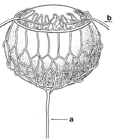
of Antoni van Leeuwenhoek, Vol V, pp 3–67 (English). Amsterdam, Swets, Zeitlinger, 1957

5. Stricker L: The Crystalline Lens System. Cincinnati, L Stricker, 1899

6. McKeehan MS: Cytological aspects of embryonic lens induction in the chick. J Exp Zool 117:31, 1951

7. Coulombre J, Coulombre AJ: Lens development: IV. Size, shape and orientation. Invest Ophthalmol 8:251, 1969

8. Coulombre AJ: Experimental embryology of the vertebrate eye. Invest Ophthalmol 4:411, 1965

9. McKeehan M: Induction of portions of the chick lens without contact with the optic 
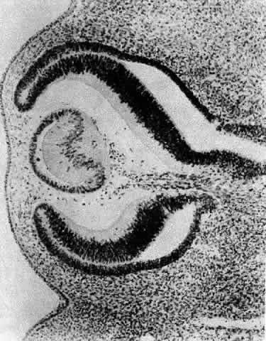
cup. Anat Rec 132:297, 1958

10. O'Rahilly R, Meyer DB: The early development of the eye in the chick: Gallus domesticus. Acta Anat 36:20, 1959

11. Cohen AI: Electron microscopic observations of the developing mouse eye: I. Basement 
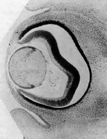
membranes during early development and lens formation. Dev Biol 3:297, 1961

12. Weiss P, Fitton-Jackson S: Fine structural changes associated with lens determination in the avian 
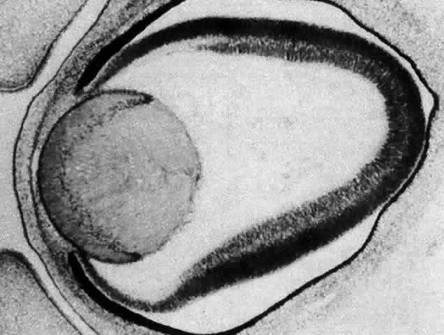
embryo. Dev Biol 3:532, 1961

13. Smelser GK: Embryology and morphology of the lens. Invest Ophthalmol 4:398, 1965

14. Saha MS, Spann CL, Grainger RM: Embryonic lens induction: More than meets the optic vesicle. Cell Differ 28:153, 1989

15. Tripathi BJ, Tripathi RC, Livingston AM, Borisuth NS: The role of growth factors in the embryogenesis and differentiation of the eye. Am J Anat 192:442, 1991

16. McAvoy JW, Chamberlain CG, deLongh RU et al: The role of fibroblast growth factor in eye lens development. Ann NY Acad Sci 638:256, 1991

17. Henry JJ, Grainger RM: Early tissue interactions leading to embryonic lens formation in Xenopus laevis. Dev Biol 141:149, 1990

18. Grainger RM, Henry JJ, Saha MS, Servetnick M: Recent progress on the mechanics of embryonic lens formation. Eye 6:117, 1992

19. Grainger RM: Embryonic lens induction: Shedding light on vertebrate tissue determination. Trends Genet 8:349, 1992

20. Zwaan J, Bryan PR, Pearce TL: Interkinetic nuclear migration during the early stages of lens formation in the chicken embryo. J Embryol Exp Morphol 21:71, 1969

21. Cuadros MA, Martin C, Rios A et al: Macrophages of hemangioblastic lineage invade the lens vesicle-ectoderm interspace during closure and detachment of the avian embryonic lens. Cell Tissue Res 266:117, 1991

22. Lerche W, Wulle KG: Electron microscopic studies on the development of the human lens. Ophthalmologica 158:296, 1969

23. Silver PHS, Wakely J: The initial stage in the development of the lens capsule in chick and mouse embryos. Exp Eye Res 19:73, 1974

24. Wang JL, Toida K, Uehara Y: The tunica vasculosa lentis; an expedient system for studying vascular formation and regression. J Electron Microsc (Tokyo) 39:46, 1990

25. Wistow GJ, Piatigorsky J: Lens crystallins: The evolution and expression of proteins for a highly specialized tissue. Annu Rev Biochem 57:479, 1988

26. van de Kamp M, Zwaan J: Intracellular localization of lens antigens in the developing eye of the mouse embryo. J Exp Zool 186:23, 1973

27. Piatigorsky J: Lens differentiation in vertebrates. A review of cellular and molecular features. Differentiation 19:134, 1981

28. McAvoy JW: Cell division, cell elongation and the coordination of crystallin gene expression during lens morphogenesis in the rat. J Embryol Exp Morphol 45:271, 1978

29. Treton JA, Jacquemin E, Courtois Y, Jeanny JC: Differential localization by in situ hybridization of specific crystallin transcripts during mouse lens development. Differentiation 47:143, 1991

30. McAvoy JW: Cell division, cell elongation and distribution of α-, ß- and Γ-crystallins in the rat lens. J Embryol Exp Morphol 44:149, 1978

31. Coulombre JL, Coulombre AJ: Lens development: Fiber elongation and lens orientation. Science 142:1489, 1963

32. Jacobson AG: Inductive processes in embryonic development. Science 152:25, 1966

33. Yamamoto Y: Growth of lens and ocular environment: Role of neural retina in the growth of mouse lens as revealed by an implantation experiment. Dev Growth Differ 18:273, 1976

34. Beebe DC, Silver MH, Belcher KS et al: Lentropin, a protein that controls lens fiber formation, is related functionally and immunologically to the insulin-like growth factors. Proc Natl Acad Sci USA 84:2327, 1987

35. McAvoy JW: Beta- and gamma-crystallin synthesis in rat lens epithelium explanted with neural retina. Differentiation 17:85, 1980

36. Zelenka PS, Pallansch L, Vatal M, Nath P: Proto-oncogene expression during in vitro differentiation of embryonic chicken lens epithelial explants. In Piatigorsky J, Shinohara T, Zelenka P (eds): Molecular Biology of the Eye, Genes, Vision and Ocular Disease, pp 249–258. New York, Alan R. Liss, 1988

37. Rinaudo JA, Zelenka PS: Expression of c-fos and c-jun mRNA in the developing chicken lens: Relationship to cell proliferation, quiescence and differentiation. Exp Cell Res 199:147, 1992

38. Kuszak JR, Bertram BA, Macsai MS, Rae JL: Sutures of the crystallin lens: A review. Scanning Electron Microsc 3:1369, 1984

39. Kuszak, Sivak JG, Weerheim JA: Lens optical function is a direct function of lens sutural architecture. Invest Ophthalmol Vis Sci 32:2119, 1991

40. Kuszak JR: Ultrastructure of mammalian lens fiber cells and epithelium. Int Rev Cytol Surv Cell Biol (in press, 1994)

41. Kuszak JR, Herbert KL, Sivak JG: The interrelationship of lens anatomy and optical quality: Primate lenses. Exp Eye Res (in press, 1994)

42. Harding JJ, Rixon KC, Marriott FHC: Men have heavier lenses than women of the same age. Exp Eye Res 25:651, 1977

43. Weekers R, Delmarcelle Y, Luyckx-Bacus J, Collignon J: Morphological changes of the lens with age and cataract. Ciba Foundation Symposium 19, The Human Lens in Relation to Cataract. Amsterdam, Associated Scientific, 1973

44. Brown N: The change in lens curvature with age. Exp Eye Res 19:175, 1974

45. Charles MW, Brown N: Dimensions of the human eye relevant to radiation protection. Phys Med Biol 20:202, 1975

46. Farnsworth PN, Shyne SE: Anterior zonular shifts with age. Exp Eye Res 28:291, 1979

47. Lowe RF, Clark BAJ: Posterior corneal curvature: Correlations in normal eyes and in eyes involved with primary angle-closure glaucoma. Br J Ophthalmol 57:464, 1973

48. Lowe RF, Clark BAJ: Radius of curvature of the anterior lens surface. Correlations in normal eyes and in eyes involved with primary angle-closure glaucoma. Br J Ophthalmol 57:471, 1973

49. Duke-Elder S, Wybar K: System of Ophthalmology, Vol 2, The Anatomy of the Visual System, p205. London, Henry Kimpton, 1961

50. Wolff E, Warwick R: Anatomy of the Eye and Orbit. Philadelphia, WB Saunders, 1976

51. Karim AKA, Jacob TJC, Thompson GM: The human anterior lens capsule: Cell density, morphology and mitotic index in normal and cataractous lenses. Exp Eye Res 45:865, 1987

52. Kuszak JR, Brown HG: Embryology and anatomy of the crystallin lens. In Albert DM, Jakoviec FA (eds): Principles and Practice of Ophthalmology, pp 82–96. Philadelphia, WB Saunders, 1994

53. Howard A: Whole-mounts of rabbit lens epithelium for cytological study. Stain Technol 27:313, 1952

54. Rothstein H: Experimental techniques for the investigation of the amphibian lens epithelium. In Prescott DM (ed): Methods in Cell Physiology, Vol III, pp 45–74. New York, Academic Press, 1968

55. Harding CV, Reddan JR, Unakar NJ, Bagchi M: The control of cell division in the ocular lens. Int Rev Cytol 31:215, 1971

56. Brown NAP, Bron AJ: An estimate of the human lens epithelial size in vivo. Exp Eye Res 44:899, 1987

57. Guggenmoos-Holtzman I, Engel B, Henke V, Naumann GOH: Cell density of human lens epithelium of human cataracts. Ophthalmology 94:875, 1987

58. Vasavada AR, Cherian M, Yadav S, Rawal UM: Lens epithelial cell density and histomorphological study in cataractous lenses. J Cataract Refract Surg 17:798, 1991

59. Konofsky K, Naumann GOH: Cell density and sex chromatin in lens epithelium of human cataracts. Ophthalmology 94:875, 1987

60. Rafferty NS: Lens morphology. In Maisel H (ed): The Ocular Lens, Structure Function and Pathology, pp 1–60. New York, Marcel Dekker, 1985

61. Brown HG, Pappas GD, Ireland ME, Kuszak JR: Ultrastructural, biochemical and immunologic evidence of receptor mediated endocytosis in the crystalline lens. Invest Ophthalmol Vis Sci 31:2579, 1990

62. Gorthy WC, Snavely MR, Berrong ND: Some aspects of transport and digestion in the lens of the normal young 
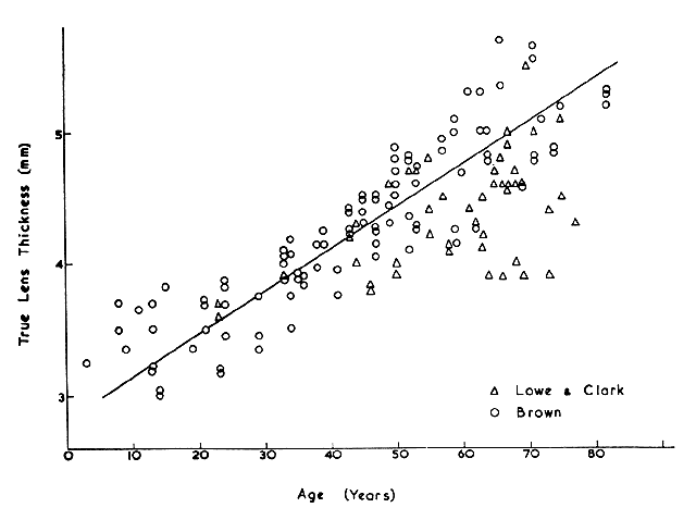
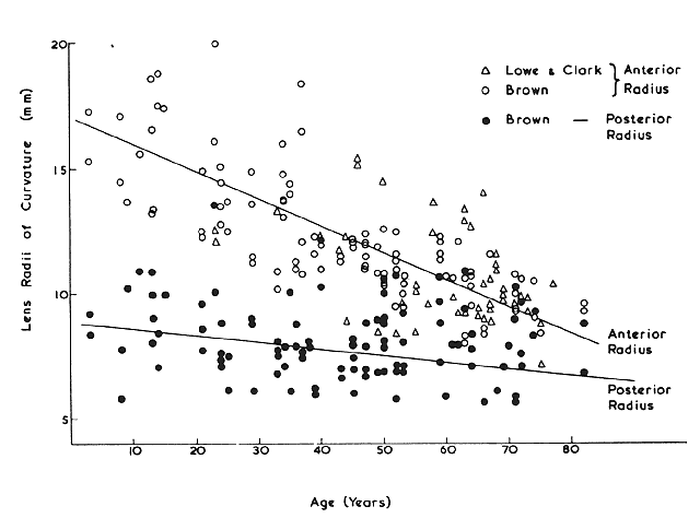
adult rat. Exp Eye Res 12:112, 1971

63. Rae JL, Stacey T: Lanthanum and procion yellow are extracellular markers in the crystalline lens of the rat. Exp Eye Res 28:1, 1979

64. Worgul BV, David J, Odrich S et al: Evidence of genotoxic damage in human cataractous lenses. Mutagenesis 6:495, 1991

65. Harding CV, Donn A, Srinivasan BD: Incorporation of thymidine by injured lens epithelium. Exp Cell Res 18:582, 1959

66. Rafferty NS: Studies of an injury induced growth in the frog lens. Anat Rec 146:299, 1963

67. Rafferty NS: Proliferative response in experimentally injured frog lens epithelium: Autoradiographic evidence for movement of DNA synthesis toward injury. J Morphol 121:295, 1967

68. Rafferty NS: The cytoarchitecture of normal mouse lens epithelium. Anat Rec 173:225, 1972

69. Rafferty NS: Mechanism of repair of lenticular wounds in Rana pipiens: I. Role of cell migration. J Morphol 133:409, 1972

70. Rothstein H, Weinsieder A, Blaiklock R: Response to injury in the lens epithelium of the bullfrog, Rana catesbeina. Exp Cell Res 35:548, 1964

71. Rothstein H, Reddan JR, Weinsieder A: Response to injury in the lens epithelium of the bullfrog: II. Spatio-temporal patterns of DNA synthesis and mitosis. Exp Cell Res 37:440, 1965

72. Weinsieder A, Rothstein H, Drebert D: Lenticular wound healing: Evidence for genomic activation. Cytobiologie 7:406, 1973

73. Gierthy JF, Rothstein H: Cell disorganization and the stimulation of mitosis in the cultured leopard frog lens. Exp Cell Res 64:170, 1971

74. Rothstein H, Worgul B: Mitotic variations in the lens epithelium of the frog: II. Possible role of temperature and hormones. Ophthalmol Res 5:151, 1973

75. van Buskirk R, Worgul BV, Rothstein H, Wainwright N: Mitotic variations in the lens epithelium of the frog: III. Somatotropin. Gen Comp Endocrinol 25:52, 1975

76. Worgul BV, Rothstein H: On the mechanism of thyroid mediated mitogenesis in adult anura: I. Preliminary analyses of growth kinetics and macromolecular syntheses, in lens epithelium, under the influence of exogenous triiodothyronine. Cell Tissue Kinet 7:415, 1974

77. Weinsieder A, Briggs R, Reddan J et al: Induction of mitosis in ocular tissue by chemotoxic agents. Exp Eye Res 20:33, 1975

78. Reddan J, Weinsieder A, Wilson D: Aqueous humor from traumatized eyes triggers cell division in the epithelia of cultured lenses. Exp Eye Res 28:267, 1979

79. Worgul BV, Merriam GR Jr: The effect of endotoxin induced intraocular inflammation on the rat lens epithelium. Invest Ophthalmol Vis Sci 18:401, 1979

80. Muggleton-Harris AL: Cellular changes occurring with age in the lens cells of the frog (Rana pipiens) in reference to the developmental capacity of the transplanted nuclei. Exp Gerontol 5:227, 1970

81. Alcala J, Maisel H: Biochemistry of lens plasma membranes and cytoskeleton. In Maisel H (ed): The Ocular Lens, pp 169–222. New York, Marcel Dekker, 1985

82. Rafferty NS, Scholz DL, Goldberg M, Lewyckyj M: Immunocytochemical evidence for an actin-myosin system in lens epithelial cells. Exp Eye Res 51:591, 1990

83. Rafferty NS, Scholz DL: Polygonal arrays of microfilaments in epithelial cell of the intact lens. Curr Eye Res 3:1141, 1984

84. Wanko T, Gavin MA: The fine structure of the lens epithelium, an electron microscopic study. Arch Ophthalmol 60:868, 1958

85. Hertl M: Kernvolumen und Nukleolarapparat wachsender Linsenzellen. Zellforsch Micro Anat Abt Histochem 43:228, 1955

86. Srinivasan BD, Iwamoto T: Electron microscopy of rabbit lens nucleoli. Exp Eye Res 16:9, 1973

87. Kuwabara T: The maturation of the lens cell: A morphologic study. Exp Eye Res 20:427, 1975

88. Eguchi G: Electron microscopic studies on lens regeneration: II. Formation and growth of lens vesicle and differentiation of lens fibers. Embryologia 8:247, 1964

89. Karasaki S: An electron microscopic study of wolffian lens regeneration in the adult newt. J Ultrastruct Res 11:246, 1964

90. Papaconstantinou J: Molecular aspects of lens cell differentiation. Science 156:338, 1967

91. Kuwabara T: Microtubules in the lens. Arch Ophthalmol 79:189, 1968

92. Rafferty NS, Goossens W: Ultrastructure of traumatic cataractogenesis in the frog: A comparison with mouse and human lens. Am J Anat 148:385, 1977

93. Maisel H: Filaments of the vertebrate lens. Experientia 33:525, 1977

94. Maisel H, Lieska N, Bradley R: Isolation of filaments of the chick lens. Experientia 34:352, 1978

95. Piatigorsky J, Webster H, Wolberg H: Cell elongation in the cultured embryonic chick lens epithelium with and without protein synthesis. J Cell Biol 55:82, 1972

96. Mousa GY, Trevithick JR: Differentiation of rat lens epithelial cells in tissue culture: II. Effects of cytochalasins B and D on actin organization and differentiation. Dev Biol 60:14, 1977

97. Worgul BV, Rothstein H: On the mechanism of radiocataractogenesis. Medikon 6:5, 1977

98. Worgul BV, Rothstein H: Congenital cataracts associated with disorganized meridional rows in a new laboratory animal: The degu (Octodon degus). Biomed Exp 23:1, 1975

99. Streeten BW, Eshaghian J: Human posterior subcapsular cataract. Arch Ophthalmol 96:1653, 1978

100. Messier B, Leblond C: Cell proliferation and migration as revealed by radioautography after injection of thymidine-H3 into male rats and mice. Am J Anat 106:247, 1960

101. Brolin SE, Diderholm H, Hammar H: An autoradiographic study on cell migration in the eye lens epithelium. Acta Soc Med Ups 66:43, 1961

102. Mikulicich A, Young R: Cell proliferation and displacement in the lens epithelium of young rats injected with tritiated thymidine. Invest Ophthalmol 2:344, 1963

103. Hammar H: An autoradiographic study on cell migration in the eye lens epithelium from normal and alloxan diabetic rats. Acta Ophthalmol 43:442, 1965

104. Worgul BV, Merriam GR Jr, Szechter A, Srinivasan BD: Lens epithelium and radiation cataract: I. Preliminary studies. Arch Ophthalmol 94:996, 1976

105. Hayden J, Rothstein H: A correlation between mitosis and cell migration in the Rana pipiens lens. J Cell Biol 79:19a, 1978

106. Moduk SP, Bollum FJ: Detection and measurement of single-strand breaks in nuclear DNA in fixed lens sections. Exp Cell Res 75:307, 1972

107. Kuwabara T, Imaizumi M: Denucleation process of the lens. Invest Ophthalmol 13:973, 1974

108. Vrensen GF, Graw J, DeWolf A: Nuclear breakdown during terminal differentiation of primary lens fibers in mice: A transmission electron microscopic study. Exp Eye Res 52:647, 1991

109. Jurand A, Yamada T: Elimination of mitochondria during wolffian lens degeneration. Exp Cell Res 46:636, 1967

110. Brown NAP: Lens change with age and cataract; slit-image photography. Ciba Foundation Symposium 19, The Human Lens in Relation to Cataract, pp 65–78. New York, Associated Scientific, 1973

111. Vinciguerra MJ, Bettelheim FA: Packing and orientation of fiber cells. Exp Eye Res 11:214, 1971

112. Tardieu A, Delaye M: Eye lens proteins and transparency: From light transmission theory to solution x-ray structural analysis. Annu Rev Biophys Chem 17:47, 1988

113. Thoft RA, Kinoshita JH: The extracellular space of the lens. Exp Eye Res 4:287, 1965

114. Paterson CA: The extracellular space of the crystallin lens. Am J Physiol 218:797, 1970

115. Yorio T, Bentley PJ: Distribution of the extracellular space of the amphibian lens. Exp Eye Res 23:601, 1976

116. Kuszak JR, Ennesser CA, Bertram BA et al: The contribution of cell-to-cell fusion to the ordered structure of the crystalline lens. Lens Eye Toxic Res 6:639, 1989

117. Dickson DH, Crock GW: Interlocking patterns on primate lens fibers. Invest Ophthalmol 11:809, 1972

118. Harding CV, Susan S, Murphy H: Scanning electron microscopy of the adult rabbit lens. Ophthalmic Res 8:443, 1976

119. Farnsworth PN, Shyne SE, Burke PA et al: Maturation of the human lens fiber. In Srivastava S (ed): The Red Blood Cell and the Lens, pp 45–48. New York, Elsevier, 1980

120. Cohen AI: The electron microscopy of the normal human lens. Invest Ophthalmol 4:433, 1965

121. Worgul BV, Iwamoto T, Merriam GR Jr: RNA-containing cytoplasmic inclusions at the termini of maturing fibers in the rat lens. Ophthalmic Res 9:388, 1977

122. Lo WK: Adherens junctions in the ocular lens of various species; ultrastructural analysis with an improved fixation. Cell Tissue Res 254:31, 1988

123. Kuszak J, Maisel H: Morphology of differentiating lens fiber cells. Invest Ophthalmol Vis Sci 17(suppl):281, 1978

124. Kuszak JR, Sheh YH, Carney KC, Rae JL: A correlate freeze-etch and electrophysiological study of communicating junctions in crystalline lenses. Curr Eye Res 4:1145, 1985

125. Nicholson BJ, Takemoto LJ, Hunkapiller MW: Differences between liver gap junction protein and lens MIP26 from rat: Implications for tissue specificity of gap junctions. Cell 32:967, 1983

126. Gorin MB, Yancy SB, Cline J et al: The major intrinsic protein (MIP) of the bovine fiber membrane: Characterization and structure based on cDNA cloning. Cell 39:49, 1984

127. Philipson BT, Hanninen L, Balazs EA: Cell contacts in human and bovine lenses. Exp Eye Res 21:205, 1975

128. Mathias RT, Rae JL: Transport properties of the lens. Am J Physiol 249:C181, 1985

129. Duncan G: Relative permeabilities of the lens membrane to sodium and potassium. Exp Eye Res 8:315, 1969

130. Rae JL: Potential profiles in the crystalline lens of the frog. Exp Eye Res 19:227, 1974

131. Young RW, Ocumpaugh DE: Autoradiographic studies on the growth and development of the lens capsule in the rat. Invest Ophthalmol 5:583, 1966

132. Wislocki GB: The anterior segment of the eye of the rhesus monkey investigated by histochemic means. Anat Rec 113:579, 1952

133. Wulle KG, Lerche W: Zur Feinstrukur der embryonalen menschlichen Linsenblase. Albrecht von Graefes Arch Klin Exp Ophthalmol 173:141, 1969

134. Farnsworth PN, Mauriello JA, Burke-Gadomski P et al: Surface ultrastructure of the human lens capsule and zonule attachments. Invest Ophthalmol 15:36, 1976

135. Hettlich HJ, Wenzel M, Janssen M, Miltermayer C: Immunohistochemische untersuchungen der menschlichen Linsenkapsel. Fortschr Ophthalmol 87:147, 1990

136. Marshall GE, Konstas AG, Bechrakis NE, Lee WR: An immunoelectron microscope study of the aged human lens capsule. Exp Eye Res 54:393, 1992

137. Kohno T, Sorgente N, Ishibashi T et al: Immunofluorescent studies of fibronectin and laminin in the human eye. Invest Ophthalmol Vis Sci 28:506, 1987

138. Parmigiani CM, McAvoy JW: The role of laminin and fibronectin in the development of the lens capsule. Curr Eye Res 10:501, 1991

139. Wolff E: Anatomy of the Eye and Orbit, 6th ed, pp 207–224. Philadelphia, WB Saunders, 1968

140. Fisher RF: The elastic constant of the human lens. J Physiol 212:147, 1971

141. Hogan MJ, Alvarado JA, Weddell JE: Histology of the Human Eye. Philadelphia, WB Saunders, 1971

142. Davanger M: The suspensory apparatus of the lens: The surface of the ciliary body. Acta Ophthalmol 53:19, 1975

143. Streeten B: The zonular insertion: A scanning electron microscopic study. Invest Ophthalmol 16:364, 1977

144. Streeten BN, Swann DA, Licari PA et al: The protein composition of the ocular zonules. Invest Ophthalmol Vis Sci 24:119, 1983

145. Streeten BN, Licari PA, Marucci AA, Dougherty RM: Immunohistochemical comparison of ocular zonules and the microfibrils of elastic tissue. Invest Ophthalmol Vis Sci 21:130, 1981

146. Farnsworth PN, Burke P: Three-dimensional architecture of the suspensory apparatus of the lens of the rhesus monkey. Exp Eye Res 25:563, 1977

147. Marshall J, Beaconsfield M, Rothery S: The anatomy and development of the human lens and zonules. Trans Ophthalmol Soc UK 102:423, 1982

148. Mann I: Developmental Abnormalities of the Eye, 1st ed. Philadelphia, JB Lippincott, 1937

149. Mann I: Developmental Abnormalities of the Eye, 2nd ed. Philadelphia, JB Lippincott, 1957

150. Baker WH: Congenital aphakia. N Y Med J 46:595, 1887

151. Mann I: Congenital absence of the lens, with special reference to an aphakic human embryo. Br J Ophthalmol 5:301, 1921

152. Duke-Elder S: System of Ophthalmology, Vol 3, Normal and Abnormal Development: Part 2. Congenital Deformities. St. Louis, CV Mosby, 1963

153. Pesme P, Montoux P: Staphylome congenital de la cornée (étude anatomique). Bull Soc Ophtalmol Fr 935, 1956

154. Farnsworth PN, Burke PA, Blanco J, Maltzman B: Ultrastructural abnormalities in a microspherical ectopic lens. Exp Eye Res 27:399, 1973

155. Dayal MB, Mehra KS: Spherophakia. Am J Ophthalmol 50:333, 1960

156. Lisch K: Uber kongenitale formanomalien der linse. Albrecht von Graefes Arch Klin Exp Ophthalmol 157:287, 1956

157. Coulombre AJ, Herrmann H: Lens development: III. Relationship between the growth of the lens and the growth of the outer eye coat. Exp Eye Res 4:302, 1965

158. Nordmann J: Biologie du Cristallin. Paris, Masson et Cie, 1954

159. Pergens E: Buphthalmus mit lenticonus posterior. Arch Augenheilk 35:1, 1897

160. Sachs E, Larsen RL: Cancer and the lens. Am J Ophthalmol 31:561, 1948

161. Franceschetti A, Rickli H: Posterior (eccentric) lenticonus. Report of first case with clinical and histological findings. Arch Ophthalmol 51:499, 1954

162. Makley TA: Posterior lenticonus. Report of a case with histologic findings. Am J Ophthalmol 39:308, 1955

163. Sand BJ, Abraham SV: Anterior lenticonus. An operated case and a review of the literature. Am J Ophthalmol 53:636, 1962

164. Krusius FF: Ueber zwei seltene anomalien des linsen systemes. Arch Augenheilk 65:233, 1910

165. Feigenbaum A: The origin of lenticonus anterior. Folia Ophthalmol Orient 1:103, 1932

166. Petronio G: Su di un caso tipico e bilaterale “di lenticono anteriore.” Arch Ottalnologia 48:153, 1941

167. Hiles DA, Kitty LA: Disorders of the lens. In Isenberg SJ (ed): The Eye in Infancy, pp 336–373. St. Louis, CV Mosby, 1994

168. Cross HE: Differential diagnosis and treatment of dislocated lenses. In Bergsma D, Bron AJ, Cotlier E (eds): The Eye and Inborn Errors of Metabolism, pp 335–346. New York, Alan R Liss, 1976

169. Burke PA, Shyne SE, Caputo AR, Farnsworth PN: Ultrastructural abnormalities in Weill-Marchesani syndrome lens. Invest Ophthalmol Vis Sci 17(suppl):279, 1978

170. Cross HE: Ectopia lentis in systemic heritable disorders. Birth Defects 10:113, 1974

171. Farnsworth PN, Burke P, Dotto ME, Cinotti AA: Ultrastructural abnormalities in a Marfan's syndrome lens. Arch Ophthalmol 95:1601, 1977

172. Lloyd IC, Goss-Sampson M, Jeffrey BG et al: Neonatal cataract: Aetiology, pathogenesis and management. Eye 6:184, 1992

173. Donaldson DD: The Crystalline Lens. St. Louis, CV Mosby, 1976

174. Phelps CD: Examination and functional evaluation of crystalline lens. In Duane TD (ed): Clinical Ophthalmology, Vol I, pp 1–23. New York, Harper & Row, 1978

175. Yanoff M, Fine BS: Ocular Pathology. New York, Harper & Row, 1975

176. Lowenstein A: Ueber die genese angeborener linsentrubungen. Klin Monatsbl Augenheilkd 87:382, 1931

177. Clapp CA: Alterations in the capsular epithelium in immature cataract. Trans Am Ophthalmol Soc 39:73, 1941

178. Samuels B: Lesions in the lens caused by purulent corneal ulcers. Trans Am Ophthalmol Soc 39:66, 1941

179. Samuels B: Lesions in the lens caused by purulent corneal ulcers. Arch Ophthalmol 27:345, 1942

180. Samuels B: Cataract complicating corneal scars after perforating ulcers. Arch Ophthalmol 29:583, 1943

181. Riedl A: Ueber Beiziehung von angeborenen Linsentrubungen zur Pupillarmembran. Klin Monatsbl Augenheilkd 69:482, 1922

182. Seefelder R: Stammbaum einer Familie mit angeborenem Star. Ber Dtsch Ophthalmol Ges 52:414, 1938

183. Waardenburg PJ: Gross remnants of the pupillary membrane, anterior polar cataract and microcornea in a mother and her children. Ophthalmologica 118:828, 1949

184. Ogata T, Matsui M: Electron microscopic studies on the capsule and epithelial cells of anterior polar cataract of human lens. Acta Soc Ophthalmol Jpn 76:1286, 1972

185. Font RI, Brownstein S: A light and electron microscopic study of anterior subcapsular cataracts. Am J Ophthalmol 78:972, 1974

186. Pellaton R: Die physiologischen linsentrubungen im kindesalter nach spaltlampenunterschung an 164 normalen kinderaugen. Albrecht von Graefes Arch Klin Exp Ophthalmol 111:341, 1923

187. Cordes FC: Types of congenital cataract. Am J Ophthalmol 30:397, 1947

188. Nettleship E, Ogilvie FM: Peculiar form of hereditary congenital cataract. Trans Ophthalmol Soc UK 26:191, 1906

189. Smith P: A pedigree of Doyne's discoid cataract. Trans Ophthalmol Soc UK 30:37, 1910

190. Vogt A: Weitere Ergebnisse der Spaltlampen-microscopic des vordern Bulbasabschnittes III. Albrecht von Graefes Arch Klin Exp Ophthalmol 107:196, 1922

191. VonPoos F: Ueber eine familiar aufgetretene besondere Schichtstarform “Cataracta Zonularis Pulverulenta.” Klin Monatsbl Augenheilkd 76:502, 1926

192. von Szily A: The contribution of pathological examinations to the elucidation of the problems of cataract. Trans Ophthalmol Soc UK 58:595, 1938

193. Francois J: Heredity in Ophthalmology, pp 364–368. St. Louis, CV Mosby, 1961

194. Falls HF: Developmental cataracts (results of surgical treatment in one hundred and thirty-one cases). Arch Ophthalmol 29:210, 1943

195. Marner E: A family with eight generations of hereditary cataract. Acta Ophthalmol 27:537, 1949

196. Nettleship E: On heredity in the various forms of cataract. R Lond Ophthalmol Hosp Rep 16:179, 1905

197. Rosenstern I: Zur wirkung des lebertrans auf rachitis und spasmophie diathese. Berl Klin Wochenschr 47:822, 1910

198. Huggert A: The connection between the position of zonular opacities in the lens and the time of their formation estimated from simultaneously occurring enamel hypoplasia. Acta Ophthalmol 26:7, 1948

199. von Szily A, Eckstein A: Vitaminmangel und Schichtstargenese Karakte als eine erscheinungsform der Avitaminose mit Storung des Kalkstuffwechsels bei saugenden Ratten, hervorgerufen durch qualitative unterernahrung der muttertiere. Klin Monatsbl Augenheilkd 71:545, 1923

200. von Goldschmidt M: Zur frage der kataraktbildung bei vitaminmangel. Klin Wochenschr 6:635, 1927

201. Adams PH: 1) A family with congenital displacement of lenses. 2) A family with congenital opacities of lenses. Trans Ophthalmol Soc UK 29:274, 1909

202. Marty F: Les couzbes de frequence en fonction de l'age, de quelques alterations congenitales ou seniles frequentes du segment anterieur. Arch Ophtal (Paris) 17:5, 1957

203. Muller O: Uber Haufigkeit und form der vorderen axialen nahtpunktierung und der vorderen axialen embryonalkatarakt. Albrecht von Graefes Arch Klin Exp Ophthalmol 124:444, 1930

204. Mann IC: The correlation of the embryology of the lens with slit-lamp appearances. Trans Ophthalmol Soc UK 45:696, 1925

205. Stocklin P: Normvarianten der morphologie der kindlichen linse. Albrecht von Graefes Arch Klin Exp Ophthalmol 158:346, 1957

206. Treacher-Collins E: Developmental deformities of the crystalline lens. Ophthalmoscope 6:577, 1908

207. Koby FE: Cataracte familiale d'un type particulier se transmettant apparement suivant le mode dominant. Arch Ophthalmol (Paris) 40:492, 1923

208. Sautter H: Die Trubungsformen der menschlichen Linse. Stuttgart, Thieme, 1951

209. Renard G, Laporte P: Un cas de cataracte congénitale a cristaux associée a des malformations craniofacialis. Bull Mem Soc Fr Ophtalmol 64:176, 1951

210. Offret G, Durand R: Cataracte en cristaux polychromes chez deux consanguins. Bull Soc Ophtalmol Fr, p 491, 1955

211. Tadros MA: Coralliform cataract. Bull Ophthalmol Soc Egypt 49:177, 1956

212. Rieger H, Stark M, von Zeynek: Zur frage der cataracta coralliformis. Albrecht von Graefes Arch Klin Exp Ophthalmol 147:426, 1944

213. Verhoeff FH: Microscopic findings in a case of coralliform cataract, with remarks on congenital cataracts in general. Arch Ophthalmol 47:558, 1918

214. Gifford SR, Puntenney I: Coralliform cataract and a new form of congenital cataract with crystals in the lens. Arch Ophthalmol 17:885, 1937

215. Parker CO: Spear cataract. Arch Ophthalmol 55:23, 1956

216. Cordes FC: Types of congenital and juvenile cataracts. In Haik GM (ed): Symposium on Diseases and Surgery of the Lens. St. Louis, CV Mosby, 1957

217. Cordes FC: Types of congenital cataract. Am J Ophthalmol 30:397, 1947

218. Lisch K: Zur Genese des vorderen Polstars. Albrecht von Graefes Arch Klin Exp Ophthalmol 145:393, 1942

219. Franceschetti A, Dieterle P: Cataracte fusiforme congénitale, (en pagode) et ses relations avec la cataracte pulvérulente centrale. Bull Mem Soc Fr Ophtalmol 72:341, 1959

220. von Szily A: Ueber angeborene familiare “Ringstarlinse” nebst Hinweisen auf ihre Entstehung. Klin Monatsbl Augenheilkd 81:145, 1928

221. Franceschetti A, Forni S, Rizzini V: Cataracte annulaire ou en bouée de sauvetage. Ophthalmologica 125:342, 1953

222. Avanza C, Balavoine C: Un nouveau biotype de la cataracte en bouée de sauvetage (cataracta umbilicata) Associée a une brachymorphie une ophtalmoplégie externe et une hyperaminoacidurie. Bull Mem Soc Fr Ophtalmol 72:331, 1959

223. Haro ES: Hereditary disk-shaped (ring) cataract. Arch Ophthalmol 36:82, 1946

224. Francois J: Total and regressive cataracts. In Francois J (ed): Congenital Cataracts, pp 175–193. Assen, Netherlands, Royal Van Gorcum, 1959

225. Jaensch PA: Anatomische Untersuchungen angeborenen Totalstars. Albrecht von Graefes Arch Klin Exp Ophthalmol 115:81, 1924

226. Gregg MM: Congenital cataracts following German measles in the mother. Trans Ophthalmol Soc NZ 3:35, 1941

227. Zimmerman LE, Font RL: Congenital malformations of the eye. Some recent advances in knowledge of the pathogenesis and histopathological characteristics. JAMA 196:684, 1966

228. Yanoff M: Pathology of cataract. In Bellows JG (ed): Cataract and Abnormalities of the Lens, pp 155–189. New York, Grune & Stratton, 1975

229. Swan C: Study of three infants dying from congenital defects following maternal rubella in early stages of pregnancy. J Pathol 56:289, 1944

230. Alford CA, Neva FA, Weller TH: Virologic and serologic studies on human products of conception after maternal rubella. N Engl J Med 271:1275, 1964

231. Bellanti JA, Artenstein MS, Olson LC et al: Congenital rubella: Clinicopathologic, virologic and immunologic studies. Am J Dis Child 110:464, 1965

232. Cooper LZ, Green RH, Krugman S et al: Rubella in contacts of infants with rubella-associated anomalies. MMWR Morb Mortal Wkly Rep 14:44, 1965

233. Karkinen-Jaaskelainen M, Saxen L, Vaheri A, Leinikki P: Rubella cataract in vitro: Sensitive period of the developing human lens. J Exp Med 141:1238, 1975

234. Menser MA, Harley JD, Hertzberg R et al: Persistence of virus in lens for three years after prenatal rubella. Lancet 3:327, 1967

235. World Health Organization. Epidemiological Vital Statistics Report 19:433, 1966

236. Jay M: Linkage and chromosomal studies in congenital cataract. Trans Ophthalmol Soc UK 102:350, 1982

237. Lubsen NH, Renwick JH, Schoenmakers JGG: Hereditary cataract: Perspective for prenatal screening. Ophthalmic Paediatr Genet 7:195, 1986

238. Lewis RA: Mapping the gene for X-linked cataracts and microcornea with facial, dental and skeletal features to Xp22: An appraisal of the Nance-Horan syndrome. Trans Am Ophthalmol Soc 87:658, 1990

239. Zhu D, Alcorn DM, Antonarakis SE et al: Assignment of the Nance-Horan syndrome to the distal short arm of the X chromosome. Hum Genet 86:54, 1990

240. Stambolin D, Lewis RA, Buetow K et al: Nance-Horan syndrome; localization within the region Xp21.1–Xp22.3 by linkage analysis. Am J Hum Genet 47:13, 1990

241. Sparkes RS, Mohandes T, Heinzmann C et al: Assignment of a human beta-crystallin gene to 17 cen-q23. Hum Genet 74:133, 1986

242. Shiloh Y, Donlon T, Bruns G: Assignment of human Γ-crystallin gene cluster (CRYG) to the long arm of chromosome 2, region q33–36. Hum Genet 73:17, 1986

243. den Dunnen JT, Jongbloed RJE, Geurts van Kessel AHM et al: Human lens Γ-crystallin sequences are located in the p12-qter region of chromosome 2. Hum Genet 70:217, 1985

244. Sparkes RS, Mohandes T, Heinzmann C et al: The gene for the major intrinsic protein (MIP) of the ocular lens is assigned to human chromosome 12 cen-q14. Invest Ophthalmol Vis Sci 27:1351, 1986

245. Barrett DJ, Sparkes RS, Gorin MB et al: Genetic linkage analysis of autosomal dominant congenital cataracts with lens specific DNA probes and polymorphic phenotypic markers. Ophthalmology 95:538, 1988

246. Eiberg H, Marner E, Rosenberg T, Mohr J: Marner's cataract (CAM) assigned to chromosome 16: Linkage to haptoglobin. Clin Genet 34:272, 1988

247. Lubsen NH, Renwick JH, Tsui L-C: A locus for a human hereditary cataract is closely linked to the Γ-crystallin gene family. Proc Natl Acad Sci USA 84:489, 1987

248. Reese PD: Autosomal dominant congenital cataract associated with chromosomal translocation [t(3;4) (p26,2;p15)]. Arch Ophthalmol 105:1382, 1987

249. Conneally PM, Wilson AF, Merritt AD et al: Confirmation of genetic heterogeneity in autosomal dominant forms of congenital cataracts from linkage studies. Cytogenet Cell Genet 22:295, 1978

250. Marner E, Rosenberg T, Eiberg H: Autosomal dominant congenital cataract, morphology and genetic mapping. Acta Ophthalmol 67:151, 1989

251. Westat: Summary and critique of available data on the prevalence and economic and social costs of visual disorders and disabilities. Quoted in Vision Research: A National Plan, Vol 2, p456. The 1977 Report of the National Advisory Eye Council, DHEW Publication No (NIH) 78–1259, February 16, 1976

Back to Top