|
|
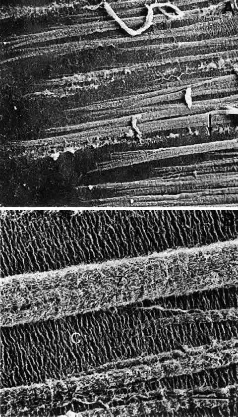
| Fig. 31. A. Scanning electron micrograph of the insertion of the zonules into the lens capsule. The area denoted by the arrow in A is shown in a higher magnification in B. The scale bar equals 5 μm, illustrating the fibrillar nature of the capsule (C) and the zonules (Z). Note the high degree of orientation of the capsular fibrils. (Farnsworth PN, Mauriello JA, Burke-Gadomski P, et al: Surface ultrastructure of the human lens capsule and zonule attachments. Invest Ophthalmol 15:36, 1976) |