|
|
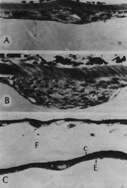
| Fig. 36. Photomicrographs of the development of anterior subcapsular cataract. A. The beginning of multistratification of the anterior epithelium, owing to localized hyperplasia of the lens epithelial cells. B. Further stratification of the epithelium, with denucleation occurring in some of the cells. C. The final stage of cataract formation showing the so-called fibrous plaque (F), bounded on the anterior and posterior sides with capsule material. Beneath the posterior capsule (C) a newly formed epithelial population can be seen (E). (Yanoff M, Fine BS: Ocular Pathology. New York, Harper & Row, 1975) |