|
|
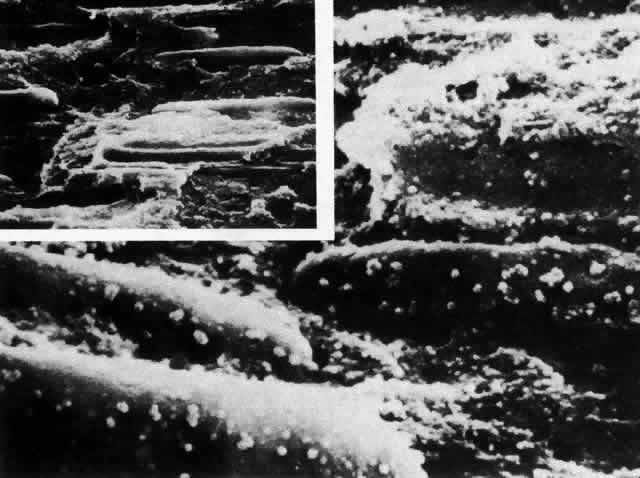
| Fig. 19. Scanning electron micrograph of the area of the bow in the rat lens following critical point drying and mechanical fracturing. Low-power scanning electron micrograph (inset) shows the elongated nuclei of internalizing cells. When viewed as increased magnification, fluffy deposits are seen on the surface. Because fracturing occurs between the double-layered membranes of the nuclei, the fluffy material comprises the remains of the outer membrane around nucleopores (× 20,000; inset × 5000). |