|
|
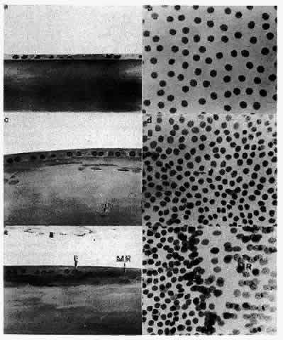
| Fig. 15. Photomicrographs showing the various regions of the human lens epithelium in sagittal section and on a whole-mount preparation. The 1-μm sagittal sections are stained by the Feulgen reaction, thus revealing only the nuclei of the cells. When viewed sagittally (a), the central zone cells are quite flat, causing the great distance seen between adjacent nuclei on a whole mount (b). In the germinative zone (c), the cells appear more cuboidal, and the nuclei are positioned nearer to the apices of the cells. Nuclei of the superficial lens cortical fibers are seen subjacent to the cells of the germinative zone (arrows). Viewed on a whole mount, this region (d) is marked by more closely packed nuclei and the presence of mitotic cells. (The arrow points to a metaphase figure.) At the equator, the cells are more columnar, and the nuclei are still located apically (e). As the cells begin to reorient in the anteroposterior direction, the nuclei maintain a more anterior (apical) position along the length of the elongating cells, thereby producing the bow pattern. The equatorial line (E) is an area of densely packed nuclei at the base of the meridional rows (MR) and is best seen on a whole mount (f). The sagittal section (e) reveals that this is a region where the nuclei of ajacent cells of the meridional rows (MR) are superimposed so that the nuclei appear to be compacted. |