|
|
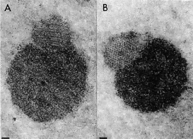
| Fig. 28. Electron micrographs of terminal bodies from the region of the posterior suture in the rat lens cortex. A difference in composition and density between the large and small subunits is clearly seen. The highly organized lattice arrangement of the smaller sphere is shown here in both longitudinal (A) and cross sections (B). The scale markers represent 0.1 μm. (Worgul BV, Iwamoto T, Merriam GR Jr: RNA-containing cytoplasmic inclusions at the termini of maturing fibers in the rat lens. Ophthalmol Res 9:388, 1977) |