|
|
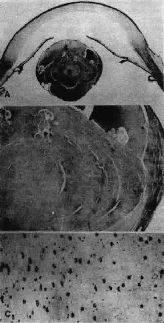
| Fig. 40. A. Rubella cataract showing advanced liquefaction of the cortex (c) and nuclear involvement (N), producing a spherophakic lens. B. Higher magnification of the lens, showing that the most superficial fibers at the equator (E) appear comparatively normal. C. Typical of rubella cataract, the nuclei of the fibers are retained deep within the nucleus of the lens, although many of these nuclei appear pyknotic and degenerating. (Zimmerman LE, Font RL: Congenital malformations of the eye. Some recent advances in knowledge of the pathogenesis and histopathological characteristics. JAMA 196:684, 1966) |