|
|
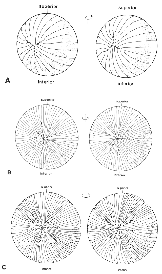
| Fig. 10. A. Diagrammatic representation of the anterior (left) and posterior (right) suture patterns of a newborn human lens. Note the offsetting anterior and posterior suture patterns as a consequence of opposite curvature of each end of the fiber cell. B. Scalar diagrammatic representation of the anterior (left) and posterior (right) suture patterns of a young adult human lens. Note the starburst pattern with nine equally spaced radial spokes. C. Scalar diagrammatic representation of the anterior (left) and posterior (right) suture patterns of an aged human lens. The number of spokes has increased, and the anterior and posterior suture patterns have become asymmetric with respect to each other. (Courtesy of Dr. Jer Kuszak, Rush Presbyterian St. Lukes Medical Center) |