Chapter 9
Hereditary Macular Dystrophies
ARTHUR D. FU, EVERETT AI, H. RICHARD MCDONALD, ROBERT N. JOHNSON and J. MICHAEL JUMPER
Main Menu   Table Of Contents

Search

INTRODUCTION
GENETIC TESTING
HISTORICAL PERSPECTIVES
SENSORY RETINA
RETINAL PIGMENT EPITHELIUM
THE PATTERN DYSTROPHIES
CHORIOCAPILLARIS
REFERENCES

INTRODUCTION
Hereditary macular dystrophies are progressive degenerations of the neurosensory retina, retinal pigment epithelium, or choroid. They affect predominantly and most profoundly the macular area. Historically the hereditary nature of these disorders was demonstrated via pedigree analysis. More recently, modern advances in genetic testing including genetic linkage analysis, karyotyping, and the use of polymerase chain reaction have allowed the further characterization and delineation of a growing number of inherited macular dystrophies.1,2

The macular dystrophies are characterized by dominance of posterior pole changes, familial and bilateral involvement, diminution of visual acuity, early age of onset, progressive course, and lack of systemic physical or laboratory abnormalities.1–4 Macular dystrophies excluded from discussion here are the vitreotapetoretinal dystrophies of Goldmann-Favre and Wagner, congenital stationary night blindness, diffuse tapetoretinal and choroidal dystrophies, metabolic disorders including the lipidoses, neurologic disorders such as the Laurence-Moon syndrome, and age-related macular degeneration. Few of the discussed dystrophies are strictly limited to the macula or even to the posterior pole. With the use of investigative techniques, including electrophysiologic and psychophysical testing, fundus photography, and fluorescein angiography, most of these dystrophies are found to involve large regions of the peripheral retina.5,6,8–14

Although many dystrophies demonstrate a gradual progressive course through discernable stages, some may begin with a functional defect and little or no visible fundus changes (e.g., rod monochromatism or Stargardt's disease), whereas others begin with visible changes and little or no functional defect (e.g., North Carolina macular dystrophy). Also, one stage of one dystrophy may appear similar to another stage of an entirely different dystrophy (e.g., adult-type foveomacular vitelliform dystrophy may resemble Best's vitelliform dystrophy).15 Therefore, the previously described investigative techniques, in addition to the fundus examination, may be needed to reach a diagnosis. Finally, despite the recent use of these genetic studies in the diagnosis of inherited macular dystrophies, the value of the family history and examination of family members in establishing the genetic pattern and correct diagnosis cannot be overemphasized.1–3,16,17

Back to Top
GENETIC TESTING
Disease genes responsible for autosomal dominant, recessive, and X-linked retinitis pigmentosa, North Carolina macular dystrophy, Best's disease, corneal dystrophy, and autosomal dominant and autosomal recessive Stargardt's macular dystrophy have been mapped to specific chromosomal regions via linkage analysis. Linkage analysis is based on the observation that alleles of two genes located close to one another on the same chromosome tend to be inherited together, or cosegregate.1,2

Therefore, based on the distance between two genes, a fundamental calculation of probability based on an observed association between the inheritance of a known chromosomal marker allele and the presence of the disease phenotype can be performed. Genetic linkage analysis and its related concepts has been applied in determining the structure and location of as part of the Human Genome Project.

Back to Top
HISTORICAL PERSPECTIVES
Numerous classifications of the macular dystrophies have been described historically based on the mode of transmission, the extent of the dystrophic process, the patient's age at onset, the fundus findings, the results of retinal function testing, and the primary involvement of the retinal and/or choroidal tissues by the disease process. The monistic theory of Behr18 classified the dystrophies according to the patient's age at the time of onset. Sorsby19 and François20 felt that the various types of macular dystrophies described by different observers constituted a single clinical entity with more than one mode of inheritance, and Duke-Elder21 adhered to this concept as late as 1967.

Falls22 classified the hereditary macular dystrophies according to the patient's age at onset and included congenital, juvenile, adult, and senile forms, although he clearly distinguished between several different entities. Blodi,23 Braley,24 and Maumenee,25 classified the dystrophies by their morphologic characteristics and their location within the retina or the choroid. Krill10 categorized the macular dystrophies morphologically into those showing absent or minimal fundus changes, discrete fundus changes present only in the macula, and macular and extramacular changes. Deutman26 has classified these dystrophies according to their primary localization within the retinal or choroidal layers as evidenced by clinical, functional, and histopathologic studies.

Although the recent uses of genetic leakage analysis and polymerase chain reaction have led to tremendous advances in the classification of many historically ambiguous and polymorphous inherited macular dystrophies, a classification based solely on these methods is still incomplete. Therefore, the classification used in this chapter characterizes the macular dystrophies according to their location within the retina and choroid as proposed by Deutman. Additional characteristics of each disorder are presented in Table 1.


TABLE 1. Hereditary Macular Dystrophies


DisorderHeredityAge at OnsetSymptomsOphthalmoscopic FeaturesFluorescein Angiography
SENSORY RETINA1st decade1st decade1st decade1st decade1st decade
X-linked juvenile retinoschisisX-linked1st decadeDecreased central vision (20/25–20/50); slowly progressiveFoveal retinoschisis—100%; peripheral retinoschisis (“vitreous veils”)—50%No abnormality within posterior pole until late in disease, then transmission defects within central macula
Familial foveal retinoschisisAR1st decadeDecreased central vision (20/30–20/50); ? slowly progressiveFoveal retinoschisis, identical to X-linked juvenile retinoschisis; no peripheral retinoschisisMay show window-like defects in foveal RPE; no intraretinal leakage
Rod monochromatism: Complete achro-matopsiaARCongenitalDecreased central vision (20/200); no color perception; congenital nystagmus, photophobiaMacula usually normal but may have hypoplastic appearance or may show mild RPE changeNormal; ± RPE transmission defects
Incomplete achromatopsiaAR or X-linked (blue cone monochromatism)CongenitalDecreased central vision (20/40–20/100) and color perception, ± nystagmus, ± photophobiaMacular changes when present the same as for complete form but less severeNormal; ± RPE transmission defects
Cone monochromatismUncertainCongenitalNormal or slightly decreased central vision; defective color perception; no nystagmus or photophobiaMacula normalNormal
Progressive cone-rod dystrophyAD (majority) AR1st–2nd decades; may not be detected early in course of diseaseDecreased central vision (20/40–20/200) defective color vision; severe photophobia; ± fine pendular nystagmusMay be normal or show nonspecific foveal granularity early; “bull's-eye” maculopathy frequent in AD type; “salt and pepper” maculopathy in AR type; ± macular and midperipheral pigmentations; ± arteriolar attenuation; ± temporal optic disc pallor“Bull's-eye” transmission pattern within macula; diffuse, irregular RPE transmission defects throughout posterior pole; ± diffuse, irregular RPE transmission defects throughout midperiphery
Pericentral (inverse) retinitis pigmentosaAR2nd–3rd decadesMay be relatively asymptomatic for long period, then gradual decrease in central visionPerifoveal RPE derangement and atrophy; ± pigmentations posterior pole and midperipheryIrregular RPE transmission defects in areas of RPE derangement and atrophy
Dominant cystoid macular dystrophyAD1st–2nd decadesDecreased central vision (20/25–20/80); progression to 20/200—CF late in diseaseEarly, cystoid macular edema; late macular RPE derangement and atrophyCystoid macular edema; increased retinal capillary permeability within posterior pole
Fenestrated sheen macular dystrophyAD1st decadeProgressive paracentral scotomas with preservation of normal central visionEarly, refractile sheen in sensory retina within fovea with red, cystlike lesions; late, RPE atrophy in annular “bull's-eye” patternRPE transmission defect in more advanced cases; normal choriocapillary filling


Hereditary Macular Dystrophies


ERGEOGDark AdaptationColor VisionVisual FieldsComments
Photopic: selective decrease in b-wave amplitudeNormal in mild cases; may become subnormal in advanced casesCone segment: normal to subnormalInitial tritan defect followed by deutantritan defect (less severe than for cone-rod dystrophy)Absolute scotomas corresponding to areas of schisisHistopathologic studies show splitting of the retina in the nerve fiber layer
Scotopic: selective decrease in b-wave amplitude Rod segment: normal to subnormal   
NormalNormalCone segment: normal to subnormalMild tritan defectRelative central scotoma 
  Rod segment: normal to subnormal   
Photopic: abnormal to nonrecordableNormalCone segment: abnormal and may be absentNo color perception; all colors appear as shades of gray± Central scotoma; normal peripheral fieldsNonprogressive; nystagmus is constant feature; histopathologic studies show many missing cones and many abnormally structured cones
Scotopic: may be subnormal Rod segment: normal   
FFF: Generally below 30 Hz     
Photopic: abnormalNormalCone segment: abnormal and may be absentSome color vision may be present± Central scotoma normal peripheral fieldsDiffers only in severity from complete form of rod monochromatism
Scotopic: normal Rod segment: normal   
NormalNormalCone segment: normalNo color perceptionUsually normal 
  Rod segment: normal   
Photopic; abnormal to nonrecordableNormal to subnormalCone segment: abnormalSevere deutan-tritan defect out of proportion to visual acuity; no color perceptionCentral scotoma; normal peripheral fields (may be midperipheral relative scotomas late)EOG is a sensitive indicator of the progress of dystrophy
Scotopic: often subnormal to abnormal Rod segment: normal (may be subnormal to abnormal later in disease)   
FFF: Reduced     
Photopic: normal to subnormalNormal to subnormalNormal to slightly subnormalColor defect progresses as central vision decreasesPericentral scotomaSome of these cases could be confused with the progressive cone-rod dystrophies
Scotopic: normal to subnormal     
NormalNormal to subnormalNormal to subnormalMild to moderate deutan-tritan defect± Central scotomaSome cases may have RPE changes in bull's-eye configuration. Remember, cases of typical retinitis pigmentosa (rod-cone dystrophy) may present with cystoid macular edema (± fluorescein leakage) before the appearance of bone-corpuscular pigmentation.
Photopic: normal to subnormalNormal to subnormalCone segment: ?Nonspecific; mild color defects± Paracentral scotomaSlowly progressive; good visual acuity despite signs of foveal change
Scotopic: normal Rod segment: normal   

CF, counting fingers; FFF, flicker fusion frequency; RPE, retinal pigment epithelium.


Hereditary Macular Dystrophies


DisorderHeredityAge at OnsetSymptomsOphthalmoscopic FeaturesFluorescein Angiography
RETINAL PIGMENT EPITHELIUM     
Fundus flavimaculatusAR1st-2nd decadesAsymptomatic if no flecks in foveaAngulated (pisciform) yellow-white flecks within posterior pole and midperipheral retinaGeneralized decreased choroidal fluorescence (dark choroid); flecks demonstrate early blockage and late hyperfluorescent staining
Stargardt's disease (Juvenile macular degeneration)AR1st-2nd decades may not be detected early in course of diseaseDecreased central vision (≥20/200) out of proportion to macular changes; moderately defective color vision; no photophobiaMay be normal early; later, “beaten bronze” macula followed by oval zone of RPE and choriocapillaris atrophy; ± flecks in posterior pole and midperipheral retinaIrregular RPE transmission within fovea; occasional “bull's-eye” pattern of transmission; nonperfusion of choriocapillaris in advanced cases; when flecks present, as for fundus flavimaculatus
Dominant progressive foveal dystrophy (autosomal dominant Stargardt's)AD1st–2nd decades may not be detected early in course of diseaseDecreased central vision (20/200) out of proportion to macular changes; moderately defective color vision; no photophobiaMay be normal early; late, “beaten bronze” macula followed by oval zone of RPE and choriocapillaris atrophy; ± flecks in posterior pole and midperipheral retinaIrregular RPE transmission within fovea; occasional “bull's-eye” pattern of transmission; nonperfusion of choriocapillaris in advanced cases. When flecks present, as for fundus flavimaculatus
Central areolar pigment epithelial (CAPE) dystrophyAD (high penetrance and variable expressivity)1st decadeUsually normal central visionFoveal pigment epithelial disturbance with progression to punctate or confluent zone of RPE atrophyRPE transmission defects corresponding to punctate or confluent lesions
North Carolina macular dystrophy (Lefler-Wadsworth-Sidbury dystrophy)AD1st decadeNormal central vision early; possible progression to 20/200 late in diseaseGrade I: drusen-like lesions and pigment dispersion in foveaStages I and II: RPE transmission defects and late staining of drusen-like lesions
    Grade II: confluent drusen-like lesions in foveaStage III: Nonperfusion of choriocapillaris
    Grade III: atrophy of RPE and choriocapillaris within central macula 
Familial drusen (Dominant drusen of Bruch's membrane, Doyne's honeycombed choroiditis, Hutchinson-Tays central guttate choroiditis, Holthouse-Batten superficial choroiditis, malattia levantineseAD (variable penetrance and expressivity)2nd–4th decadesAsymptomatic unless degenerative change occurs in maculaRound, yellow-white depositions within posterior pole and midperiphery tending to coalesce with time; may later show associated degenerative change within macula (RPE pigmentary disturbance, RPE detachment, and subretinal neovascularization)Early blockage of transmission and possible late staining; irregular dye transmission, leakage, and pooling within macula depending on degree of associated degenerative change
Vitelliform dystrophy (Best's disease, vitelliruptive macular dystrophy, polymorphic macular degeneration of Braley)AD (variable penetrance and expressivity)1st decade (may not be detected until much later in course of disease)Decreased central vision (20/30–20/50), May remain relatively stable for many yearsVariable; “egg yolk” lesion in fovea is classic but not frequently seen; ± multifocal vitelliform lesions in posterior pole; lesions eventually degenerate with associated RPE disturbance and pigment clumping; may develop subretinal neovascularization and scarringBlockage by vitelliformlesion; transmission when cyst raptures; irregular RPE transmission and staining seen depending on presence of pigmentary disturbance, subretinal neovascularization and scarring


Hereditary Macular Dystrophies


ERGEOGDark AdaptationColor VisionVisual FieldsComments
Photopic; normal to subnormalSubnormalUsually normalGenerally normal color perceptionNormalHistopathologic studies show that the yellow flecks are groups of enlarged RPE cells packed with a granular substance with ultrastructural, autofluorescent and histochemical properties consistent with lipofuscin
Scotopic: normal     
Photopic: normal to subnormalNormal to subnormalNormal to subnormalMild to moderate deutan-tritan defectCentral scotomaMany similar features of Stargardt's disease and fundus flavimaculatus have led investigators to believe that they are variants of the same entity
Scotopic: normal     
Photopic; normal to subnormalNormal to subnormalNormal to subnormalMild to moderate deutan-tritan defectCentral scotomaFindings identical to Stargardt's disease except for AD inheritance pattern, later onset, and generally less progressive course
Scotopic: normal     
NormalNormalNormalNormal?Typically nonprogressive, unless complicated by subretinal neovascularization; probably represents a variant of North Carolina macular dystrophy
NormalNormalNormalNormal in patients tested± Central scotoma normal peripheral fieldsTypically nonprogressive; visual acuity normal in patients with grade 1 lesions; grade 3 lesions may resemble a coloboma; initial reports of associated aminoaciduria later refuted
Normal± SubnormalNormalGenerally normal color perceptionNormal; central scotoma if macular degenerationGenerally agreed that this condition represents an early manifestation of senile macular degeneration. In this regard, it is also believed that Sersby's pseudoinflammatory dystrophy and hereditary hemorrhagic macular dystrophy are yet other atypical manifestations on this same spectrum
NormalAbnormal (even in otherwise normal-appearing “carriers”)NormalColor defects proportional to degree of visual lossRelative central scotoma early; more dense scotomas may be noted following degeneration and organization of lesionVariable ophthalmoscopic appearance of macula often necessitates search for other affected relatives with more classic macular lesion. This is one disorder with an abnormal EOG and a normal ERG.


Hereditary Macular Dystrophies


DisorderHeredityAge at OnsetSymptomsOphthalmoscopic FeaturesFluorescein Angiography
Pattern dystrophy of the pigment epithelium of Marmor and ByersAD2nd–4th decadesAsymptomatic or slight decrease in central vision (20/20–20/30)Granular appearance of pigmentation within central macula, which appears to coalesce slowly into a reticular pattern; + pigment slippling in peripheral retinaHypofluorescent pattern corresponding to areas of hyperpigmentation
Foveomacular vitelliform dystrophy: Adult type (peculiar foveomacular dystrophy of Gass; adult-onset foveomacular pigment epithelial dystrophy)Probably AD4th–6th decadesAsymptomatic or slight decrease in central vision in the 20/30–20/40 range; ± metamorphopsiaSymmetric, round or oval, slightly elevated yellow subretinal lesions, one third disc diameter in size with a central pigmented spot within the fovea; ± small drusen in paracentral regionSmall, irregular ring of hyperfluorescence surrounding a central hypofluorescent spot; no evidence of perifoveal fluorescein dye leakage
Dominant slowly progressive macular dystrophy of Singerman-Berkow-PatzAD5th–6th decadesAsymptomatic or slight decrease in central vision in the 20/25–20/40 rangeSubtle RPE mottling in younger patientsAn ovoid area of hyperfluorescence and late staining surrounding a central nonfluorescent spot; ± hyperfluorescence of posterior pole flecks
    Type I macular lesion: an ovoid zone of RPE defect less than I disc diameter with a central, round, grayish yellow spot without associated flecks or spots within posterior pole 
    Type II macular lesion: similar to type I lesions but associated with definite flecks in posterior pole 
Butterfly-shaped pigment dystrophy of the foveaProbably AD2nd–5th decadesAsymptomatic or slight decrease in central vision in the 20/20–20/25 rangeReticular (so-called butterfly-shaped) pattern of pigmentation within the central macula; ± pigment stippling in peripheral retinaReticular hypofluorescent pattern corresponding to areas of hyperpigmentation
Macrorcticular dystrophy of the RPEProbably AD (variable penetrance and expressivity)5th decadeMild to moderate decrease in central vision in the 20/30–20/70 rangeVariably pigmented bands within the central macula arranged in a reticular, propeller-like or hot-cross-bun pattern; ± pigment slippling in peripheral retinaReticular hypofluorescent pattern corresponding to areas of hyperpigmentation; intervening spaces are hyperfluorescent and larger and more irregular than those seen in Sjögren's reticular dystrophy of the RPE
Sjögren's reticular dystrophy of the RPEAR1st decadeAsymptomaticEarly, irregular central hyperpigmented patch of RPE (may have “bull's eye”); later, network of pigmented lines throughout posterior pole resembling “knotted fishnet”Netlike hypofluorescent pattern corresponding to reticular hyperpigmentation
Pigment epithelial dystrophy of Noble-Carr-SiegelAD (complete penetrance and variable expressivity)Congenital or occurs shortly after birthDecreased central vision (20/40-HM): ± pendular nystagmusGeneralized RPE and choriocapillary atrophy; ± pigment clumping within posterior poleHypofluorescent pattern corresponding to areas of hyperpigmentation, and hyperfluorescent areas related to RPE atrophy


Hereditary Macular Dystrophies


DisorderHeredityAge at OnsetSymptomsOphthalmoscopic FeaturesFluorescein Angiography
Benign concentric annular macular (bull's eye) dystrophyAD2nd decadeAsymptomatic or slight decrease in central vision (20/20–20/25)“Bull's eye” macula (circular or ovoid zone of RPE atrophy surrounding an intact central area); ± pigment stippling in peripheral retinaRPE transmission defect corresponding to areas of RPE atrophy
CHORIOCAPILLARIS     
Central areolar choroidal dystrophy (central areolar choroidal selerosis, central areolar choroidal atrophy)AD3rd–5th decadesDecreased central vision (20/25–20/200)Mild nonspecific foveal granularity early; later develops circumscribed zone of neurosensory RPE and choriocapillaris atrophy within central maculaEarly lesions may show faint RPE transmission defects within the fovea; later lesions demonstrate a well-circumscribed zone of choriocapillaris atrophy within the central macula


Back to Top
SENSORY RETINA

X-LINKED JUVENILE RETINOSCHISIS

Synonyms: Congenital hereditary retinoschisis, congenital vascular veils, anterior dialysis of the young, cystic disease of the retina, inherited retinal detachment

Introduction

Initially described in two patients by Haas27 in 1898, X-linked juvenile retinoschisis is an X-linked recessive condition with 100% penetrance, but varying expressivity. The fundamental disorder in X-linked juvenile retinoschisis is a splitting of the superficial layers of the retina. The pathogenesis is unknown, but histopathologic and electrophysiologic studies have suggested an underlying defect in the Müller's cells.28,29 The gene locus for this condition is located on the distal short arm of the X chromosome localized to the p22 region.30 There is little heterogeneity with this disorder. X-linked juvenile retinoschisis has been described worldwide, and the large series have been reported from Finland, The Netherlands, Germany, and the United States.30–33 Most cases have been in Caucasians, but other races, including those of African and Asian descent, have also been reported.32,33 Only males are affected and female carriers have normal vision and are normal on clinical examination and electrophysiologic testing. A rare female homozygote has been reported.34

The characteristic histopathologic feature of juvenile retinoschisis is a split between the nerve fiber and ganglion cell layers. This is to be distinguished from the split between the outer plexiform and adjacent nuclear layers seen in senile or acquired retinoschisis.27,33 Juvenile retinoschisis also differs in that no acid mucopolysaccharide is present in the retinoschisis cavity. Some authors postulate that an inherited defect in the inner core of the cytoplasm of Müller's cells may result in the retinoschisis.27 Ultrastructural studies have demonstrated the presence of extracellular fine filaments 7 to 16 nm in diameter in the schisis cavity and adjacent retina. These filaments may be the product of the defective Müller's cells.28 Extracellular accumulation of these filaments within the retina is postulated to lead to further degeneration of Müller's cells and formation of the schisis cavity. When the retina splits, the inner aspect of Müller's cell is damaged and can no longer contribute to the formation of the inner limiting membrane, which is thinned. Vascular defects have been postulated in the pathogenesis of juvenile retinoschisis.36 Degenerative changes in the retinal pigment epithelium occur only where there is substantial photoreceptor cell degeneration and probably represent a secondary phenomenon.

Clinical Findings

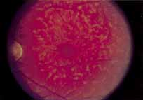
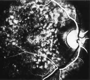
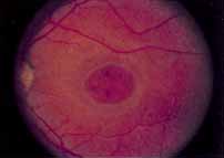
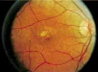
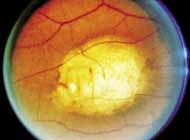
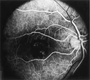
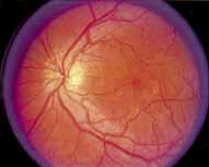
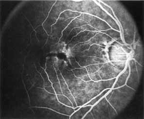
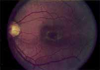
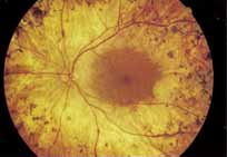
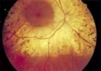
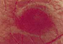
Although there is no reported genetic heterogeneity, there is wide phenotypic variation within the disorder. The major finding within the macula is a classic radial cystic maculopathy. Although retinal signs have been described in infants as young as 3 months, foveal schisis may be difficult to detect, leading to underdiagnosis. The diagnosis is usually not made until the affected male reaches school age (4 to 8 years of age) and encounters visual problems secondary to foveal involvement. Typical foveal schisis findings have been reported in 68% to 100% of eyes within various series.37,38 Foveal schisis is the only finding in about half the cases. It is characterized by the presence of radiate perifoveal microcysts located in the nerve fiber layer (Fig. 1) with radiate plications of the overlying internal limiting membrane that are seen especially well on monochromatic (red-free) photography (Fig. 2). The microcystoid change may slowly progress to form a macular cyst or hole. Foveal schisis has been reported in association with Goldmann-Favre vitreotapetoretinal dystrophy and rarely may be seen in rod-cone dystrophy or as an autosomal dominant or recessive condition.39–43

Fig. 1 X-linked juvenile retinoschisis. Central foveal schisis, presumably within the inner nerve fiber layer, surrounded by a fine radiate plication of the internal limiting membrane.

Fig. 2 X-linked juvenile retinoschisis. Red-free photography is well suited for demonstrating both the central foveal schisis and the fine radiate plication of the internal limiting membrane.

Peripheral features of this disorder include peripheral schisis, vitreous changes, perivascular sheathing, dendritiform patterns, retinal dragging, subretinal exudates, pigment lines, and a generalized tapetal-like reflex. Forsius and colleagues examined 268 eyes and classified severity of involvement into three cases, mild, moderate, and severe.31 Mild cases in 57% of eyes in this series demonstrated a typical schisis that was no longer readily distinguished except for pigment layer degeneration after 40 to 50 years of age. Moderate cases in 37% of patients in the series demonstrated similar macular findings with peripheral schisis or veils with retinal holes. Severe cases demonstrated schisis extending to the optic nervehead and severe anomalies involving retinal vessels and vitreous. In half of the cases, peripheral schisis is present which is usually bilateral, symmetric, and in the inferior temporal quadrant. The peripheral schisis only rarely extends to the ora serrata. The inner layer is usually elevated and may have multiple holes. Dendritic, opacified retinal vessels may be present within the retinal periphery, and fluorescein angiography in these cases reveals the presence of vascular leakage and capillary nonperfusion.36 Vitreous veils, which may contain branching retinal vessels, are sometimes prominent. Other vitreous changes include vitreous liquefaction, vitreous strands, traction bands, and vitreous detachment. Other changes associated with this disease include vitreous hemorrhage, optic atrophy, pseudopapilledema, ectopic maculae, iridocorneal angle anomalies, cataracts, disc and peripheral retinal neovascularization, neovascular glaucoma, and hemorrhagic retinal cyst.44–48 In end-stage disease, the inner wall of the schisis cavity may be a focus for glial proliferation and vitreous condensation, forming a retrolental membrane that results in leukocoria as a possible clinical presentation.35

Vision in this disease is variable and cannot be predicted based on the fundus findings alone. On occasion, it has been shown to be normal even in the presence of a foveal schisis. Visual acuity is usually in the 20/60 range in the affected young adult and may remain stationary for many years, with gradual deterioration to about 20/100 by the sixth decade. Most affected patients are legally blind by the seventh decade.31

Laser photocoagulation, cryopexy, and scleral buckling have been used with varying degrees of success to treat peripheral schisis that is rapidly advancing and threatening the posterior pole.49–53 The incidence of retinal detachment has varied from 0% to more than 20%.54 For these patients, scleral buckling may be used to treat retinal detachment that may arise in areas of peripheral retinoschisis with inner and outer layer holes. The redetachment rate may be as high as 40% regardless of scleral buckling versus vitrectomy.45 Vitrectomy may also be needed if significant vitreous traction or vitreous hemorrhage is present.54

Ancillary Tests

The posterior pole most often appears normal on fluorescein angiography and this may be helpful in the clinical differentiation of this entity from cystoid macular edema. In some cases there is evidence of pigment epithelial atrophy at the site of the macula. Kellner reported macular focal hyperfluorescence in the early phase, more distinct in older patients, paralleling pigment derangements found on ophthalmoscopy.44 Early in the course of the disease, color testing using the Farnsworth-Munsell 100-hue test shows a tritan defect.55,56 Later, a progressive deutan defect is found, correlating well with the visual acuity. Absolute visual field defects corresponding to zones of peripheral retinoschisis are present, as are relative central scotomas.

Electrophysiologic Tests

There is an overlapping of the fundus, electrophysiologic, and psychophysical findings among X-linked juvenile retinoschisis and the dominant (Wagner's) and recessive (Goldmann-Favre) types of vitreotapetoretinal dystrophies.55 In X-linked retinoschisis, the electroretinogram (ERG) is often abnormal. Both the photopic and scotopic b-wave, which arise from the bipolar cell layer, are usually reduced, whereas the a-wave, which arises from the inner segments of the photoreceptors, is intact, suggesting an inner retinal localization for these diseases. A demonstrated decrease in amplitude over several years indicates progressive disease, with nonrecordable ERG responses in advanced disease. The electrooculogram (EOG) is normal in mild cases, but may become subnormal in advanced cases. Dark adaptometry generally shows normal-to-subnormal cone and rod segments.

Differential Diagnosis

The diagnosis of the female carrier has been problematic because these females have normal visual function and electrophysiologic testing reveals no abnormalities. The fundus examination is also normal, although one case of a female carrier with radial wrinkling of the inner limiting membrane around the fovea, but no retinoschisis, has been reported.57 Recent studies of rod-mediated cone flicker thresholds and genetic linkage studies using restriction fragment length polymorphisms show promise in identifying the fundus-copically normal female carrier.57–59

X-linked juvenile retinoschisis must be differentiated from Goldmann-Favre disease and the other forms of inherited juvenile retinoschisis described in the next section, senile or acquired retinoschisis, cystoid macular edema, idiopathic macular hole formation, Wagner's disease, Stickler's syndrome, and other types of peripheral vitreoretinal abnormality.

FAMILIAL FOVEAL RETINOSCHISIS

Introduction

Lewis and Lee40 have described foveal retinoschisis in three daughters of funduscopically normal parents in a nonconsanguineous marriage. A male sibling was not affected. The condition is believed to be autosomal recessive. All three patients exhibited a mild degree of visual loss in the 20/30 to 20/50 range. Fundus changes were limited to the fovea. The pathology was identical to that seen in the early stages of X-linked retinoschisis. Fluorescein angiography revealed a barely discernible area of hyperfluorescence within the fovea of the oldest girl and the fovea was normal in the other two girls. Each patient showed a relative central scotoma on visual field testing.

Electrophysiologic testing (ERG and EOG) was normal in all girls tested. Dark adaptometry showed normal-to-subnormal cone and rod segments, and color testing revealed a mild tritan defect. The authors suggested that a deficiency of macular xanthophyll or a reduced sensitivity of foveal blue cones may exist in these patients.40 The complete natural history of this disorder has not yet been elucidated.

Subsequently, two families who demonstrated autosomal dominant transmission of juvenile retinoschisis have been reported. Yassur and investigators41 described eight affected members (five males and three females) in a three-generation pedigree with peripheral retinoschisis observed in all affected members. Of these individuals, only three had evidence of foveal schisis.41 The ERG was normal in mild cases, and subnormal b-waves were reported in the more severely affected individuals. Shimazaki and co-workers42 described a female with foveal and peripheral schisis whose mother also had foveal schisis. They proposed that an autosomal dominant pattern of inheritance was probably present in their limited pedigree.

Autosomal juvenile peripheral retinoschisis without foveal retinoschisis and autosomal juvenile retinoschisis associated with generalized rod and cone dysfunction probably represent yet other entities that are distinct from those described above.43,60,61

CONE DYSTROPHIES

Introduction

St. Albertus Magnus has been credited with the first description of cone dystrophy in the thirteenth centrury.62 The cone dystrophies represent a heterogenous group of disorders that can be inherited as an autosomal recessive, autosomal dominant, or X-linked recessive trait. The stationary cone dystrophies present as congenital disorders with with various degrees of cone dysfunction but normal rod function.63 In contrast, the progressive cone dystrophies often present in childhood or even early adulthood. These patients often develop rod dysfunction in later life and therefore overlap in clinical features with cone-rod dystrophies64,65

Based on an analysis of the cone dysfunction syndromes by Goodman and collaborators,65 and on work published by Krill and Deutman,66 the classification of cone dystrophies listed below can be used as an aid in the understanding of these conditions.

  1. Stationary forms:
    1. Rod monochromatism
      1. Autosomal recessive Complete
      2. Autosomal recessive Incomplete

    2. X-linked Blue Cone monochromatism
    3. Miscellaneous forms
      1. Achromatopsia with normal visual acuity
      2. Oligocone trichomacy


  2. Progressive forms: cone-rod dystrophies

The two forms, complete and incomplete rod monochromatism,66–68 probably reflect the total number of abnormal and missing cones. The presence of both forms of rod monochromatism in members of several affected families suggests that the complete and incomplete forms of rod monochromatism may represent different phenotypic expressions of the same genotype. Krill and Deutman66 have combined progressive cone dystrophy and progressive cone-rod dystrophy into one entity and refer to it as dominant macular degeneration. However, we shall refer to these dystrophies as progressive cone-rod dystrophies.

ROD MONOCHROMATISM: COMPLETE FORM

Synonyms: Typical monochromatism, complete achromatopsia

Introduction

Complete rod monochromatism is a rare form of congenital color defect. It is characterized by the presence of a marked generalized cone abnormality or complete absence of cones associated with a low level of visual acuity.64,65,69,70 The inheritance pattern is autosomal recessive. The mutations associated with rod monochromatism have not yet been completely identified. Arbour and colleagues72 have demonstrated linkiage of the disorder in an Iranian-Jewish pedigree to a centromeric region of chromosome 2. Rod monochromacy has also been reported with mutations in chromosome 14.73 Histopathologic study of donor eyes from patients with rod monochromatic has demonstrated the presence of cone-like structures within the retina.73 There is some controversy as to the nature and distribution of these cones. Two separate reports, one by Larsen74 and a second by Falls and co-workers75 found a normal total number of cones but with abnormal shapes. Harrison and colleagues76 reported abnormally shaped cones, reduced in total number throughout the retina.

Clinical Findings

The dystrophy is first detected in early youth as an absence of color vision with associated nystagmus, photophobia, and hemeralopia.74 Both the photophobia and the nystagmus tend to disappear as the patient becomes older, especially after 15 years of age. The patients often present with a high degree of hypermetropic refractive error. The clinical findings include a pronounced abnormality of color perception found on all color tests and a visual acuity in the 20/200 range. The vision is sometimes better for near distances and in dim illumination and worse in bright illumination. Visual fields may be normal or slightly constricted, particularly to colored targets, and a central scotoma can sometimes be demonstrated as the patient grows older. Nystagmus may make testing difficult. The nystagmus is pendular in youth, may decrease at near distances, and tends to decrease or disappear with time. The pupils respond slowly to light and paradoxic responses have been recorded during dark adaptation.76 Fundus changes are absent or minimal and, when present, are limited to the fovea. When present, these changes may include an abnormality or absence of the foveal reflex and, occasionally, a discrete, fine, granular disturbance of the retinal pigment epithelium within the central macula. The fovea has also been described as being hypoplastic.

No medical treatment is available, although tinted glasses help to decrease photophobia in bright illumination and may aid in slightly improving the visual acuity. Red-tinted contact lenses have also been tried with some success. Though central acuity remains diminished, the dystrophy is not progressive.

Ancillary Tests

Fluorescein angiography may show mild retinal pigment epithelium (RPE) transmission defects when a disturbance of the retinal pigment epithelium is present. Rod monochromats fail to recognize any plates on the common plate tests such as the Ishihara and Hardy-Rand-Ritller (HRR) tests. Rarely, residual cone function in rod monochromats can be demonstrated. A Stiles Crawford effect may be demonstrated and the dark adaptation curve maybe biphasic. Although Sharpe and Nordby reported residual cone function with ERG testing, these patients may have occult cases of incomplete achromatopsia or even progressive cone rode dystrophy.

Electrophysiologic Tests

The diagnosis is based on the findings of a congenital complete color defect with a nonprogressive subnormal visual acuity in the 20/200 range, normal scotopic ERG, absent or markedly subnormal photopic ERG, and a normal EOG.66–68

ROD MONOCHROMATISM: INCOMPLETE FORM

Synonym: Incomplete achromatopsia

Introduction

The clinical findings in incomplete achromatopsia are the same as those in the complete form, but they are usually less severe.64,66,69,70 Patients often have some residual color discrimination. The inheritance pattern is autosomal recessive. Photophobia and nystagmus may be absent, and visual acuity is less severely impaired than in individuals with the complete form. Unlike complete rod monochromats, individuals with the incomplete form have residual cone function mediated by photosensitive pigments other than rhodopsin, which provide the basis for some color perception.67,68

Clinical Findings

The diagnosis is based on the clinical findings of a congenital incomplete color defect with a nonprogressive subnormal visual acuity in the 20/60 to 20/200 range.66–68 Fundus changes are absent or minimal and, when present, are similar to those found in the complete form of rod monochromatism.

Ancillary Tests

Patients with incomplete rod chromatism demonstrate normal scotopic ERG response, nonrecordable or subnormal photopic ERG response, and a normal EOG.

BLUE CONE MONOCHROMATISM

Synonym: Atypical monochromatism

Introduction

Blue cone monochromatism, which demonstrates an X-linked recessive pattern of inheritance, is similar to the autosomal recessive form of rod monochromatism in its functional defects and clinical characteristics, although the two forms may be differentiated on the basis of spectral sensitivity and color matching. Female carriers of blue cone monochromatism may have an abnormal macular appearance and demonstrate abnormal photopic and flicker ERGs.

Clinical Findings

These male patients present with reduced acuity, nystagmus, and photophobia. Affected individuals are myopic, with fundus findings reflecting a tilted optic disc with a normal macular appearance.

Ancillary Tests

Under low photopic illumination levels, the blue cone monochromat will show dichromatic color vision. Hansen short wavelength perimetry can also be used to distinguish between blue cone and rod monochromats.

MISCELLANEOUS FORMS OF CONE MONOCHROMATISM

Synonyms: Atypical achromatopsia, achromatopsia with normal visual acuity, oligocone trichromacy.

Achromatopsia with normal visual acuity is a rare form of total color defect present in youth, reported in approximately 1 in every 100 million people64,69,77,78 It is characterized by minimal or no evidence of cone abnormality and relatively normal or only slightly decreased vision. The inheritance pattern is uncertain. Clinically, there is no photophobia or nystagmus, and the visual fields are usually normal. The fundus examination is also normal. Patients frequently have some degree of color vision, depending on the saturation of colors used, the luminance level, and the size of the test field. On routine color testing, however, these patients are found to be totally color defective. The ERG and the EOG are both normal.

Diagnosis is based on the clinical findings of congenital complete color blindness without amblyopia and a normal ERG and EOG. The differential diagnosis includes other forms of complete color defect without amblyopia, that is, the protan and deutan defects. No medical treatment is available, although the prognosis is good because the dystrophy is nonprogressive.

Oligocone trichromacy, first recognized by van Lith80 in a patient with reduced acuity, reduced photopic electroretinogram, and a normal appearing fundus, show good color discriminination. There appears to be a decreased photopigment concentration in these patients with normal regeneraration rates. Thus, it has been proposed that this disorder arises from a reduced cone population of all cone types. Its classification is ambiguous because many feel these patients should be classified as incomplete achromats versus oligocone trichromats.

PROGRESSIVE CONE-ROD DYSTROPHIES

Synonym: Progressive cone dystrophy

Introduction

The progressive cone-rod dystrophies constitute a relatively rare group of cone disorders that, for now, may be regarded as a single entity demonstrating variable expressivity.65,66,80–82 An attempt, however, has been made by one investigator to separate the group into two types based on electrophysiologic and psychophysical testing.71

This dystrophy is characterized by the presence of progressive color defects and a progressive decrease in visual acuity. There is usually a gradual decrease in visual acuity during the first or second decades of life, followed by a gradual loss of color vision. Frequently, these patients may go undiagnosed for years and they may be mistakenly believed to have a functional disorder.

The inheritance pattern is usually autosomal dominant, although autosomal recessive and X-linked recessive pedigrees have been described.83,84 Even within these inheritance modes, multiple different locations of mutations have been documented. Autosomal dominant progressive cone dystrophy has been mapped to chromosome 19q13.3, 17q12-p13, and to specific mutations of the peripherin/RDS gene on chromosome 6p.85–87 In patients with X-linked recessive progressive cone dystrophy, several loci have been mapped, including Xp21-p11.1, Xq27, and Xq28.88

No consistent metabolic or systemic abnormality has been found in patients with the progressive cone or cone-rod dystrophies, although there have been isolated reports of an association with low α-L-fucosidase activity in serum and leukocytes and chromosomal translocation.89 Histopathologic study reveals a complete disappearance of the photoreceptors in the macular and paramacular regions, with associated degeneration and disappearance of the retinal pigment epithelium.90 Likewise, there may be atrophy of the outer retinal layers in the periphery. These findings, together with the results of electrophysiologic and psychophysical testing, indicate that this dystrophy may be a form of outer segment photoreceptor degeneration and may be regarded, in some ways, to be the cone-rod counterpart to the rod-cone dystrophies (retinitis pigmentosa).

Clinical Findings

The progressive cone-rod dystrophies are characterized by selective involvement of the cones with the gradual development of color defects and a decrease in visual acuity. Photophobia is extremely common, along with hemeralopia. Night blindness (nyctalopia) is relatively rare but, when present, is usually associated with the more advanced stages of the dystrophy. Patients may develop total achromatopsia with visual acuities in the 20/200 range.

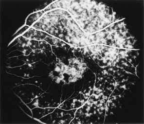
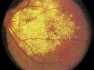
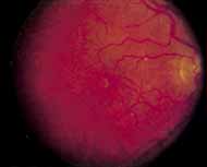
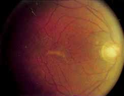
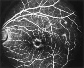
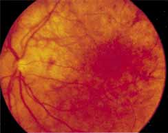
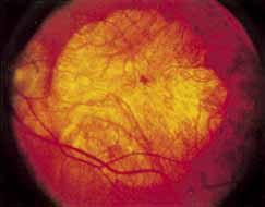
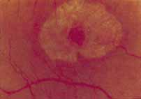
During the early stage of the dystrophy, when patients demonstrate a slight-to-moderate decrease in visual acuity and minimal color defects, there are minor or no visible fundus abnormalities. At most, the foveal reflex may be absent and there may be some increased granularity of the retinal pigment epithelium in the macula. Later, there is a decrease of visual acuity to the 20/400 range, oval atrophy of the macular retinal pigment epithelium (“beaten bronze” atrophy), and associated choroidal atrophy (Fig. 3). A characteristic bull's-eye maculopathy, similar to that seen in patients with chloroquine retinopathy, may also be seen.90 Photophobia, occasional nyctalopia, incomplete-to-complete color defects, and a central scotoma are often present. The symmetry of the process in both eyes is remarkable.

Fig. 3 Progressive cone-rod dystrophy. An oval zone of pigment epithelial derangement and atrophy is seen within the central macula. A faint bull's-eye pattern can be appreciated.

In the early stages of the dystrophy, the optic disc, retinal vessels, and peripheral retina are usually normal. The patient may have a visual acuity of 20/200 and normal color vision by standard testing and, yet, generalized cone dysfunction can be demonstrated by color field testing. In the late stages, there may be temporal optic disc pallor, attenuation of the retinal arterioles, and granular or bone spicule peripheral retinal pigmentary changes. Not infrequently, the peripheral pigmentary abnormalities may be regional. This probably reflects regional rod involvement, as evidenced by ERG, which may demonstrate a reduction in amplitude without a change in latency of the rod responses.72,80 At this stage, contracted peripheral fields may be found on both white and color target testing.

No medical treatment is available. The prognosis for vision is guarded and poorer than for the other cone dystrophies because both cone and rod function is impaired in the macula and retinal periphery. Accordingly, central vision is decreased and color vision impaired in association with contraction of the peripheral visual fields. As the dystrophy progresses, vision may decrease to the 20/400 level or less and may also be accompanied by contracted peripheral fields and a total color defect. Patients with poor central vision should be referred to low visual aids. Severe photophobia in adults and children has been reduced with the use of appropriately tinted spectacles or contacts. The tint will often depend on the dystrophy as some patients with relatively well-preserved color vision require neutral tints and other more severely affected patients require red tints similar to those in rod monochromats. Miotic drops have been used in some patients with severe photophobia but are not generally well tolerated or advised.

Ancillary Tests

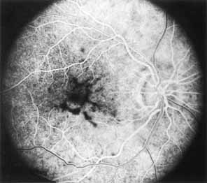
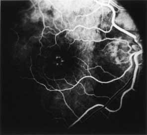
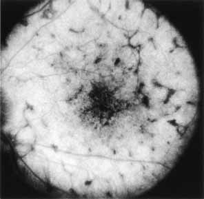
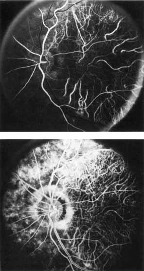
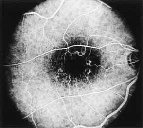
Fluorescein angiography demonstrates increased transmission of choroidal fluorescence in the macula during early phases of the study, without late leakage of dye or fluorescein staining. In addition, an annular pattern of hyperfluorescence is often seen in the macula, highlighting the bull's-eye pattern seen on fundus examination (Fig. 4). Visual field defects include central scotoma, peripheral field loss, and ring scotoma.

Fig. 4 Progressive cone-rod dystrophy. Fluorescein angiography in mid arteriovenous phase demonstrates an annular retinal pigment epithelium (RPE) transmission window defect accentuating the bull's-eye pattern seen in Fig. 3.

Electrophysiologic Testing

In the early stages of the dystrophy, electrophysiologic testing reveals normal photopic and scotopic ERG responses. With progression of the disease, there is a moderate-to-marked loss of cone function, associated with a slight-to-moderate loss of rod function, markedly decreased or absent photopic responses and mildly decreased scotopic responses. Unusual variants with supernormal scotopic responses to suprathreshold stimuli have been described in some pedigrees.91,92 The EOGin the early stage of the dystrophy is normal. With more widespread involvement of the retina, a reduction of the light rise develops and the EOG becomes a sensitive indicator of the progress of the dystrophy. The EOG first becomes subnormal, and is then followed by the presence of a subnormal ERG. Dark adaptation curves late in the course of the dystrophy are monophasic with slightly elevated rod thresholds.

Differential Diagnosis

The diagnosis is based on the findings of progressive loss of central and color vision beginning in the first or second decades of life, with associated photophobia, a characteristic bull's-eye macular lesion, markedly decreased or absent photopic ERG responses, mildly decreased scotopic ERG responses, a mildly abnormal dark adaptation curve and, in the late stages, peripheral retinal pigmentary changes with retinal arteriolar attenuation.

The differential diagnosis includes rod monochromatism, pericentral and sine pigmento forms of retinitis pigmentosa (rod-cone dystrophy), Stargardt's disease, central areolar choroidal dystrophy, chloroquine retinopathy, and congenital optic atrophy. Finally, certain syndromes are associated with cone dystrophies including Bardet-Biedl syndrome, Alstroms's syndrome, and Pierre-Marie ataxia with cone rod dystrophy.

PERICENTRAL (INVERSE) RETINITIS PIGMENTOSA (ROD-CONE DYSTROPHY)

Synonyms: Pericentral Pigmentary retinopathy, pericentral Pigmentary dystrophy, Peripapillary Pigmentaryretinal degeneration, pericentral Pigmentary degeneration

Introduction

Pericentral retinitis pigmentosa (rod-cone dystrophy) is characterized by pigmentary disturbances similar to those occurring in classic retinitis pigmentosa (rod-cone dystrophy), but which occur solely in the pericentral retina with no involvement of the peripheral retina.20 Grondahl94 described twenty-eight patients from four families, some demonstrating an autosomal dominant pattern of inheritance. Other previous studies from Traboulsi, O'Neill and Maumenee have reported an autosomal recessive mode of inheritance.95

Clinical Findings

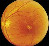
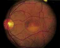
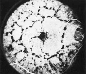
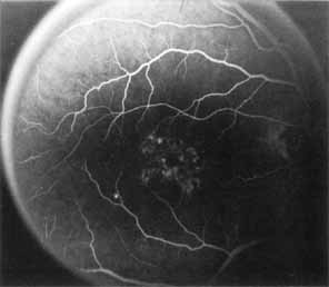
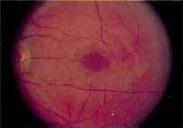
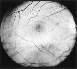
Vision is usually normal in the early stage and patients are often diagnosed on routine fundus examinations in the second and third decades of life. In later stages, vision may be markedly reduced to the 20/200 level or worse. Color vision is normal early on, but frequently becomes defective and may be accompanied by a progressive decrease in central vision. Nyctalopia is often present. The pigmentary changes may take the form of bone spicules or scattered black dots and are the earliest signs of the dystrophy (Fig. 5 and 6). Because the fovea is lacking in vessels, no bone spicules are present in this region. Atrophy of the retinal pigment epithelium may also be present. In contradistinction to retinitis pigmentosa (rod-cone dystrophy), the disc and retinal vessels are usually normal until late in the course of the disease.

Fig. 5 Pericentral retinitis pigmentosa. Pigment epithelial atrophy and associated pigmentations surround an intact central macula. As the disease in this patient is fairly advanced, the retinal vessels show moderate attenuation. (Courtesy of Dr. Roland Sabutes: Fenestrated sheen macular dystrophy: A new autosomal dominant maculopathy. Arch. Ophthalmol. 97:1293, 1979. Copyright © 1979 American Medical Association.)

Fig. 6 Pericentral retinitis pigmentosa. Fellow eye of patient photographed in Fig. 5 demonstrates the large annular zone of pigment epithelial atrophy and pigmentations surrounding an intact central macula. Note the unaffected retina peripheral to this annular zone of involvement. (Courtesy of Dr. Roland Sabutes: Fenestrated sheen macular dystrophy: A new autosomal dominant maculopathy. Arch. Ophthalmol. 97:1293, 1979. Copyright © 1979 American Medical Association.)

Durlu and colleagues96 described macular complications occurring in two patients including a full thickness macular hole and central retinal artery occlusion. As is the case with retinitis pigmentosa, many forms of therapy have been attempted, none of which have been successful.

Ancillary Tests

Tritan color defects have been reported on Ishihara and Farnsworth-Munsell testing. Despite the common presenting symptom of nyctalopia, the dark adaptometry is normal or slightly delayed.96 The angiogram in the early course of the disease is often unremarkable without evidence of retinal pigmentary derangement.

Electrophysiologic Tests

The primary lesion is thought to reside in the photoreceptors and the pigment epithelium, but due to the localized nature of the process, the EOG and ERG are often normal or only slightly subnormal.11,21,96

Differential Diagnosis

The differential diagnosis includes classic retinitis pigmentosa (rod-cone dystrophy), progressive cone-rod dystrophy, the late stage of fundus flavimaculatus, and inflammatory and infectious causes of chorioretinitis. The ERG should enable one to differentiate this dystrophy from the progressive cone-rod dystrophies and classic retinitis pigmentosa (rod-cone dystrophy). The retinopathy of neuronal ceroid lipofuscinosis and olivo- pontoceerebeallar ataxia potentially involves the central macula in a similar presentation.96,97

DOMINANT CYSTOID MACULAR DYSTROPHY

Introduction

Deutman and Pinckers reported a 4-year-old female with severe hyperopia and diminished acuity.98 In addition to a pigmentary retinopathy, cystoid edema was a prominent feature.98–102 Deutman and colleagues98 further characterized three of five families with the disorder. An additional report describes an affected Greek family.101 Since that time, the gene for dominant cystoid macular dystrophy has been localized to chromosome 7p.102

Clinical Findings

Dominantly inherited cystoid macular edema is characterized in the first and second decades with a prolonged course of macular cystoid change and progressive loss of central vision. The pathogenesis and prevalence of this dystrophy are as yet undetermined. Hyperopia from +2.00 to +10.00 D is seen along with hypopigmentation within the central macula. Macular atrophy is a fairly prominent finding late in the disease. Occasionally, a bull's-eye pattern of atrophic change may be seen. Vitreous cells, strands, and veils, and peripheral retinal pigmentary disturbances ranging from a slight hyperpigmentation and depigmentation to the formation of bone spicule changes have also been reported. Late in the disease, vision may be reduced to the 20/200 to finger counting range and a relative-to-absolute central scotoma may be present. No known treatment is available.

Ancillary Tests

Fluorescein angiography reveals leakage from perifoveal capillaries with dye pooling in a cystoid pattern. Later in the disease, transmission defects are seen in areas of RPE atrophy. In some cases, fluorescein leakage from optic disc capillaries has also been noted. Color testing in some patients often demonstrated blue yellow defects likely secondary to macular edema and in chronic cases red green deficiencies secondary to photoreceptor damage.

Electrophysiologic Studies

The ERG has been normal in all patients studied to date. The EOG and results of dark adaptometry, however, are subnormal in most, but not all, patients. The subnormal EOG would indicate widespread dysfunction of the retinal pigment epithelium.

Differential Diagnosis

The condition should be differentiated from other hereditary retinal disorders that are associated with cystoid macular edema, especially retinitis pigmentosa (rod-cone dystrophy), and dystrophies characterized by the presence of foveal retinoschisis (which may be mistaken for cystoid macular edema) such as X-linked juvenile retinoschisis and familial foveal retinoschisis. Cystoid macular edema is seen after ophthalmic surgeries, pars planitis, and other ocular inflammatory disorders. Vascular disorders such as diabetic retinopathy, Coat's disease, and parafoveal telangectasis also cause cystoid macular edema. Finally, high dose nicotinic acid has been reported to cause cystoid edema without obvious leakage from perifoveal capillaries.103

FENESTRATED SHEEN MACULAR DYSTROPHY

Introduction

Fenestrated sheen macular dystrophy is an autosomal dominant condition with high penetrance. This is a rare disorder and only four affected pedigrees have been reported in the literature.104–106

Clinical Findings

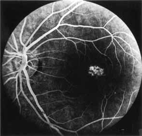
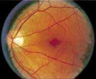
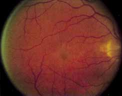
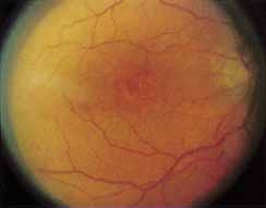
Fenestrated sheen macular dystrophy is a slowly progressive disease that develops in childhood.103–107 The youngest reported case was in a 4-year-old girl. In the early stages, small, red demarcated lesions can be seen within the deep neurosensory retina106 It is characterized by the presence of a yellowish refractile sheen with red fenestrations within the macula (Fig. 7). The sheen, which is located between the retinal pigment epithelium and the retinal vessels, is also present within the foveal avascular zone and may be related to the macular luteal pigment. The fenestrations appear to be tiny windows within the sheen that allow increased visualization of the underlying pigment epithelium. The changes within the sensory retina are slowly progressive and are eventually accompanied by changes of the retinal pigment epithelium. By the third decade, an annular zone of hypopigmentation of the retinal pigment epithelium appears around the area of sheen and progressively enlarges to form a bull's-eye macular lesion (Fig. 8). An abnormal degree of RPE granularity in the posterior pole and the peripheral retina has also been reported in some affected individuals.106 During this period of change, visual acuity usually remains normal, although paracentral scotomas develop by the sixth decade of life. The prognosis for maintenance of good central vision is excellent and no treatment is available. Despite the ophthalmoscopically evident macular changes, central visual acuity remains normal.

Fig. 7 Fenestrated sheen macular dystrophy. A yellowish retractile sheen and red fenestrations are present within the central macula in this relatively early case of the disease. (Courtesy of O'Donnell, FE, Jr., Welch RB: Fenestrated sheen macular dystrophy: A new autosomal dominant maculopathy. Arch. Ophthalmol. 97:1293, 1979. Copyright © 1979 American Medical Association.)

Fig. 8 Fenestrated sheen macular dystrophy. An annular zone of hypopigmentation of the pigment epithelium appears around the central area of sheen creating a bull's-eye pattern. (Courtesy of O'Donnell, FE, Jr., Welch RB: Fenestrated sheen macular dystrophy: A new autosomal dominant maculopathy. Arch Ophthalmol 97:1293, 1979. Copyright © 1979 American Medical Association.)

Ancillary Tests

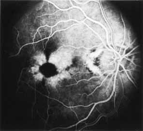
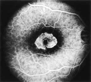
The fluorescein angiographic features of this dystrophy are dependent on chronicity of the disorder. Younger patients have essentially normal studies. Older patients demonstrate multiple punctate window defects in an annular zone ringed by an area of slightly subnormal choroidal fluorescence due to increased pigmentation of the retinal pigment epithelium (Fig. 9). The oldest reported patient, a 56-year-old male, had a large confluent annular transmission defect of the retinal pigment epithelium with normal perfusion of the underlying choriocapillaris (Fig. 10). This individual demonstrated progression of his lesion over an 18-month period.

Fig. 9 Fenestrated sheen macular dystrophy. Fluorescein angiography in arteriovenous phase reveals multiple punctate RPE transmission window defects surrounding a central area of somewhat increased hypofluorescence. (O'Donnell FE Jr, Welch RB: Fenestrated sheen macular dystrophy: a new autosomal dominant maculopathy. Arch Ophthalmol 97:1292, 1979. Copyright © 1979 American Medical Association.)

Fig. 10 Fenestrated sheen macular dystrophy. Fluorescein angiography in arteriovenous phase reveals a large, confluent, annular RPE transmission window defect with normal perfusion of the choriocapillaris. (O'Donnell FE Jr, Welch RB: Fenestrated sheen macular dystrophy: a new autosomal dominant maculopathy. Arch Ophthalmol 97:1292, 1979. Copyright © 1979 American Medical Association.)

Electrophysiologic Tests

Retinal function studies correlate roughly with age. Younger patients have normal ERGs and EOGs and a mild red-green color defect. Older patients have subnormal photopic ERGs and normal-to-subnormal EOGs.

Differential Diagnosis

The small red macular fenestrations in the early course of the disease appear similar to those described in acute macular neuroretinopathy. Patients with that disorder are often symptomatic, however. The differential diagnosis includes diseases with retinal pigment epithelial changes in an annular pattern including Stargardt's disease, progressive cone-rod dystrophy, benign concentric annular macular dystrophy, central areolar pigment epithelial dystrophy, and central areolar choroidal dystrophy.

Back to Top
RETINAL PIGMENT EPITHELIUM

FUNDUS FLAVIMACULATUS

Introduction

Although historically described as two separate entities, investigators have come to believe that, apart from their ophthalmoscopic appearance, there is no clear distinction between fundus flavimaculatus and Stargardt's disease.108–111 This is supported by reports of the presence of both entities within the same pedigrees, and evolution of one entity into the other in the same individual over time.111–116 These conditions, however, are still often described as though they were two separate entities because of the distinctions made in the past literature. Together, these dystrophies account for 7% of all retinal dystrophies and are noted to be among the most common causes for macular diseases in children.113

Fundus flavimaculatus was initially described by Franceschetti in 1963115 as a condition in which yellow subretinal lesions were scattered throughout the posterior pole in 30 patients, half of whom were older than 25 years of age. Fundus flavimaculatus is an autosomal-recessive dystrophy now localized to the short arm chromosome 1p.116 It is is usually diagnosed before the third and fourth decades of life and affects both genders equally. It may be discovered on routine ophthalmoscopic examination, or the patient may present with a slowly progressive decrease in vision secondary to macular involvement. Histopathologic examination of an eye with fundus flavimaculatus has demonstrated that the yellow flecks are dense accumulations of periodic acid–Schiff (PAS)–positive lipofuscin-like material within engorged RPE cells.117 The “dark choroid” may be caused by diffuse deposition of this lipofuscin-like material in the RPE cells rather than a defect in choroidal perfusion. The presence of this lipofuscin-like material within RPE cells has even been demonstrated in the eye of a 16-month-old infant with incipient fundus flavimaculatus enucleated because of retinoblastoma. The fellow eye developed findings typical of fundus flavimaculatus years later.118 Another ultrastructural study has revealed elevated aggregates of enlarged RPE cells with apices distended by tubulovesicular lipid membranes in the absence of a demonstrable abnormal accumulation of lipofuscin.119 These differing histopathologic reports indicate that the clinical appearance of fundus flavimaculatus may be the result of more than one specific metabolic disorder of the retinal pigment epithelium.

Clinical Findings

The bilaterally symmetric fundus picture is characterized by the presence of multiple angulated or fishtail-shaped yellow-white lesions confined to the retinal pigment epithelium of the posterior pole (Fig. 11). The size, shape, and confluency of the yellow flecks vary considerably, and new clusters may appear periodically as old ones fade. The optic disc, retinal vessels, and periphery are normal in the early stage of the disease.

Fig. 11 Fundus flavimaculatus. Numerous, yellowish, angulated flecks tending to be confluent are present within the posterior pole at the level of the pigment epithelium. This patient has sparing of the central fovea.

In the typical case, there are no atrophic macular lesions and flecks may be present within the posterior pole without significant accompanying visual loss. Occasionally, however, a fleck may happen to be present in the fovea resulting in secondary involvement of photoreceptors with variable visual consequences.120 An atrophic macular process may occur many years after the typical flavimaculatus spots are first observed. Rare cases of subretinal neovascularization, retinal neovascularization, and cystoid macular change have also been reported in association with fundus flavimaculatus.121–123 Visual acuity may be in the 20/40 level range for many years, but once visual acuity drops below this level, more rapid deterioration of vision occurs. Vision eventually stabilizes at the 20/200 level depending on the extent and degree of macular involvement. There is presently no therapy for this dystrophy.

Ancillary Tests

Fluorescein angiography reveals that recently formed fundus flavimaculatus lesions do not fluoresce, possibly because the RPE cells are still intact.108,124,125 In fact, in the early phase of fluorescein angiography, many lesions, particularly the earliest lesions, block background fluorescence (Fig. 12). Later, multiple well-defined hyperfluorescent areas corresponding to the ophthalmoscopically visible lesions develop. In general, more lesions can be detected on fluorescein angiography than are seen on clinical examination. The hyperfluorescence is believed to be the result of transmission defects in the deranged and atrophic retinal pigment epithelium, although some flecks may also be taking up fluorescein dye late in the study (Fig. 13). There is often a generalized decreased visualization of chroidal fluorescence, which has been referred to as a dark choroid.125,126 An acquired red-green dyschromatopsia, as well as a central scotoma, may occur in patients with atrophic macular lesions.

Fig. 12 Fundus flavimaculatus. Fluorescein angiography in midarteriovenous phase shows many flecks blocking background fluorescence while many others transmit background fluorescence.

Fig. 13 Fundus flavimaculatus. Fluorescein angiography in late phase shows RPE transmission window defects corresponding to many of the flecks.

Electrophysiologic Tests

Electrophysiologic studies suggest primary involvement of the retinal pigment epithelium. The ERG may be normal and show mild abnormalities, but it is never nonrecordable as is the case with the diffuse tapetoretinal dystrophies. The EOG is usually subnormal, suggesting a widespread defect in the retinal pigment epithelium. However, it is never as abnormal as is found in Best's vitelliform dystrophy123,126

Differential Diagnosis

The differential diagnosis of fundus flavimaculatus includes familial drusen, fundus albipunctatus, and fleck retina of Kandori, all of which have been grouped together and referred to as the flecked retina syndromes.127,128 A funduscopic appearance simulating fundus flavimaculatus has also been reported in association with non-Hodgkin's lymphoma.129 Patients with Kjellin's syndrome, an autosomal recessively inherited disorder characterized by the presence of spastic paraparesis and dementia, may also demonstrate bilaterally symmetric, yellow-white, round or dumbbell-shaped, perifoveal lesions at the level of the retinal pigment epithelium that may resemble the lesions seen in patients with fundus flavimaculatus.130

Cases of fundus flavimaculatus have been described in association with tapetoretinal degenerative changes, Laurence-Moon-Bardet-Biedl syndrome, cerebral atrophy, cerebellar atrophy, incontinentia pigmenti, myotonic dystrophy, and other ocular and systemic conditions. It is not clear whether these have any connection with the pathogenesis of fundus flavimaculatus.130–132 The postulated enzymatic defect in the retinal pigment epithelium remains unknown.

STARGARDT'S DISEASE

Synonym: Juvenile macular degeneration

Introduction

Originally described by Stargardt in 1909, Stargardt's disease is a bilaterally symmetric, autosomal recessively inherited condition that is characterized by a progressive loss of central vision.133–137 The locus is identical to that described in fundus flavimaculatus, the short arm chromosome 1.137 A dominant form, which may represent a separate entity, is described in the next section.

Clinical Findings

The appearance of the fundus and the decrease in acuity may not always parallel each other and one should be cautioned against prematurely dismissing a patient with decreased vision and normal-appearing fundi as having “hysteria.” The macula is generally involved first, usually between the ages of 6 and 20. The process may remain central or involve the periphery, producing an ophthalmoscopic picture similar to that seen in fundus flavimaculatus. Many transitional forms exist, so that a long period of follow-up may be needed to delineate the full extent of involvement.

Clinically, the most important features are the progressive loss of vision and the fundus appearance. The age at onset, which is used in some classifications, is much less reliable than these other characteristics and should not be used in defining this disease. A classification incorporating a system of stages in the development and resorption of flecks and in the progression of RPE atrophy is of much greater value and allows quantitative evaluation of the extent of retinal disease.114

As the disease progresses, a horizontal oval area of pigment epithelial atrophy develops, and the foveal region takes on a characteristic “beaten-bronze” appearance (Fig. 14) that is virtually identical to that seen in the progressive cone-rod dystrophies. Parafoveal and midperipheral yellow-white flecks may occur late in the disease, representing the large degree of heterogeneity between Stargardt's disease and fundus flavimaculatus. When the periphery is involved, it may demonstrate the presence of round, granular black spots of increased pigmentation with surrounding depigmented areas. As the atrophic process extends deeper, the choroid becomes exposed and, in the later stages, the geographic atrophy within the posterior pole may resemble that seen in central areolar choroidal dystrophy. Meanwhile, the peripheral retina may develop an appearance simulating retinitis pigmentosa (rod-cone dystrophy) with bone spicule pigmentation and irregular zones of pigment atrophy.

Fig. 14 Stargardt's disease. An oval area of pigment epithelial derangement and atrophy is present within the central macula. Early pigmentations within this zone are also apparent. Scattered yellowish flecks identical to those seen in fundus flavimaculatus surround this central zone but are quite subtle and poorly visualized. They can be much better appreciated on fluorescein angiography.

Visual acuity usually diminishes to the 20/200 level or worse if peripheral involvement is extensive. Central scotomas are present, while peripheral fields remain little affected until late in the course of the disease.

Ancillary Tests

The classic dark or silent choroid is a diagnostic angiographic finding that has been postulated as a secondary effect of blockage of the choroidal fluorescence from the aforementioned accumulation of lipufuscin. Fishman and colleagues reported the dark choroid effect was seen in 85% of patients with clinically diagnosed Stargardts and fundus flavimaculatus, though genetic testing was not performed on all patients.110 Fluorescein angiography otherwise may also show expected pigment epithelial and choroidal transmission defects also described in fundus flavimaculatus (Fig. 15). Wrobleski and associates134 have also described the use of indocyanine green angiography in patients with Stargardt's and fundus flavimaculatus. An acquired red-green dyschromatopsia may be characteristic in these patients with color testing.

Fig. 15 Stargardt's disease. Fluorescein angiography in arteriovenous phase shows an oval zone of retinal pigment epithelium (RPE) transmission window defect within the central macula. The hypofluorescent spots correspond to pigmentations developing within this area (see Fig. 14). Transmission pigment epithelial window defects within the remainder of the posterior pole relate to many of the flecks not well seen on Fig. 14.

Electrophysiologic Tests

Both the ERG and the EOG are normal with purely central involvement, but they are often abnormal with more diffuse involvement extending into the periphery. Lee and Heckenlively135 reported a greater likelihood of normal ERG findings in Stargardt's disease versus fundus flavimaculatus. Visual-evoked responses are often subnormal, even with good vision and minimal fundus changes and may be helpful in establishing an early diagnosis.

Histopathologic studies reveal a complete disappearance of the visual elements in the macula.22 However, it is uncertain whether this represents a primary defect in the photoreceptors themselves or if it is secondary to a defect in the retinal pigment epithelium. It is probable that the defects in the photoreceptors are secondary to the RPE abnormalities.12

There is no known enzymatic defect associated with Stargardt's disease and there is no effective therapy.

AUTOSOMAL DOMINANT FUNDUS FLAVIMACULATUS

Synonyms: Dominant progressive foveal dystrophy, dominant Stargardt's disease

This clinical entity presents a picture similar to that just described for Stargardt's disease with retinal flecks occasionally being present.138,139 It has, in fact, been called a dominant form of Stargardt's disease.135–137 Other authors point out that since all of Stargardt's cases were recessively inherited, a strict differentiation between the two entities should be made because each could involve a different pathologic gene.

Dominant progressive foveal dystrophy is much less common than autosomal recessive Stargardt's disease. Three large pedigrees have been described in the literature as well as multiple smaller pedigrees in larger studies including all forms of Stargardt's disease and fundus flavimaculatus.138,139 In pedigree, mapping of the autosomal dominant form was located at chromosome 6q and in another, chromosome 13q.138,139 Patients may present at a later age with decreased vision and/or decreased color discrimination. Peripheral retinal involvement is usually less extensive, the course of the disease is usually less progressive, and any accompanying macular changes may be more subtle. Central scotomas may accompany the decrease in vision. Dark adaptometry and the ERG are normal. The EOG, however, may be abnormal. These findings tend to place the primary level of involvement as that of the retinal pigment epithelium and photoreceptors.

CENTRAL AREOLAR PIGMENT EPITHELIAL DYSTROPHY

Synonyms: North Carolina macular dystrophy, Lefler-Wadsworth-Sidbury dystrophy

Introduction

Dobbie and colleagues140 in 1975 first described this autosomal dominant disorder with a high degree of penetrance and variable expressivity. Characteristics of this dystrophy include onset in the first decade, lack of visual symptoms, normal visual acuity, and a nonprogressive course. Subsequent reports of this rare dystrophy have confirmed the autosomal dominant inheritance of this entity140–144 Central areolar pigment epithelial (CAPE) dystrophy, together with the autosomal dominant central pigment epithelial and choroidal degenera-tion of Leveille-Morse-Kiernan, now represent variants of North Carolina macular dystrophy described be-low.144,145 CAPE dystrophy shared similarities with North Carolina macular dystrophy, and since their initial description, Small and associates have since reported both diseases were found in patients descended from the same three Irish brothers who lived in Spartanbur, South Carolina in 1790.146,147 Although North Carolina macular dystrophy was initially thought to be progressive, later reports have emphasized its stationary nature, a situation similar to that seen with CAPE dystrophy. For the purpose of additional historical perspective, we have elected to include a separate discussion of North Carolina Macular dystrophy. The North Carolina Macular dystrophy locus (MCDR1) has been mapped to 6q14-q16.2.148

Clinical Findings

The fundus lesions of CAPE dystrophy consist of an RPE disturbance within the fovea which may progress to become a confluent zone of RPE atrophy (Fig. 16).

Fig. 16 Central areolar pigment epithelial (CAPE) dystrophy. Irregular pigment epithelial derangement and atrophy is present within the fovea. This zone is surrounded by fine drusen-like lesions. (Fetkenhour CL, Gurney N, Dobbie JG, et al: Central areolar pigment epithelial dystrophy. Am J Ophthalmol 81:745, 1976)

Ancillary Tests

Fluorescein angiography reveals RPE transmission defects corresponding to the punctate and confluent lesions within the fovea (Fig. 17). In the occasional instance when the disorder is complicated by subretinal neovascular membrane formation, subretinal hemorrhage, retinal edema, dye leakage, and fluorescein pooling may be seen within the macula depending on the degree of associated degenerative change.

Fig. 17 Central areolar pigment epithelial (CAPE) dystrophy. Fluorescein angiography in arteriovenous phase demonstrates retinal pigment epithelium (RPE) transmission window defects corresponding to lesions within the central macula (see Fig. 16). (Fetkenhour CL, Gurney N, Dobbie JG, et al: Central areolar pigment epithelial dystrophy. Am J Ophthalmol 81:745, 1976)

Electrophysiologic Tests

ERG and EOG studies performed on patients with CAPE dystrophy are normal, as are the results of dark adaptometry and color testing. The normality of these results helps to distinguish this entity from most other macular dystrophies.

Differential Diagnosis

The differential diagnosis includes dominant progressive foveal dystrophy (dominant Stargardt's), familial drusen, central areolar choroidal dystrophy, and congenital macular colobomas.149 However, visual acuity is frequently decreased in the presence of minimal, if any, foveal changes in dominant progressive foveal dystrophy and drusen usually extend beyond the central macula in individuals with familial drusen.

NORTH CAROLINA MACULAR DYSTROPHY

Synonyms: Lefler-Wadsworth-Sidbury dystrophy, CAPE dystrophy

Introduction

In 1971, Lefler and colleagues147 described an autosomal dominant foveal dystrophy found in 25 of 70 members of a family spanning four generations. By 1974, this pedigree had been expanded to include 545 individuals spanning seven generations; 130 were studied ophthalmoscopically, and 50 were found to be affected by the dystrophy.149 Because this pedigree was traced back to a family originating in North Carolina, it is now referred to as North Carolina macular dystrophy.

Gass,151 who examined two families with this entity who originated from North Carolina, also emphasized the generally stationary nature of this dystrophy. He noted the presence of staphylomatous areas of chorioretinal atrophy in severe disease and the frequent presence of peripheral drusen. He also reported one patient with a disciform scar in one eye. It is now believed that dominant progressive foveal dystrophy of Frank-Landers-Williams-Sidbury,147 CAPE dystrophies of Hermsen and Judisch,144 as well as of Fetkenhour-Gurney-Dobbie-Choromokos,142 represent cases of what we now refer to as North Carolina macular dystrophy. In addition, it is likely that the central pigment epithelial and choroidal degeneration of Leveille-Morse-Kiernan145 and the central retinal pigment epithelial dystrophy of Klein and Bresnick143 also represent the same entity.

There is no known underlying enzymatic disorder, although a transport type of aminoaciduria was noted in many members of the family studied, but was found to be unrelated genetically to the foveal dystrophy.147

Clinical Findings

The dystrophy develops during the first year of life, and the foveal lesions were thought to progress throughout childhood to reach their final stage by late puberty. The final visual acuity is also reached by late puberty. Marked variations are noted in the final stage achieved by different siblings. Some individuals have foveal lesions arresting at an early or intermediate stage, whereas the lesions of others progress to a final atrophic stage.

Ophthalmoscopically, the macular changes can be divided into three stages:

Stage 1. Scattered drusen and pigment dispersion within the fovea (Fig. 18). Visual acuity is usually 20/20, and central or paracentral scotomas are not present.

Fig. 18 Lefler-Wadworth-Sidbury foveal dystrophy. The stage 1 lesion is characterized by scattered drusen and pigment dispersion within the fovea. (Courtesy of Dr. Michael E. Barricks; G from Marmor, MF, Byers B: Pattern dystrophy of the pigment epithelium. Am J Ophthalmol. 84:32, 1977)

Stage 2. Confluent drusen with or without pigment clumping within the central macula. RPE atrophy and partial choroidal atrophy may be seen (Fig. 19). The visual acuity is usually in the 20/50 range, and small central or paracentral scotomas may be present.

Fig. 19 Lefler-Wadworth-Sidbury foveal dystrophy. The stage 2 lesion is characterized by confluent drusen with early pigment clumping within the central macula. In this case early pigment epithelial atrophy is also seen. (Courtesy of Dr. Michael E. Barricks; G from Marmor, MF, Byers B: Pattern dystrophy of the pigment epithelium. Am J Ophthalmol. 84:32, 1977)

Stage 3. Choroidal atrophy. Total RPE and choriocapillary atrophy of the fovea, parafovea, or entire macula may develop (Fig. 20).

Fig. 20 Lefler-Wadworth-Sidbury foveal dystrophy. The stage 3 lesion is characterized by total pigment epithelial and choriocapillary atrophy within the macula. (Courtesy of Dr. Michael E. Barricks; G from Marmor, MF, Byers B: Pattern dystrophy of the pigment epithelium. Am J Ophthalmol. 84:32, 1977)

Visual acuity is usually in the 20/50 to 20/200 range and large central or paracentral scotomas are present. The foveal lesion is always moderately severe before any decrease in visual acuity occurs. Thereafter, the decrease in visual acuity closely parallels the severity of the foveal lesion. Likewise, the size and development of central scotomas correspond closely to the stage and size of the foveal lesions. There is no known therapy for this dystrophy.

Ancillary Tests

Stage 1. Fluorescein angiography reveals transmission defects within the fovea corresponding to the drusen.

Stage 2. Fluorescein angiography shows a more confluent pattern of transmission defects with no evidence of dye leakage or pooling.

Stage 3. Angiography demonstrates diffuse transmission defects secondary to total RPE and choriocapillary atrophy (Fig. 20).

Electrophysiologic Testing

The ERG and EOG are normal, as are dark adaptometry and color vision testing.

Differential Diagnosis

The aforementioned differential diagnosis includes dominant progressive foveal dystrophy (dominant Stargardt's), familial drusen, central areolar choroidal dystrophy, and congenital macular colobomas.148

FAMILIAL DRUSEN

Synonyms: Dominant drusen of Bruch's membrane, Doyne's honeycomb choroiditis, Hutchinson-Tay central guttate choroiditis, Holthouse-Batten superficial choroiditis, Malattia-leventinese, Sorsby's macular dystrohy

Introduction

The name drusen is derived from the German word druse (plural, drusen), meaning “gland,” indicating that early observers were reminded of glandular structures by these yellow-white deposits. Gass152 described druse as “nodule”, pertaining to areas of clear crystallization within stones. All of the synonyms noted above apply to the same disease. This entity demonstrates an autosomal dominant pattern of inheritance which was isolated to Chromosome 2p16-p21, with the onset often between the third and fourth decades.153 However, since patients are generally asymptomatic in the early stages of the condition, the diagnosis may not be made until later in life.

Retinal function studies point toward localization of the dystrophic process at or near the level of the retinal pigment epithelium. Histopathologically, drusen are focal collections of eosinophilic homogeneous material lying between the basement membrane of the retinal pigment epithelium and the collagenous portion of Bruch's membrane. As a result, they represent focal detachments of the retinal pigment epithelium, which may show varying degrees of thinning and depigmentation. Electron microscopic studies of drusen demonstrate that they consist of conglomerates of denatured mitochondria, cytoplasmic debris, pigment granules, photoreceptor remnants, and residual bodies that have been extruded into Bruch's membrane from degenerating retinal pigment epithelium.154 Histochemically, drusen consist of cerebrosides and sialic acid, either occurring alone or bound to a mucin or ganglioside.155

This disease is distinct from drusen occurring in later life, which they consider to be part of senescence.155,156 The criteria for making this distinction, however, is not clear. In 1965, Krill and Klein128 presented data for classifying familial drusen, fundus flavimaculatus, fundus albipunctatus, and fleck retina of Kandori under the single term flecked retina syndrome. This classification, however, is now avoided because of the many dissimilarities between these three entities and because it fails to encompass a wide variety of other entities characterized by the presence of white flecks in the posterior pole.

Clinical Findings

Drusen are bilaterally symmetric, multiple, deep, yellow lesions of various sizes and configurations (Fig. 21). Early in life, they may be relatively difficult to detect on ophthalmoscopy, although they can be seen with slit-lamp retroillumination as semitranslucent depositions. As the pigment epithelium overlying the deposits becomes thinned, the drusen turns yellow and are more easily detected. Drusen change in size, shape, distribution, and consistency over years, with some drusen eventually becoming calcified and demonstrating a crystalline appearance ophthalmoscopically, while others may disappear and leave behind small areas of geographic atrophy of the overlying pigment epithelium.

Fig. 21 Familial drusen. Numerous round, yellow depositions are present beneath the pigment epithelium within the macula. There is associated pigment epithelial derangement and, in areas, pigment clumping.

Ancillary Tests

Fluorescein angiography reveals RPE transmission defects corresponding to the areas of drusen deposition and thinning of the overlying retinal pigment epithelium.15,124 The fluorescent areas usually remain constant in size, reach peak fluorescence within the first minute following dye injection, and rapidly begin to fade along with the background choroidal fluorescence. In some instances, however, the lesions continue to show fluorescence even after the background choroidal fluorescence has faded, indicating fluorescein dye staining of the deposition material itself. In addition, fluorescein angiography often reveals the presence of more drusen than are apparent on fundus examination alone.

Dark adaptation, color testing, and visual field studies are usually normal.

Electrophysiologic Tests

The ERG is generally normal, while the EOG may be subnormal, especially in advanced cases, indicating a more widespread defect than would be revealed based on ophthalmoscopy or angiography.

Differential Diagnosis

The differential diagnosis includes age-related macular degeneration, fundus flavimaculatus, retinitis punctata albescens and Best's vitelliform dystrophy with multifocal vitelliform lesions (polymorphic macular degeneration of Braley). It has been suggested that familial drusen represents an early manifestation of age-related macular degeneration. In this regard, it is also felt that Sorsby's pseudoinflammatory dystrophy156,157 and hereditary hemorrhagic macular dystrophy158 are atypical manifestations of this same spectrum.

VITELLIFORM DYSTROPHY

Synonyms: Best's disease, vitelliruptive macular dystrophy, polymorphic macular degeneration of Braley, central exudative detachment of the retina, hereditary macular pseudocysts

Introduction

The first familial cases of vitelliform dystrophy were reported by Best159 in 1905, who found the disease in 8 of 59 examined members of one family. It is an autosomal-dominant disorder of variable penetrance and expressivity initially mapped to chromosome 11q13 in 1992.160 The age of onset ranges from 3 to 15 years (average, 6 years). However, this may vary widely (one case has been reported 1 week after birth), and the condition may not be detected until much later in life.161,162 Although most commonly described in Caucasians, Best's has also been described in isolated case reports with Asians.163 In African Americans, an association with sickle cell trait has been described.164

Most histopathologic descriptions of vitelliform dystrophy involve eyes from elderly individuals who exhibited late manifestations of the disease. Frangieh and collaborators,165 in a report of the eyes of an 80-year-old woman with documented vitelliform dystrophy, described a diffuse deposition of abnormal lipofuscin within the retinal pigment epithelium and the deposition of a finely granular, electron-dense material within the inner segments of photoreceptor cells and in Müller's cells. They postulated that the sensory retina may be the primary site of the disease process because the severity and extent of the photoreceptor degenerative changes exceeded the changes in the retinal pigment epithelium.

Weingeist and associates166 reported that the retinal pigment epithelium was diffusely abnormal in the eye of a 29-year-old patient with well-documented vitelliform dystrophy and a scrambled-egg lesion in the macula. They reported the accumulation of PAS-positive granular material believed to be lipofuscin within the cytoplasm of the retinal pigment epithelium, between the sensory retina and the retinal pigment epithelium, within macrophages in the subretinal space, and free within the choroid. The retinal pigment epithelium was postulated as the primary site of the disease process. This is supported by reports of vitelliform dystrophy associated with a pattern dystrophy of the retinal pigment epithelium (both a butterfly dystrophy and a reticular dystrophy) within a pedigree, and one report of the evolution of a vitelliform lesion (with a consistently abnormal EOG) into lesions resembling butterfly, reticular, and macroretinal dystrophy.164–170

Clinical Findings

The visual acuity may be surprisingly good, despite the apparently severe macular disturbance, and often remains relatively stable in the 20/30 to 20/50 range for many years. Despite relatively good visual acuity testing, patients have noted visual field defects.169 Progressive severe visual loss worse than 20/200 is unusual and is often associated with the development of atrophic macular changes or fibrous scarring. Hyperopia, esotropia, and strabismic amblyopia are commonly encountered in this disorder. One case of vitelliform dystrophy complicated by a macular hole and retinal detachment has also been reported.171 Despite these reports, prognosis for vision is generally good. There is no treatment available, but asymptomatic carriers (heterozygotes with no penetrance) can be detected by the presence of an abnormal EOG, which provides a valuable tool for genetic counseling.172 Molecular testing via blood sample for the chromosome 11q13 gene can be used within those families affected with Best's.

A classification of the variable ophthalmoscopic appearance of vitelliform dystrophy has been proposed by Deutman, and subsequently modified by Mohler and and Fine.172,173 In stage 0, the macula may be relatively normal in appearance with a mild degree of foveal hypoplasia and an abnormal EOG. In stage I, a speckled fine pigmentary disturbance in the macula is seen. In stage II, the typical vitelliform or egg-yolk lesion composed of a round, homogeneous, opaque yellow lesion with discrete margins measuring approximately 1 disc diameter in size is present. This lesion may degenerate, leading to a scrambled-egg appearance (stage IIa). In stage III, the yellow material within the vitelliform cyst develops a fluid level, resulting in the appearance of a pseudohypopyon (Fig. 22). Stage IV represents advanced disease with an atrophic macular pigment epithelium (stage IVa), fibrous scarring (stage IVb), or subretinal neovascularization (stage IVc). Finally, this entity may present with multifocal vitelliform lesions, with the so-called polymorphic macular degeneration of Braley being one presentation of this group.24 It should be stressed that the variability of the clinical findings makes staging the progression of the disease difficult. In addition the age at which patients present with vitelliform lesions varies widely.174

Fig. 22 Vitelliform dystrophy. A large, well defined, partially filled, egg-yolk lesion occupies the entire fovea. Pigment epithelial change with pigment clumping is also seen in the superior portion of the lesion not filled by the yellow deposition material.

Ancillary Tests

Fluorescein angiography demonstrates hypofluorescence of the vitelliform lesion due to blockage of dye transmission by the yellow deposition material (Fig. 23).175 When this material partially or completely dis-appears, hyperfluorescence due to RPE transmission defects and staining may be seen, depending on the presence of RPE pigmentary changes, subretinal neovascularization, and scarring. Visual field testing demonstrates relative central scotomas early in the disease, with more dense scotomas being noted after degeneration and organization of the macular lesions.

Fig. 23 Vitelliform dystrophy. Fluorescein angiography in early arteriovenous phase of fellow eye of patient seen in Fig. 22 showing clearly the area of dense hypofluorescence corresponding to the yellow deposition material within the fovea. In addition, retinal pigment epithelium (RPE) transmission defects can also be seen within the fovea, indicating early RPE derangement and atrophy.

Electrophysiologic Tests

The a-waves and b-waves of the ERG are normal and it is the EOG that demonstrates the most important electrophysiologic findings. The EOG is markedly depressed, a finding that may precede any ophthalmoscopic or clinical evidence of abnormality.175 This indicates that the degenerative process is not confined to the macula, but probably includes more widespread involvement of the retinal pigment epithelium, Bruch's membrane, or the potential space between these two layers. A nonrecordable or reduced c-wave of the ERG177 (produced by the retinal pigment epithelium–photoreceptor complex), support this conclusion. Dark adaptometry is normal, while color defects proportional to the degree of visual loss may be noted.

The differential diagnosis of macular vitelliform lesions includes pattern dystrophies, Stargardt's disease, age-related macular degeneration, foveomacular vitelliform dystrophy: adult type (adult-onset foveomacular pigment epithelial dystrophy [AOFPED]), confluent drusen, and acquired RPE detachments.

Back to Top
THE PATTERN DYSTROPHIES
The pattern dystrophies were initially isolated reports of pedigrees demonstrating various retinal pigment abnormalities. Finally, a family with a spectrum of these described patterns including macroreticular dystrophy and butterfly dystrophy allowed investigators to propose a unified heading of pattern dystrophies.178,179 In addition, the finding of mutations in the peripherin/RDS gene in patients with various pattern dystrophies has allowed these previously separate dystrophies to be unified.180,181. The peripherin/RDS gene is a associated with a photoreceptor membrane glycoprotein postulated to affect outer-segment morphogenesis.182 In addition to these pattern dystrophies, some investigators have found mutations in the peripherin/RDS complex in patients with AOFPED.183

PATTERN DYSTROPHY OF THE PIGMENT EPITHELIUM OF MARMOR AND BYERS

Introduction

In 1977, Marmor and Byers184 reported on three generations of a family with an unusual pigmentary disturbance of the macula. This dystrophy, with its autosomal dominant pattern of inheritance, onset in the second to fourth decades, and normal or slight decrease in central vision in the 20/20 to 20/30 range is believed to represent the earliest manifestation of a group of disorders which can be referred to as pattern dystrophies of the retinal pigment epithelium.

Clinical Findings

The pigmentary changes seen in patients with the pattern dystrophy of the pigment epithelium of Marmor and Byers184 are similar to those noted in the other pattern dystrophies. In younger patients, the macular pigmentation is primarily granular, while in older individuals the pigment seems to coalesce into branching patterns or a partial network (Fig. 24). The retinal periphery is less involved, but does show diffuse depigmentation and a dusting of granular pigment. This dystrophy is believed to be slowly progressive with only minimal changes in the fundus appearance and visual acuity being noted over a 3-year period. No known treatments are available for any of the pattern dystrophies of the retinal pigment epithelium.

Fig. 24 Pattern dystrophy of the pigment epithelium. An irregular branching or dendritiform pattern of pigment clumping is seen within the central macula. This pattern becomes far more apparent on fluorescein angiography.

Ancillary Tests

Fluorescein angiography demonstrates a hypofluorescent pattern corresponding to the areas of hyperpigmentation within the central macula. As in the other pattern dystrophies of the retinal pigment epithelium, the pigmentary changes are much more striking on fluorescein angiography (Fig. 25) than on clinical examination.

Fig. 25 Pattern dystrophy of the pigment epithelium. Fluorescein angiography in mid arteriovenous phase of fellow eye of patient seen in Fig. 26 demonstrates the dendritiform or reticular hypofluorescent pattern corresponding to pigment clumping within the central macula. (Marmor MF, Byers B: Pattern dystrophy of the pigment epithelium. Am J Ophthalmology 84:32, 1977)

Electrophysiologic Tests

The major physiologic abnormality in this dystrophy is a subnormal EOG, which suggests the presence of a diffuse disturbance of the retinal pigment epithelium consistent with the anatomic findings. The ERG is normal, as is dark adaptometry, color vision testing, and visual field studies.

FOVEOMACULAR VITELLIFORM DYSTROPHY: ADULT TYPE

Synonyms: Peculiar foveomacular dystrophy of Gass, AOFPED, pseudovitelliform macular degeneration, adult vitelliform macular degeneration

Introduction

This autosomal dominant condition was first described by Gass185 in 1974, who noted a “peculiar foveomacular dystrophy” in patients between 30 and 50 years of age who were either visually asymptomatic or who noted some blurring and metamorphopsia in one or both eyes. Since then, numerous reports of this dystrophy have been published.186–192 Linkage studies of a five-generation pedigree, in which 43 out of 101 members of a family were affected, suggest that the locus for this disorder in that particular family may be near the GPT1 locus on the short arm of chromosome 16.189,193

The first histopathologic description of this dystrophy demonstrated focal loss of the photoreceptors and atrophy of the retinal pigment epithelium within the fovea. The central pigmented spot seen ophthalmoscopically resulted from a clump of large pigment-laden cells and extracellular pigment lying between the retina and Bruch's membrane. The yellowish material seen ophthalmoscopically corresponded to a ringlike zone of slightly granular, eosinophilic, PAS-positive material lying between the thinned atrophic pigment epithelium and Bruch's membrane. The paracentral yellow spots seen ophthalmoscopically proved, on histopathologic examination, to be typical drusen.194 These findings were subsequently confirmed in a histopathologic report by Jaffe and Schatz,195 who also noted more extensive photoreceptor damage and serous retinal detachment in their patient. Patrinely and co-workers,196 who examined the eyes of a patient with this dystrophy without the central hyperpigmented foveal spot, described the presence of extensive photoreceptor degeneration overlying a focal region of atrophic retinal pigment epithelium which was surrounded by hypertrophic RPE cells.The RPE cells in the posterior pole were found to have large amounts of PAS-positive lipofuscin granules, a feature that was not present in the other two reports.196

Clinical Findings

Visual acuity is usually better than 20/80, with most patients being in the 20/30 to 20/60 range. Despite the obvious ophthalmoscopic and fluorescein angiographic changes, a mild degree of slowly progressive deterioration may occur. However, several cases of severe deterioration to the level of legal blindness have been documented, with some individuals having developed subretinal neovascularization.186–188

The ophthalmoscopic appearance of this dystrophy consists of bilaterally symmetric, round or oval, slightly elevated, yellow subretinal lesions measuring one-third disc diameter in size with a central pigmented spot within the fovea of each eye (Fig. 26). However, large lesions may occasionally be seen and may be misdiagnosed as the egg-yolk lesion of Best's vitelliform dystrophy. In addition, small paracentral, discrete yellow lesions resembling drusen may be seen at the level of the retinal pigment epithelium in some patients. Subretinal neovascularization and perifoveal capillary leakage have been reported, but are uncommon.

Fig. 26 Foveomacular vitelliform dystrophy: Adult type. A small, round yellow, subretinal lesion is present within the central fovea. A hyperpigmented spot is apparent at its center.

Ancillary Tests

Fluorescein angiography during the early stage of the disease demonstrates either a hypofluorescent lesion or, more typically, a small irregular ring of hyperfluorescence surrounding a central hypofluorescent spot (Fig. 27), a finding we have called the corona sign. While somewhat irregular in appearance, the areas of surrounding hyperfluorescence usually display a definite radial or spoke-wheel pattern visible in at least one eye. It is this angiographic pattern in particular that indicates that this entity shares significant characteristics in common with the other pattern dystrophies of the retinal pigment epithelium. Color testing demonstrates a mild tritan defect, while visual fields may reveal a small central scotoma.

Fig. 27 Foveomacular vitelliform dystrophy: adult type. Fluorescein angiography in mid arteriovenous phase in patient seen in Fig. 26 shows clearly the hypofluorescence corresponding to the pigment clumping within the central fovea. Typically, an irregular halo or corona, as is seen here, surrounds the hypofluorescent spot.

Pierro and associates197 performed optical coherence tomography studies on 72 eyes with this disorder demonstrating thickening of the retinal pigment epithelium. A corresponding reduction in neurosensory retina thickness was associated in reduced visual acuity in these patients.

Electrophysiologic Tests

The ERG is normal and EOG is normal to subnormal. Burgess and Colleagues performed electrophysiologic testing in 42 eyes and found a reduction in the EOG Arden ration below 1.8 in 57% of eyes.198

Differential Diagnosis

The differential diagnosis includes age-related macular degeneration, Best's vitelliform dystrophy, and adult vitelliform macular degeneration associated with basal laminar drusen of Gass et al.199

DOMINANT SLOWLY PROGRESSIVE MACULAR DYSTROPHY OF SINGERMAN-BERKOW-PATZ

Introduction

In 1977, Singerman and collaborators200 reported a dominant pedigree with a slowly progressive macular dystrophy affecting 18 members of one family. In contradistinction to Deutman's use of the term dominant progressive foveal dystrophy (which refers to a dominant form of Stargardt's disease), Singerman and collaborators named their entity dominant slowly progressive macular dystrophy. It is believed by some that this dystrophy may also predispose affected individuals to age-related macular degeneration, not unlike those cases of foveomacular vitelliform dystrophy: adult type, that have been complicated by subretinal neovascularization.186 Cortin and coinvestigators201 have described what they call “a patterned macular dystrophy with yellow plaques and atrophic changes” that bears a significant degree of resemblance to this dystrophy.

Clinical Findings

This dystrophy manifests itself in the fifth to sixth decades, and patients are generally asymptomatic or show a slight decrease in central vision in the 20/25 to 20/40 range. Younger patients demonstrate only a subtle degree of pigment epithelial mottling within the fovea. Older patients have macular lesions that can be divided into one of two types. Type 1 lesions consist of an ovoid RPE defect measuring less than 1 disc diameter in size with a central spot of grayish yellow deposition material and no associated flecks or spots within the posterior pole (Fig. 28). Type 2 lesions are smilar to type 1 lesions but are associated within definite flecks in the posterior pole (Fig. 29).

Fig. 28 Dominant slowly progressive macular dystrophy. Type 1 lesion demonstrates a zone of yellowish subretinal deposition material at the level of the pigment epithelium within the fovea.

Fig. 29 Dominant slowly progressive macular dystrophy. Type 2 lesion demonstrates an irregular zone of yellow-gray subretinal deposition material at the level of the pigment epithelium within the fovea. Additional yellow deposition flecks are seen scattered throughout the superotemporal and temporal posterior pole.

Ancillary Tests

Fluorescein angiography demonstrates an ovoid area of hyperfluorescence and late staining corresponding to the RPE defect surrounding a central hypofluorescent spot related to the grayish yellow deposition material (Fig. 30). The posterior pole flecks seen in the type 2 lesions may demonstrate some hyperfluorescence and, possibly, late staining (Fig. 31).

Fig. 30 Dominant slowly progressive macular dystrophy. Fluorescein angiography in midarteriovenous phase of patient seen in Fig. 28 demonstrates a fluorescent pattern intermittent between foveomacular vitelliform dystrophy: adult type (see Fig. 27) and macroreticular dystrophy (see Fig. 34).

Fig. 31 Dominant slowly progressive macular dystrophy. Fluorescein angiography in mid arteriovenous phase of patient seen in Fig. 29 demonstrates central hypofluorescence with surrounding hyperfluorescent corona similar to that seen in foveomacular vitelliform dystrophy: adult type. Additional hyperfluorescence relates to the flecks within the superotemporal and temporal posterior pole.

Electrophysiologic Tests

A consistent pattern of electrophysiologic abnormalities was not observed in the original description. However, the ERG usually reveals normal-to-subnormal photopic and scotopic responses, suggesting that the retinal abnormality in this disorder may be more diffuse than would have been suspected on the basis of the ophthalmoscopic appearance alone. The EOG is usually normal, as is color vision testing. Visual field studies performed on 10 affected family members demonstrated paracentral scotomas in 3 patients and a generalized field constriction in 2 patients.

BUTTERFLY-SHAPED PIGMENT DYSTROPHY OF THE FOVEA

Introduction

Deutman and associates202 in The Netherlands first described this autosomal dominant dystrophy with a peculiar pigmentary pattern in the central macula in 1970. As of this date, histopathologic studies have not been performed. The clinical findings, however, suggest a primary dystrophy of the retinal pigment epithelium. Butterfly-shaped pigment dystrophy of the fovea, as well as pattern dystrophy of the pigment epithelium of Marmor and Byers, foveomacular vitelliform dystrophy: adult type, dominant slowly progressive macular dystrophy of Singerman-Berkow-Patz, macroreticular dystrophy, fundus pulverulentus, the reticular dystrophies of Benedikt-Werner and of Kingham-Fenzl-Wilierson-Aaberg, and autosomal dominant dystrophy of the retinal pigment epithelium of O'Donnell-Schatz-Reid-Green demonstrate remarkably similar characteristics, both clinically and pathophysiologically.201,203–206 Accordingly, we chose to refer to this group of disorders as pattern dystrophies of the retinal pigment epithelium. This is supported by a report of a single family demonstrating the presence of a reticular dystrophy, macroreticular dystrophy, and butterfly dystrophy. Another family with RPE changes resembling reticular dystrophy, butterfly dystrophy, and fundus pulverulentus highlights the spectrum of changes that may be seen with the pattern dystrophies of the retinal pigment epithelium.207 The presence of vitelliform macular lesions and a pattern dystrophy of the pigment epithelium (including butterfly-shaped lesions) within the same family has also been reported.208

Clinical Findings

This disorder manifests itself in the second to fifth decades and is accompanied by normal or only slightly decreased vision in the 20/20 to 20/25 range. The predominant feature of this dystrophy is the presence of a bilaterally symmetric reticular pattern of pigmentation (the so-called butterfly-shape) within the central macula (Fig. 32) which is best seen with fluorescein angiography. Pigment stippling in various configurations and drusen-like changes may also be noted in the peripheral retina. Reported patients thus far have visual acuities of

Fig. 32 Butterfly-shaped pigment dystrophy of the fovea. Yellowish and yellow-gray deposition material lying at the level of the pigment epithelium within the fovea. A reticular pattern of pigmentation is not well seen in this patient; however, the fluorescein study dramatically reveals this pattern.

20/25 or better. Whether deterioration will eventually occur in older patients is, as yet, unclear.

Ancillary Studies

Fluorescein angiography demonstrates a reticular hypofluorescent pattern corresponding to the areas of hyperpigmentation with no obvious hyperfluorescence or dye leakage (Fig. 33). In cases in which the pigmented structures are barely visible on fundus examination, the lesions can be seen clearly on fluorescein angiography, a characteristic shared by all of the pattern dystrophies of the retinal pigment epithelium. Dark adaptometry and color testing are normal, while visual field studies may reveal a relative central scotoma with normal peripheral fields.

Fig. 33 Butterfly-shaped pigment dystrophy of the fovea. Fluorescein angiography in mid arteriovenous phase of patient seen in Fig. 32 shows clearly reticular or propeller-like hypofluorescent pattern corresponding to the yellow and yellow-gray deposition material. Pigment epithelial transmission hyperfluorescence is present within this pattern.

Electrophysiologic Studies

The ERG is normal, while the EOG demonstrates subnormal-to-abnormal values, suggesting a more widespread disturbance of the retinal pigment epithelium than is appreciable ophthalmoscopically.

Differential Diagnosis

The differential diagnosis of butterfly-shaped pigment dystrophy of the fovea includes myotonic dystrophy, because some patients with this condition have macular lesions that are clinically identical to butterfly-shaped dystrophy. However, the pattern dystrophies of the retinal pigment epithelium are not associated with systemic abnormalities.209,210

MACRORETICULAR DYSTROPHY OF THE RETINAL PIGMENT EPITHELIUM

Synonym: Spider dystrophy

Introduction

In 1970, Mesker and investigators211 described a Dutch pedigree consisting of three individuals from one generation who demonstrated a peculiar pigment structure within the central macula. Because the fundus picture was different from Sjögren's reticular dystrophy of the retinal pigment epithelium, the name dystrophia macroreticularis laminae pigmentosae was proposed. Kingham and co-workers212 in 1978 reported an apparently autosomal dominant pedigree, several members of which demonstrated a striking reticular pattern of pigmentation within the posterior pole similar in many respects to Sjögren's reticular dystrophy.213 As previously noted, we regard this dystrophy as another disorder in the spectrum of the pattern dystrophies of the retinal pigment epithelium. While the mode of inheritance of this entity is uncertain, an autosomal dominant pattern of inheritance with variable penetrance and expressivity is probable. To our knowledge, no histopathologic studies have thus far been performed on patients with this entity.

Clinical Findings

This disorder manifests itself in the fifth decade and affected individuals may present with a mild-to-moderate decrease in central vision in the 20/30 to 20/70 range. The fundus appearance of this entity is somewhat variable, although it often demonstrates the presence of more or less pigmented bands arranged in a reticular, propeller-like, or hot–cross-bun pattern within the posterior pole. In one of the patients described in the original report, pigment migration reminiscent of bone spicule formation was also noted in the peripheral retina. This dystrophy appears to be very slowly progressive. However, central vision may ultimately be threatened.

Ancillary Tests

Fluorescein angiography demonstrates a reticular hypofluorescent pattern corresponding to the zones of hyperpigmentation (Fig. 34). The intervening spaces are large, hyperfluorescent, and more irregular than those seen in Sjögren's reticular dystrophy. In all cases described in the original report, the pigmentary abnormalities were much more clearly visible on fluorescein angiography than on clinical examination.

Fig. 34 Macroreticular dystrophy of the pigment epithelium. Fluorescein angiography in late arteriovenous phase reveals the reticular hypofluorescent pattern corresponding to the reticular hyperpigmentation seen ophthalmoscopically. (Kingham JD, Fenzl RE, Willerson D, et al: Reticular dystrophy of the retinal pigment epithelium. Arch Ophthalmol 96:1177, 1978. Copyright © 1978 American Medical Association)

Electrophysiologic Tests

The ERG is normal, as is dark adaptometry. Visual field studies may reveal a relative central scotoma with normal peripheral fields.

SJÖGREN'S RETICULAR DYSTROPHY OF THE RETINAL PIGMENT EPITHELIUM

Introduction

In 1950, Sjögren213 reported a macular dystrophy affecting five offspring of a consanguineous mating which he called dystrophia reticularis laminae pigmentosae retinae. This rare dystrophy has an autosomal recessive mode of inheritance, distinguishing it from the autosomal dominant pattern dystrophies of the retinal pigment epithelium just discussed. Two additional patients, also offspring of a consanguineous mating, were subsequently reported by Deutman and Rumke in 1969.214 The deafness and spherophakia reported in Sjögren's original pedigree were not present in Deutman's patients, and probably represent a separate genetic entity.

Clinical Findings

This condition probably manifests itself at approximately 5 years of age. At this stage, patients are asymptomatic and have normal visual acuities. However, some loss of vision may occur later in the disease process. The initial lesion is a dark pigment spot within the central macula measuring approximately 1 disc diameter in size. A hyperpigmented network first forms around the central accumulation of pigmented granules with a gradual extension of the pattern toward the midperiphery (Fig. 35). Pigmented knobs at the intersection of the dark lines give the fundus the appearance of a fishnet with knots.212 This network may assume a horizontally oval shape with a vertical dimension of about 5 disc diameters and a horizontal dimension of approximately 7 disc diameters. In advanced stages of the disease, the shape of the network tends to become irregular and its appearance becomes bleached. Drusen are frequently present during this stage. While this dystrophy is generally believed to progress at a slow rate,66 patients with advanced stages of the disorder may manifest markedly decreased visual acuities and show evidence of abnormal retinal function.214 The retinal changes may, therefore, not be as benign as was once thought.

Fig. 35 Sjögren's reticular dystrophy of the pigment epithelium. Reticular hyperpigmentation can be seen in the posterior pole. Numerous polygonal units are formed by alternate areas of pigment epithelial hypopigmentation and hyperpigmentation. Note knoblike formations at polygonal interfaces. (From Fishman GA, Woolf MB, Goldberg MF, et al: Reticular tapeto-retinal dystrophy. Br J Ophthalmol 60:35, 1976)

Ancillary Tests

Fluorescein angiography demonstrates a netlike hypofluorescent pattern corresponding to the reticular pattern of increased pigmentation with hyperfluorescence of the meshes of the network (Fig. 36). No leakage or staining is observed in the later phases of the fluorescein angiogram. Dark adaptometry is normal-to-abnormal, while color testing and visual field studies are normal.

Fig. 36 Sjögren's reticular dystrophy of the pigment epithelium. Fluorescein angiography reveals a fluorescent pattern within the posterior pole remarkably similar to that seen in macroreticular dystrophy (see Fig. 34). (Fishman GA, Woolf MB, Goldberg MF, et al: Reticular tapeto-retinal dystrophy. Br J Ophthalmol 60:35, 1976)

Electrophysiologic Tests

The ERG is normal, while the EOG is normal-to-subnormal, suggesting the presence of diffuse involvement at the level of the retinal pigment epithelium.

Differential Diagnosis

The differential diagnosis includes the dominant reticular dystrophies of Werner-Benedikt204 and of Kingham-Fenzl-Willerson-Aaberg,205 macroreticular dystrophy, fundus flavimaculatus, senile reticular peripheral degeneration, and the other pattern dystrophies of the retinal pigment epithelium.

PIGMENT EPITHELIAL DYSTROPHY OF NOBLE-CARR-SIEGAL

Introduction

In 1977, Noble and colleagues216 studied a family exhibiting a hereditary syndrome of myopia, nystagmus, and an RPE dystrophy. This disorder demonstrates an autosomal dominant pattern of inheritance with complete penetrance and variable expressivity and is believed to occur early in life, perhaps even being present at birth.

Clinical Findings

Several features that characterize this entity include decreased central vision in the 20/40 to hand motions range, pendular nystagmus (noted in 6 to 12 affected individuals), and a moderate-to-severe degree of myopia (seen in 9 of 12 patients reported). Changes in the retinal pigment epithelium were observed in all affected individuals. This ranged from a mild pigmentary disturbance with irregularity or loss of the foveal reflex to an advanced derangement that involved loss of the retinal pigment epithelium and choriocapillaris with increased visibility of the choroidal vasculature and pigment clumping. These changes were noted both focally within the posterior pole and diffusely throughout the retina. Examination of family members ranging from 11 to 70 years of age would appear to indicate that this dystrophy is stationary or slowly progressive. Poor vision, when present, is noted in infancy, and the mild pigmentary changes observed in younger, asymptomatic individuals can still be seen in patients in their fifth decade of life.

Ancillary Tests

Fluorescein angiography demonstrates an irregular pattern of hypofluorescence corresponding to areas of increased pigmentation, and hyperfluorescence related to zones of RPE atrophy. The single reported fluorescein angiographic study revealed what appeared to be a normal choriocapillaris.

Electrophysiologic Tests

Abnormalities in the ERG were invariably present in the reported cases and ranged from a moderate decrease in scotopic b-wave amplitudes to a nonrecordable response. A direct relationship between the retinal changes and electrophysiologic results was noted; patients manifesting greater losses in their scotopic b-wave amplitudes showed more extensive fundus lesions. The EOG was normal, and visual field studies demonstrated a central scotoma when the posterior pole alone was involved.

BENIGN CONCENTRIC ANNULAR MACULAR DYSTROPHY

Introduction

In 1974, Deutman217 described an autosomal dominant macular dystrophy with an onset in the second decade in four individuals from three generations of one family. The characteristic lesion was a bull's-eye macular lesion consisting of a ringlike zone of atrophic retinal pigment epithelium surrounding an intact central fovea (Fig. 37). Affected individuals had no history of chloroquine ingestion. Because visual acuity was in the 20/20 to 20/25 range, even in older individuals, he called this dystrophy benign concentric annular macular (bull's-eye) dystrophy. A subsequent report 10 years later described this entity in a total of seven affected individuals from the original pedigree and found that some of these individuals complained of a deterioration of night vision and color vision.218

Fig. 37 Benign concentric annular macular dystrophy. An irregular ringlike zone of atrophic pigment epithelium surrounds an intact central fovea and resembles somewhat a bull's-eye maculopathy.

Clinical Findings

Decreased visual acuity to the 20/40 to 20/60 range in 2 individuals was reported, with the remaining 5 patients maintaining vision in the 20/20 to 20/25 range. Varying degrees of peripheral pigmentary disruption were described, and ranged from a subtle granular change to obvious bone spicule pigmentary changes. Varying degrees of retinal arteriolar attenuation, waxy optic disc pallor, and peripapillary atrophy were also described.

Ancillary Tests

Fluorescein angiography demonstrates a pattern of hyperfluorescence corresponding to the area of RPE atrophy with hypofluorescence of the central intact zone (Fig. 38). Scattered areas of extramacular hyperfluorescence secondary to RPE transmission defects may also be seen. Dark adaptometry reveals normal-to-abnormal cone and rod segments, color vision testing demonstrates an acquired tritan defect, and visual field studies reveal ring scotomas with varying degrees of peripheral contraction.

Fig. 38 Benign concentric annular macular dystrophy. Fluorescein angiography in arteriovenous phase of patient seen in Fig. 37 demonstrates irregular retinal pigment epithelium (RPE) transmission window defects corresponding to the retinal pigment epithelium (RPE) atrophy.

Electrophysiologic Tests

Retinal function studies reveal changes that parallel the fundus findings and demonstrate the presence of a progressive photoreceptor dysfunction with equal involvement of the rod and cone systems. The ERG is initially normal to subnormal; most individuals eventually develop subnormal scotopic and photopic responses, with early involvement of the photopic system. One patient had nonrecordable photopic and scotopic responses. The EOG is normal in less severely affected individuals, and is subnormal-to-markedly abnormal in severely affected patients. This dystrophy may be due to a primary abnormality of the retinal pigment epithelium with secondary nonselective dysfunction of the photoreceptors, or it may represent a mild benign variant of the progressive cone-rod dystrophies. No known treatment is available.

Back to Top
CHORIOCAPILLARIS

CENTRAL AREOLAR CHOROIDAL DYSTROPHY

Synonyms: Central areolar choroidal sclerosis, central areolar choroidal atrophy

Introduction

Central areolar choroidal dystrophy is an autosomal dominant disorder, as was first reported by Sorsby and Crick,219 and later confirmed by others.219–222 Hoyng et al.223 described an autosomal dominant form caused by a mutation in codon 142 in the peripherin/RDS gene while Lotery et al.224 localized the gene to chromosome 17p. An autosomal recessive pattern of inheritance has also been demonstrated in some families.

The nomenclature of this disorder was initially controversial. It was originally thought to represent choroidal atrophy, but this was later changed to choroidal sclerosis based on the ophthalmoscopic appearance of yellowish white, apparently thickened choroidal vessels. However, histopathologic studies by Ashton225 revealed a well-circumscribed zone of RPE and choriocapillary atrophy within the central macula with no evidence of vessel wall sclerosis. This has been confirmed by a more recent histopathologic study.226 Therefore, the term choroidal atrophy has regained favor.227 The term choroidal dystrophy is more appropriate in that it indicates the familial occurrence and progressive nature of this entity.

Clinical Findings

The onset of symptoms usually occurs in the second decade, with the initial symptom being a mild-to-moderate decrease in central vision in the 20/25 to 20/200 range, depending on the location of the changes within the macula. This may be followed by a progressive decrease in visual acuity, occasional patients experiencing decreases in vision to the 20/200 level or worse.

The earliest fundus changes seen in patients with central areolar choroidal dystrophy consist of a mild degree of nonspecific granularity within the fovea. Only after many years does the pathognomonic round or oval zone of neurosensory, RPE, and choriocapillary atrophy appear within the posterior pole (Fig. 39). When this occurs, the underlying choroid becomes much more clearly visualized, with the larger choroidal vessels appearing yellowish white simulating the appearance of heavily sheathed, fine red lines.

Fig. 39 Central areolar choroidal dystrophy. A relatively large, fairly circular, atrophic zone of pigment epithelium and choriocapillaris involves most of the macula. Underlying choroidal vessels are visible, some appearing heavily sheathed.

Ancillary Studies

Fluorescein angiography of the early lesions may show faint areas of hyperfluorescence within the fovea corresponding to RPE transmission defects (Fig. 40). Lesions seen later in the course of the dystrophy demonstrate findings consistent with a sharply demarcated zone of chorioretinal atrophy with no choroidal fluorescence in the early phases of the study. However, persistent remnants of the choriocapillaris may be present and demonstrate some fluorescein dye leakage. Color vision testing reveals the presence of moderate protan-deutan defects. Visual field studies demonstrate large central scotomas in those patients manifesting zones of RPE and choriocapillary atrophy within the central macula. As would be expected, patients with such advanced lesions usually have a marked reduction in visual acuity to the 20/200 level or worse.

Fig. 40 Central areolar choroidal dystrophy. Top. Early venous phase. Bottom. Midvenous phase. Fluorescein angiography in arteriovenous phase reveals the diffuse epithelial and choriocapillary atrophy and lack of choriocapillary filling and hyperfluorescence within the area of macular involvement. Elsewhere, the choriocapillaris does show filling, although it is not entirely normal.

Electrophysiologic Studies

Retinal function studies are normal except in advanced cases. The ERG may demonstrate normal-to-slightly subnormal photopic responses and normal scotopic responses, while the EOG may reveal normal-to-slightly subnormal results. Multifocal electroretinogram has shown localized areas of depressed retinal function despite normal full-field electroretinograms.228

Differential Diagnosis

The differential diagnosis includes CAPE dystrophy, dominant progressive foveal dystrophy (dominant Stargardt's), North Carolina macular dystrophy, Best's disease, and age-related macular degeneration with geographic atrophy within the central macula. Central areolar choroidal dystrophy is usually not associated with any systemic disorders, although the coexistence of pseudoachondroplastic spondyloepiphyseal dysplasia has been reported in one family.229

Back to Top
REFERENCES

1. Zhang K, Nguyen TH, Crandall A, et al: Genetic and Molecular Studies of Macular Dystrophies: Recent Developments. Surv Ophthalmol 40:51, 1995

2. Mosarella MA: Gene mapping of ocular disease. Surv Ophthalmol 36:285, 1992

3. Merin S. Inherited macular disease. In Inherited Eye Diseases: Diagnosis and Clinical Management. New York: Dekker, 1991:137–175.

4. Fishman GA: Inherited macular dystrophies: a clinical overview. Aust NZ J Ophthalmol 18:123, 1990

5. Carr RE, Siegel IM: The vitreo-tapeto-retinal degenerations. Arch Ophthalmol 84:436, 1970

6. Fishman GA: Electrophysiology and inherited retinal disorders. Doc Ophthalmol 30:107, 1985

7. Deleted in proof

8. François J: The diagnostic importance of functional electro-oculography in some hereditary dystrophies of the eye fundus. Ann Ophthalmol 3:929, 1971

9. Gouras P: Relationship of the electro-oculogram to the electroretinogram. In Francois J, ed. Clinical Value of Electroretinography. Basel: Karger, 1968:66

10. Krill AE: The electroretinographic and electro-oculographic findings in patients with macular lesions. Trans Am Acad Ophthalmol Otolaryngol 70:1063, 1966

11. Krill AE: The electroretinogram and electro-oculogram: clinical applications. Invest Ophthalmol 9:600, 1970

12. Potts AM: Clinical usefulness of electrophysiological measurements. In Straatsma BR, et al., eds. The Retina: Morphology, Function and Clinical Characteristics. Berkeley: University of California Press, 1969:545

13. Krill AE, Fishman FA: Acquired color vision defects. Trans Am Acad Ophthalmol Otolaryngol 75:1095, 1971

14. Maumenee AE: Doyne Memorial Lecture. Fluorescein Angiography in the diagnosis and treatment of lesions of the ocular fundus. Trans Opthalmol Soc UK. 88:529, 1969

15. Morse PH, MacLean AC: Fluorescein fundus studies in hereditary vitelliruptive macular degeneration. Am J Ophthalmol 66:486, 1968

16. Norton EWD, Gass JDM: Diagnosis: Fluorescein in the study of macular disease. Trans Am Acad Ophthalmol Otolaryngol 69:631, 1965

17. François J: Heredity in Ophthalmology. St. Louis: CV Mosby, 1961

18. Behr C: Die Heredodegeneration der Makula. Klin Monatsbl Augenheilkd 65:465, 1920

19. Sorsby A: The dystrophies of the macula. Br J Ophthalmol 24:469, 1940

20. François J: Nouvelle contribution a 1'etude des heredodegenerescences tapeto-retiniennes peripheriques et centrales. Acta XVI Concilium Ophthalmol Br 1950:498

21. Duke-Elder S: System of Ophthalmology, Vol 10, Diseases of the Retina. St. Louis, CV Mosby, 1967

22. Falls HF: A classification and clinical description of hereditary macular lesions. Trans Am Acad Ophthalmol Otolaryngol 70:1034, 1966

23. Blodi FC: The pathology of central tapeto-retinal dystrophy (hereditary macular degenerations). Trans Am Acad Ophthalmol Otolaryngol 70:1047, 1966

24. Braley AE: Dystrophy of the macula. Am J Ophthalmol 61:1, 1966

25. Maumenee AE, Emery JM: An anatomic classification of diseases of the macula. Am J Ophthalmol 74:594, 1972

26. Deutman AF, Houng Carel B: Macular dystrophies. In Ryan SJ, ed. Retina, 3rd ed. St. Louis, Mosby, 3:1211, 2001

27. Haas, J. Ueber das Zusammenvorkommen von Veranderungun der Retina and Choroidea. Arch Augenheilkd 37:343, 1898

28. Yanoff M, Rahn EH, Zimmerman LE: Histopathology of juvenile retinoschisis. Arch Ophthalmol 79:49, 1968

29. Condon GP, Brownstein S, Wang NS, et al: Congenital hereditary (juvenile X-linked) retinoschisis: Histopathologic and ultrastructural findings in three eyes. Arch Ophthalmol 104:576, 1986

30. Sieving PA, Bingham EL, Roth MS, et al: Linkage relationship of X-linked juvenile Retinoschisis with Xp22.1–22.3 probes. Am J Hum Genet 47:616, 1990

31. Forsius H, Krause U, Helve J, et al: Visual acuity in 183 cases of X-linked chromosomal Retinoschisis. Can J Ophthalmol 8:385, 1973

32. Kraushar MF, Schepens CL, Kaplan JA, Freemen HM: Congenital Retinoschisis. In: Bellows JG, ed. Contemporary Opthalmology Honoring Sir Sterwart Duke-Elder. Baltimore: Williams and Wilkins, 1972:265–290

33. Constantaras AA, Dobbie JG, Choromakis EA, et al: Juvenile sex-linked recessive retinoschisis in a black family. Am J Ophthalmol 74:1166, 1972

34. Ide CH, Wilson RJ: Juvenile retinoschisis. Br J Ophthalmol 57:560, 1973

35. Forsius H, Vainio-Mattila B, Erikson A: X-linked hereditary retinoschisis. Br J Ophthalmol 46:678, 1962

36. Manschot WA: Pathology of hereditary juvenile retinoschisis. Arch Ophthalmol 88:131, 1972

37. Deutmann AF. Sex-linked juvenile Retinoschisis. In: Deutman AF, ed. The Hereditary Dystrophies of the Posterior Pole of the Eye. Assen, The Netherlands: Van Gorcum, 1971:48–98

38. Green JL, Jampol LM: Vascular opacification and leakage in X-linked retinoschisis. Br J Ophthalmol 63:368, 1979

39. George ND, Yates J, Moore AT. Clinical features in affected males with X-linked retinoschisis. Arch Ophthalmol 114:274, 1996

40. Lewis RA, Lee GB, Martonyi CL, et al: Familial foveal retinoschisis. Arch Ophthalmol 95:1190, 1977

41. Yassur Y, Nissenkorn I, Ben-Sira I, et al: Autosomal dominant inheritance of retinoschisis. Am J Ophthalmol 94:338, 1982

42. Shimazaki J, Matsuhashi M: Familial retinoschisis in female patients. Doc Ophthalmol 65:393, 1987

43. Noble KG, Carr RE, Siegel IM: Familial foveal retinoschisis associated with a rod-cone dystrophy. Am J Ophthalmol 85:551, 1978

44. Kellner U, Brummer S, Foerster MH, Wessing A: X-linked congenital Retinoschisis. Graefes Arch Clin Exp Ophthalmol 228:432, 1990

45. Lewis RA: Juvenile hereditary macular dystrophies. In Newsome DA, ed. Retinal Dystrophies and Degenerations. New York: Raven Press, 1988:126

46. Pearson R, Jagger J: Sex-linked juvenile retinoschisis with optic disc and peripheral retinal neovascularisation. Br J Ophthalmol 73:311, 1989

47. Hung JY, Hilton GF: Neovascular glaucoma in a patient with X-linked juvenile retinoschisis. Ann Ophthalmol 12:1054, 1980

48. Conway BP, Welch RB: X-chromosome-linked juvenile retinoschisis with hemorrhagic retinal cyst. Am J Ophthalmol 83:853, 1977

49. Okun E, Cibis PA: The role of photocoagulation in the management of retinoschisis. Arch Ophthalmol 72:309, 1964

50. Brockhurst RJ: Photocoagulation in congenital retinoschisis. Arch Ophthalmol 84:158, 1970

51. Harris GS: Retinoschisis: Pathogenesis and treatment. Can J Ophthalmol 3:312, 1968

52. François P, Turut P, Leguern JY, et al: Results of peripheral retinoschisis treatment in sex-linked congenital retinoschisis. Bull Soc Ophthalmol Fr 89:1239, 1989

53. Krausher MF, Schepens CL, Kaplan JA, et al: Congenital retinoschisis. In Bellows JG, ed. Contemporary Ophthalmology Honoring Sir Steward Duke-Elder, Baltimore: William and Wilkins, 1972:265

54. Schulman J, Peyman GA, Jednock N, et al: Indications for vitrectomy in congenital retinoschisis. Br J Ophthalmol 69:482, 1987

55. Harris GS, Yeung JW-S: Maculopathy of sex-linked juvenile retinoschisis. Can J Ophthalmol 11:1, 1976

56. Husted RC, Cavender JC: Retinal function abnormalities in X-linked juvenile retinoschisis. Invest Ophthalmol Vis Sci 17(Suppl 2):203, 1978

57. Wu G, Cotlier E, Brodie S: A carrier state of X-linked juvenile retinoschisis. Ophthalmic Paediatr Genet 5:13, 1985

58. Arden GB, Gorin MB, Polkinghorne PJ, et al: Detection of the carrier state of X-linked retinoschisis. Am J Ophthalmol 105:590, 1988

59. Dahl N, Pettersson U: Use of linked DNA probes for carrier detection and diagnosis of X-linked juvenile retinoschisis. Arch Ophthalmol 106:1414, 1988

60. Yamaguchi K, Hara S: Autosomal juvenile retinoschisis without foveal retinoschisis. Br J Ophthalmol 73:470, 1989

61. Hirose T, Schepens CL, Brockhurst RJ et al: Congenital retinoschisis with nightblindness in two girls. Ann Ophthalmol 12:848, 1980

62. Theiss P, Grusser O: Vision and cognition in the natural philosophy of Albert the Great (Albertus Magnus). Doc Ophthalmol 86:2:123, 1994

63. Ruddock KH: Psychophytsics of inherited colour vision deficiencies. In: Foster D, ed. Inherited and Acquired Colour Vision Deficiencies. Basingstoke: Macmillan Press, 1991:88–114

64. Nathans J, Sung CH, Weitz CJ, et al: Visual pigments and inherited variation in human vision. Soc Gen Physiol Serv 47:109, 1992

65. Goodman G, Ripps H, Siegel IM: Cone dysfunction syndromes. Arch Ophthalmol 70:214, 1963

65a. Nordby K, Shape LT: The dimensional sensitivity of the photoreceptors in the human achromats. J Physiol 399:267–281, 1988

66. Krill AE, Deutman AF: Dominant macular degenerations: the cone dystrophies. Am J Ophthalmol 73:352, 1972

67. Krill AE: Hereditary Retinal and Choroidal Diseases, Vol. 2, Clinical Characteristics. Hagerstown MD:, Harper & Row, 1977:359

68. Smith VC, Pokorny J, Newell FW: Autosomal recessive incomplete achromatopsia with deutan luminosity. Am J Ophthalmol 87:393, 1979

69. Smith VC, Pokorny J, Newell FW: Autosomal recessive incomplete achromatopsia with protan luminosity function. Ophthalmologica 177:197, 1978

70. Rubin ML: Congenital defects of color vision. Trans Am Acad Ophthalmol Otolaryngol 75:1091, 1971

71. Krill AE: Congenital color vision defects. In Symposium on Surgical and Medical Management of Congenital Anomalies of the Eye. Transactions of the New Harrison Orleans Academy of Ophthalmology. St. Louis: CV Mosby, 1968:422

72. Arbour NC, Zlotogora J, Knowiton RG, et al: Homozygosity mapping of achromatopsia to chromosome 2 using DNA pooling. Hum Mol Genet 6:689, 1997

73. Pentao L, Lewis RA, Ledbetter DH, et al: Maternal uniparental isodisomy of chromosome 14; association with autosomal recessive rod monochromacy. Am J Hum Genet 50:690 1992

74. Larssen H: Deomnstration microscopischer Prapparate von einem monochromatischen. Auge. Klin Monatabl Augenheilkd 67:301, 1921

75. Falls HF, Wolter JR, Alpern M: Typical total monochromacy. Arch Ophthalmol 74:610, 1965

76. Harrison R, Hoefnagel D, Hayward JN: Congenital total color blindness: a clinicopathological report. Arch Ophthalmol 64:685, 1960

77. Kazarian EL, Flynn JT, Barricks ME: The paradoxical pupillary response in congenital achromatopsia. Invest Ophthalmol Vis Sci 17(Suppl 2):161, 1978

78. Spivey BE: The X-linked recessive inheritance of atypical monochromatism. Arch Ophthalmol 74:327, 1965

79. Carr RE, Siegel IM: Cone dysfunction in man. Trans Am Acad Ophthalmol Otolaryngol 81:653, 1976

80. van G: General cone dysfunction without achromatopsia. In: Verriest G, ed. Doc Ophthalmol Proc Ser X, ISCERG International Society of Clinical Electroretinography (Symposium), Los Angele: 175–180, 1973

81. Berson EL, Gouras P, Gunkel RD: Progressive cone degeneration, dominantly inherited. Arch Ophthalmol 80:77, 1968

82. Krill AE, Deutman AF, Fishman M: The cone degenerations. Doc Ophthalmol 35:1, 1973

83. Fishman GA: Progressive human cone-rod dysfunction (dystrophy). Trans Am Acad Ophthalmol Otolaryngol 81:716, 1976

84. Heckenlively JR, Weleber RG: X-linked recessive cone dystrophy with tapetal-like sheen: A newly recognized entity with Mizuo-Nakamura phenomenon. Arch Ophthalmol 104:1322, 1986

85. Pinckers A, Deutman AF: X-linked cone dystrophy. Int Ophthalmol 10:241, 1987

86. Freund CL, Gregory-Byans CY, Furukawa T, et al: Cone-rod dystrophy due to mutations in a novel photoreceptor-specific hoeobox gene (CRX) essential for mainatenance of the photoreceptor. Cell 91:543, 1997

87. Payne AM, Downes SM, Bessant DAR, et al: A mutation in guanylate cyclase activator 1A (GUCA1A) in autosomal dominat cone dystrophy mapping to a new locus on chromosome 6p21.1. Hum Mol Genet 7:273, 1998

88. Meire F, Bergan AA, De Rouck A, et al: X-linked to progressive cone dystrophy: localization of the gene locus to Xp21-p11.1 by linkage anyalysis. Br J Ophthalmol 78:103, 1994

89. Hayasaka S, Nakazawa M, Okabe H, et al: Progressive cone dystrophy associated with low alpha--fucosidase activity in serum and leukocytes. Am J Ophthalmol 99:681, 1985

90. Tranebjaerg L, Sj O, Warburg M: Retinal cone dysfunction and mental retardation associated with a de novo balanced translocation 1;6(q44;q27). Ophthalmic Paediatr Genet 7:167, 1986

91. Gray RHB, Flach RK, Barnard WM: Bull's eye maculopathy with early cone degeneration. Br J Ophthalmol 61:702, 1977

92. Alexander KR, Fishman GA: Supernormal scotopic ERG in cone dystrophy. Br J Ophthalmol 68:69, 1984

93. Gouras P, Eggers HM, Mackay CJ: Cone dystrophy, nyctalopia, and supernormal rod responses: a new retinal degeneration. Arch Ophthalmol 101:718, 1983

94. Grondahl J: Pericentral retinal dystrophy. Acta Ophthalmol (Copenh) . 65:3:344, 1987

95. Traboulsi El, O'Neill JF, Maumenee IH: Autosomal recessive pericentral pigmentary retinopathy. Am J Ophthalmol 106:551, 1988

96. Durlu YK, Burumcek E, Devranoglu K, et al: Associated ocular findings in pericentral Pigmentary retinopathy. Acta Ophthalmol Scand 75:101, 1997

97. François J, DeRouck A, Cambie E, et al: Visual functions in pericentral and central pigmentary retinopathy. Ophthalmologica 165:38, 1972

98. Deutman AF, Pinckers AJ, Aan de Kerk AL: Dominantly inherited cystoid macular edema. Am J Ophthalmol 82:540, 1976

99. Pinckers A, Deutman AF, Netting JG: Retinal function in dominant cystoid macular dystrophy. Acta Ophthalmol 54:579, 1976

100. Netting JG, Pinckers JL: Dominant cystoid macular dystrophy. Am J Ophthalmol 83:234, 1977

101. Fishman GA, Goldberg MF, Trautmann JC: Dominantly inherited cystoid macular edema. Ann Ophthalmol 11:21, 1979

102. Kremer H, Pinchers A, van den Helm B, et al: Localization of the gene for dominant cystoid macular dystrophy on chromosome 7. Hum Mol Genet 2:299, 1994

103. Gass JD: Nicotinic acid maculopathy. Am J Ophthalmol. 76:88, 1972

104. O'Donnell FE Jr, Welch RB: Fenestrated sheen macular dystrophy: a new autosomal dominant maculopathy. Arch Ophthalmol 97:1292, 1979

105. Slagsvold JE: Fenestrated sheen macular dystrophy: a new autosomal dominant maculopathy. Acta Ophthalmol 59:683, 1981

106. Daily MJ, Mets MB: Fenestrated sheen macular dystrophy. Arch Ophthalmol 102:855, 1984

107. Sneed SR, Sieving PA: Fenestrated sheen macular dystrophy. Am J Ophthalmol 112:1, 1991

108. Irvine AR, Wergland FL: Stargardt's hereditary progressive macular degeneration. Br J Ophthalmol 56:817, 1972

109. Hadden OB, Gase JDM: Fundus flavimaculatus and Stargardt's disease. Am J Ophthalmol 82:527, 1976

110. Fishman GA: Fundus flavimaculatus: a clinical classification. Arch Ophthalmol 94:2061, 1976

111. Noble KG, Carr RE: Stargardt's disease and fundus flavimaculatus. Arch Ophthalmol 97:1281, 1979

112. Isashiki Y, Ohba N: Fundus flavimaculatus: polymorphic retinal change in siblings. Br J Ophthalmol 69:522, 1985

113. Aaberg TM: Stargardt's disease and fundus flavimaculatus: evaluation of morphologic progression and intra-familial co-existence. Trans Am Ophthalmol Soc 84:453, 1986

114. Weleber RG, Carr RE, Murphey WH, et al: Phenotypic variation including Retinitis pigmentosa, pattern dystrophy, and fundus flamvimaculatus in a single family with a deletion of codon 153 or 154 or the peripheryin/RDS gene. Arch Ophthalomology 111:1531, 1993

115. Franceschetti A: Uber tapeto-retinale degnerationen im kindesalter. In von Sautter H, ed. Entwicklung und Fortschritt in der Augenheilkunde. Stuttgart: Ferdinand Enk Verlag, 1963:107–120

116. Kaplan J, Gerber S, Larget-Piet D, et al. A gene for Stargardt's disease (fundus flavimaculatus) maps to the short arm of chromosome 1. Nat Genet 5:308, 1993

117. Smith BF, Smith JB, Low J: Stargardt's disease: the evolution of a diagnosis. J Pediatr Ophthalmol Strabismus 24:259, 1987

118. Eagle RC Jr, Lucier AC, Bernardino VB Jr, et al: Retinal pigment epithelial abnormalities in fundus flavimaculatus: A light and electron microscopic study. Ophthalmology 87:1189, 1980

119. Steinmetz RL, Garner A, Maguire JI, et al: Histopathology of incipient fundus flavimaculatus. Ophthalmology 98:953, 1991

120. McDonnell PJ, Kivlin JD, Maumenee IH, et al: Fundus flavimaculatus without maculopathy: a clinicopathologic study. Ophthalmology 93:116, 1986

121. Klein R, Lewis RA, Meyers SM, et al: Subretinal neovascularization associated with fundus flavimaculatus. Arch Ophthalmol 96:2054, 1978

122. Coscas G, Gaudric A, Barthelemy F: Un cas de fundus flavimaculatus avec neovaisseaux preretiniens. J Fr Ophthalmol 3:27, 1980

123. Morse PH, Smith VC, Pokorny JM, et al: Fundus flavimaculatus with cystoid macular changes and abnormal Stiles-Crawford effect. Am J Ophthalmol 91:190, 1981

124. Klein BA, Krill AE: Fundus flavimaculatus: clinical, functional and histopathological observations. Am J Ophthalmol 64:3, 1967

125. Ernest JT, Krill AE: Fluorescein studies in fundus flavimaculatus and drusen. Am J Ophthalmol 62:1, 1966

126. Uliss AE, Moore AT, Bird AC: The dark choroid in posterior retinal dystrophies. Ophthalmology 94:1423, 1987

127. Newell FW, Krill AE, Farkas TG: Drusen and fundus flavimaculatus: clinical, functional and histological characteristics. Trans Am Acad Ophthalmol Otolaryngol 76:88, 1972

128. Krill AE, Klein BA: Flecked retina syndrome. Arch Ophthalmol 74:496, 1965

129. Gass JDM, Weleber RG, Johnson DR: Non-Hodgkin's lymphoma causing fundus picture simulating fundus flavimaculatus. Retina 7:209, 1987

130. Farmer SG, Longstreth WT Jr, Kalina RE, et al: Fleck retina in Kjellin's syndrome. Am J Ophthalmol 99:45, 1985

131. Fiore C, Santoni G, Lupidi G, et al: Associated retinitis pigmentosa and fundus flavimaculatus. Ophthalmologica 194:111, 1987

132. Charney RE, Kraushar MF, Appel N: Stargardt's type macular dystrophy associated with retinitis pigmentosa. Ann Ophthalmol 14:118, 1982

133. van Meel GJ, Winkelman JE: Dystrophia retinae pigmentosa, fundus flavimaculatus and Stargardt's disease in one family. Ophthalmologica 184:13, 1982

134. Wroblewski JJ, Gitter KA, Cohen G, et al: Indocyanin green angiography in Stargardt's flavimaculatus. Am J Ophthalmol 120:208, 1995

135. Lee BL, Heckenlively, JR: Stargardt's Disease and Fundus Flavimaculatus: In Gyer DR, Yanuzzi LA, Chang S, et al., ed. Retina Vitreous Macula, Vol. 2. Philadelphia: W.B. Saunders, 1999:978–979

136. Cibis GW, Morey M, Harris DJ: Dominantly inherited macular dystrophy with flecks (Stargardt). Arch Ophthalmol 98:1785, 1980

137. Pearce WG: Hereditary macular dystrophy: a clinical and genetic study of two specific forms. Can J Ophthalmol 10:319, 1975

138. Cavender JC: Fundus flavimaculatus: variable fundus findings in a dominant pedigree. Invest Ophthalmol Vis Sci 17(Suppl 2):204, 1978

139. Stone EM, Nichols BE, Kimura AE, et al: Clinical features of a Stargardt-like dominat progressive macular dystrophy with genetic linkage to chromosome 6q. Arch Ophthalmol 112:765, 1994

140. Dobbie JG, Fetkenhour CL, Shoch D: Central areolar pigment epithelial (CAPE) dystrophy. Trans Am Ophthalmol Soc 73:141, 1975

141. Zhang K, Bither PP, Park R, et al. A dominant Stargardt's macular dystrophy locus maps to chroimosome 13q34. Arch Ophthalmol 112:759, 1994

142. Fetkenhour CL, Gurney N, Dobbie JG, et al: Central areolar pigment epithelial dystrophy. Am J Ophthalmol 81:745, 1976

143. Klein R, Bresnick G: An inherited central retinal pigment epithelial dystrophy. Birth Defects 18:281, 1982

144. Hermsen VM, Judisch GF: Central areolar pigment epithelial dystrophy. Ophthalmologica 189:69, 1984

145. Leveille AS, Morse PH, Kiernan JP: Autosomal dominant central pigment epithelial and choroidal degeneration. Ophthalmology 89:1407, 1982

146. Small KW, Hermsen V, Gurney N, et al: North Carolina macular dystrophy and central areolar pigment epithelial dystrophy: one family, one disease. Arch Ophthalmol 110:515, 1992

147. Lefler WH, Wadsworth JAC, Sidbury JB Jr: Hereditary macular degeneration and amino-aciduria. Am J Ophthalmol 71:224, 1971

148. Small KW, Weber J, Roses A, et al: North Carolina macular dystrophy (MCDR1). A review and refined mapping to 6q14-q16.2. Ophthalmic Paediatr Genet. 14:143, 1993

149. Miller SA, Bresnick G: Familial bilateral macular colobomata. Br J Ophthalmol 62:261, 1978

150. Frank HR, Landers MB, Williams RJ, et al: A new dominant progressive foveal dystrophy. Am J Ophthalmol 78:903, 1974

151. Gass JDM: Stereoscopic Atlas of Macular Disease, 3rd ed, Vol 1., St. Louis: CV Mosby, 1987:98

152. Gass JDM: Stereoscopic Atlas of Macular Disease, 3rd ed, Vol 1, p 98. St. Louis, CV Mosby, 1987

153. Heon E, Piguet B, Munier F, et al. Linkage of autosomal dominant radial drusen (malattia leventinese) to chromosome 2p16–21. Arch Ophthalmol 114:193, 1996

154. Farkas TG: Drusen of the retinal pigment epithelium. Surv Ophthalmol 16:75, 1971

155. Farkas TG, Sylvester V, Archer D, et al: The histochemistry of drusen. Am J Ophthalmol 71:1206, 1971

156. Sorsby A, Mason MEJ, Gardener N: A fundus dystrophy with unusual features. Br J Ophthalmol 33:67, 1949

157. Fraser HB, Walace DC: Sorsby's familial pseudoinflammatory macular dystrophy. Am J Ophthalmol 71:1216, 1971

158. Carr RE, Noble KG, Nasaduke I: Hereditary hemorrhagic macular dystrophy. Am J Ophthalmol 85:318, 1978

159. Best F: Uber eine hereditare Maculaaffektion: Beitraage zur Verebungslehre. Z Augenheilkd 13:199, 1905

160. Stone EM, Nichols BE, Streb LM, et al: Genetic linkage vitelliform macular degeneration (Best's disease) to chromosome 11q13. Nat Genet 1:246, 1992

161. Barkman Y: A clinical study of a central tapetoretinal degeneration. Acta Ophthalmol 39:553, 1961

162. Wiznia RA, Perina B, Noble KG: Vitelliform macular dystrophy of late onset. Br J Ophthalmol 65:866, 1981

163. Shibuya Y, Hayasaka S: Various fundus manifestations in a Japanese family with Best's vitelliform macular dystrophy. Jpn J Ophthalmol 37:478, 1993

164. Galinos SO, Birrer RB, Tsamparlakis J, et al: Multifocal Best's disease and sickle cell trait. Ann Opthalmol 13:1181, 1981

165. Frangieh GT, Green WR, Fine SL: A histopathologic study of Best's macular dystrophy. Arch Ophthalmol 100:1115, 1982

166. Weingeist TA, Kobrin JL, Watzke RC: Histopathology of Best's macular dystrophy. Arch Ophthalmol 100:1108, 1982

167. Gutman I, Walsh JB, Henkind P: Vitelliform macular dystrophy and butterfly-shaped epithelial dystrophy: a continuum? Br J Ophthalmol 66:170, 1982

168. Giuffre G, Lodato G: Vitelliform dystrophy and pattern dystrophy of the retinal pigment epithelium: Concomitant presence in a family. Br J Ophthalmol 70:526, 1986

169. Cardillo Piccolino F, Singirian M: Pattern dystrophy of the retinal pigment epithelium with vitelliform macular lesion: evolution in ten years. Int Ophthalmol 11:207, 1988

170. Horiguchi M, Miyake Y, Yagasaki K: Local macular ERG in patients with Best's disease. Doc Ophthalmol 63:325, 1986

171. Schachat AP, de la Cruz Z, Green WR, et al: Macular hole and retinal detachment in Best's disease. Retina 5:22, 1985

172. Deutman AF: Electrooculography in families with vitelliform dystrophy of the fovea: Detection of the carrier state. Arch Ophthalmol 81:305, 1969

173. Mohler CW, Fine SL: Long-term evaluation of patients with Best's vitelliform dystrophy. Ophthalmology 88:688, 1981

174. Clemett R. Vitelliform dystrophy: long-term observations on New Zealand pedigrees. Aust NZ J Ophthalmol 19:221, 1991

175. Curry HF, Moorman LT: Fluorescein photography of vitelliform macular degeneration. Arch Ophthalmol 79:705, 1968

176. François J, De Rouck A, Fernandez-Sasso D: Electrooculography in vitelliform degeneration of the macula. Arch Ophthalmol 77:726, 1967

177. Skoog K, Nilsson SEG: The c-wave of the electroretinogram in vitelliruptive macular degeneration. Acta Ophthalmologica 59:756, 1981

178. Hsieh RC, Fine BS, Lyons JS. Patterned dystrophies of the retinal pigment epithelium. Arch Ophthalmol 95:429, 1977.

179. Deutman AF, van Blommested JDA, Henkews HE, et al. Butterfly-shaped pigment dystrophy of the fovea. Arch Ophthalmol 83:558, 1970

180. Nichols BE, Sheffield VC, Vandenburgh K, et al: Butterfly-shaped pigment dystrophy of the fovea caused by a point mutation in codon 167 of the RDS gene. Nat Genet 3:3202, 1993.

181. Nichols BE, Drack AV, Vandenburgh K, et al: A 2 base pair deletion in the RDS gene associeated with butterfly-shaped pigment dystrophy of the fovea. Hum Mol Genet 2:601. 1993

182. Travis GH, Sutcliffe G, Bok D: The retinal degeneration slow (rds) gene product is a photoreceptor disc membrane-associated glycoprotein. Neuron 6:61, 1991

183. Wells J, Wroblewski J, Keen J, et al: Mutations in the human retinal degeneration slow (RDS) gene can cause either Retinitis pigmentosa or macular dystrophy. Nat Genet 3:213, 1993

184. Marmor MF, Byers B: Pattern dystrophy of the pigment epithelium. Am J Ophthalmol 84:32, 1977

185. Gass JDM: A clinicopathologic study of a peculiar foveomacular dystrophy. Trans Am Ophthalmol Soc 72:139, 1974

186. Fishman GA, Trimble S, Rabb M et al: Pseudovitelliform macular degeneration. Arch Ophthalmol 95:73, 1977

187. Vine AK, Schatz H: Adult-onset foveomacular pigment epithelial dystrophy. Am J Ophthalmol 89:680, 1980

188. Bloom LH, Swanson DE, Bird A: Adult vitelliform macular degeneration. Br J Ophthalmol 65:800, 1981

189. Hittner HM, Ferrel RE, Borda RP, et al: Atypical vitelliform macular dystrophy in a 5-generation family. Br J Ophthalmol 68:199, 1984

190. Hodes BL, Feiner LA, Sherman S et al: Progression of pseudovitelliform macular dystrophy. Arch Ophthalmol 102:381, 1984

191. Epstein GA, Rabb MF: Adult vitelliform macular degeneration: diagnosis and natural history. Br J Ophthalmol 64:733, 1980

192. Burgess DB, Oik RJ, Uniat LM: Macular disease resembling adult foveomacular vitelliform dystrophy in older adults. Ophthalmology 94:362, 1987

193. Ferrell RE, Hittner HM, Antoszyk JH: Linkage of atypical vitelliform macular dystrophy (VMD–1) to the soluble glutamate pyruvate transaminase (GPT1) locus. Am J Hum Genet 35:78, 1983

194. Gass JDM: Stereoscopic Atlas of Macular Diseases: Diagnosis and Treatment, 2nd ed. St. Louis, CV Mosby, 1977

195. Jaffe GJ, Schatz H: Histopathologic features of adult-onset foveomacular pigment epithelial dystrophy. Arch Ophthalmol 106:958, 1988

196. Patrinely JR, Lewis RA, Font RL: Foveomacular vitelliform dystrophy, adult type: A clinicopathologic study including electron microscopic observations. Ophthalmology 92:1712, 1985

197. Pierro L, Tremolada G, Introini U, et al: Optical coherence tomography findings in adult-onset foveomacular vitelliform dystrophy. Am J Ophthalmol. 134:675, 2002

198. Burgess DB, Olk RJ, Uniat LM: Macular disease resembling adult foveomacular vitelliform dystrophy in older adults. Ophthalmology 94:362, 1987

199. Gass JDM, Jallow S, Davis B: Adult vitelliform macular detachment occurring in patients with basal laminar drusen. Am J Ophthalmol 99:445, 1985

200. Singerman LJ, Berkow JW, Patz A: Dominant slowly progressive macular dystrophy. Am J Ophthalmol 83:680, 1977

201. Cortin P, Archer D, Maumenee IH, et al: A patterned macular dystrophy with yellow plaques and atrophic changes. Br J Ophthalmol 64:127, 1980

202. Deutman AF, Van Blommenstein JDA, Henkes HE, et al: Butterfly-shaped pigment dystrophy of the fovea. Arch Ophthalmol 83:558, 1970

203. Prensky JG, Bresnick GH: Butterfly-shaped macular dystrophy in four generations. Arch Ophthalmol 101:1198, 1983

204. Benedikt O, Werner W: Retikulare Pigmentdystrophie der Netzhaut. Klin Monatsbl Augenheilkd 159:794, 1971

205. Kingham JD, Fenzl RE, Willerson D, et al: Reticular dystrophy of the retinal pigment epithelium: a clinical and electrophysiological study of three generations. Arch Ophthalmol 96:1117, 1978

206. O'Donnell FE, Schatz H, Reid P, et al: Autosomal dominant dystrophy of the retinal pigment epithelium. Arch Ophthalmol 97:680, 1979

207. deJong PTVM, Delleman JW: Pigment epithelial pattern dystrophy: four different manifestations in a family. Arch Ophthalmol 100:1416, 1982

208. Giuffre F, Lodato G: Vitelliform dystrophy and pattern dystrophy of the retinal pigment epithelium: concomitant presence in a family. Br J Ophthalmol 70:526, 1986

209. Hayasaka S, Kiyosawa M, Katsumata S, et al: Ciliary and retinal changes in myotonic dystrophy. Arch Ophthalmol 102:88, 1984

210. Chew EY, Deutman AF, Cruysberg JRM: Macular changes in myotonic dystrophy. In Ryan SJ, Dawson AK, Little HL, eds. Retinal Diseases. Orlando, FL: Grune & Stratton, 1985:141

211. Mesker RP, Oosterhuis JA, Delleman JW: A retinal lesion resembling Sjögren's dystrophia reticularis laminae pigmentosae retinae. In Perspectives in Ophthalmology, Vol. 2. Amsterdam: Excerpta Medica, 1970:40

212. Kingham JD, Fenzl RE, Willerson D, et al: Reticular dystrophy of the retinal pigment epithelium. Arch Ophthalmol 96:1177, 1978

213. Sjögren H: Dystrophia reticularis laminae pigmentosae retinae: an earlier not described hereditary eye disease. Acta Ophthalmol 28:279, 1950

214. Deutman AF, Rumke AML: Reticular dystrophy of the retinal pigment epithelium: dystrophia reticularis laminae pigmentosae retinae of H. Sjögren. Arch Ophthalmol 82:4, 1969

215. Fishman GA, Woolf MB, Goldberg MF, et al: Reticular tapeto-retinal dystrophy: a possible late stage of Sjögren's reticular dystrophy. Br J Ophthalmol 60:35, 1976

216. Noble KG, Carr RE, Siegel IM: Pigment epithelial dystrophy. Am J Ophthalmol 83:751, 1977

217. Deutman AF: Benign concentric annular macular dystrophy. Am J Ophthalmol 78:384, 1974

218. van den Biesen PR, Deutman AF, Pinckers AJLG: Evolution of benign concentric annular macular dystrophy. Am J Ophthalmol 100:73, 1985

219. Sorsby A, Crick RP: Central areolar choroidal sclerosis. Br J Ophthalmol 37:129, 1953

220. Sandvig K: Central areolar choroidal atrophy. Acta Ophthalmol 37:325, 1959

221. Carr RE: Central areolar choroidal dystrophy. Arch Ophthalmol 73:32, 1965

222. Noble KG: Central areolar choroidal dystrophy. Am J Ophthalmol 84:310, 1977

223. Hoyng CB, Heutink P, Testers L, et al: Autosomal dominat central areolar choroidal dystrophy caused by by a mutation in codon 142 in the peripherin/RDS gene. Am J Ophthalmol June;121(6);623–9, 1996

224. Lotery AJ, Ennis KT, Silvestri G, Nicholl S, et al: Localization of a gene for central areolar chroidal dystrophy to chromosome 17p. Hum Mol Genet 5:705, 1996

225. Ashton N: Central areolar choroidal sclerosis: a histopathological study. Br J Ophthalmol 37:140, 1953

226. Ferry AP, Llovera I, Shafer DM: Central areolar choroidal dystrophy. Arch Ophthalmol 88:39, 1972

227. Klein BA: Some aspects of classification and differential diagnosis of senile macular degeneration. Am J Ophthalmol 58:927, 1964

228. Hartley KL, Blodi B, VerHoeve JN: Use of the mutlifocal electroretinogram in the evaluation of a patient with central areolar choroidal dystrophy. Am J Ophthalmol 133;852, 2002

229. Mansour AM: Central areolar choroidal dystrophy in a family with pseudoachondroplastic spondyloepiphyseal dysplasia. Ophthalmic Paediatr Genet 9:57, 1988

Back to Top