|
|
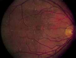
| Fig. 29 Dominant slowly progressive macular dystrophy. Type 2 lesion demonstrates an irregular zone of yellow-gray subretinal deposition material at the level of the pigment epithelium within the fovea. Additional yellow deposition flecks are seen scattered throughout the superotemporal and temporal posterior pole. |