|
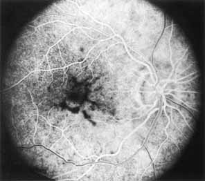
Fig. 25 Pattern dystrophy
of the pigment epithelium. Fluorescein angiography in mid arteriovenous
phase of fellow eye of patient seen in Fig.
26 demonstrates
the dendritiform or reticular hypofluorescent pattern corresponding to pigment
clumping within the central macula. (Marmor MF, Byers B: Pattern dystrophy
of the pigment epithelium. Am J Ophthalmology 84:32, 1977)
|