|
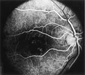
Fig. 23 Vitelliform
dystrophy. Fluorescein angiography in early arteriovenous phase of fellow
eye of patient seen in Fig. 22
showing clearly the area of dense hypofluorescence corresponding to the
yellow deposition material within the fovea. In addition, retinal pigment
epithelium (RPE) transmission defects can also be seen within the fovea,
indicating early RPE derangement and atrophy. |