|
|
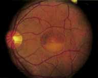
| Fig. 22 Vitelliform dystrophy. A large, well defined, partially filled, egg-yolk lesion occupies the entire fovea. Pigment epithelial change with pigment clumping is also seen in the superior portion of the lesion not filled by the yellow deposition material. |