|
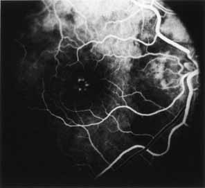
Fig. 27 Foveomacular
vitelliform dystrophy: adult type. Fluorescein angiography in mid arteriovenous
phase in patient seen in Fig. 26
shows clearly the hypofluorescence corresponding to the pigment clumping
within the central fovea. Typically, an irregular halo or corona, as is
seen here, surrounds the hypofluorescent spot. |