|
|
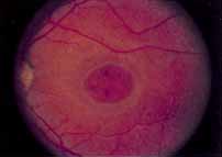
| Fig. 14 Stargardt's disease. An oval area of pigment epithelial derangement and atrophy is present within the central macula. Early pigmentations within this zone are also apparent. Scattered yellowish flecks identical to those seen in fundus flavimaculatus surround this central zone but are quite subtle and poorly visualized. They can be much better appreciated on fluorescein angiography. |