|
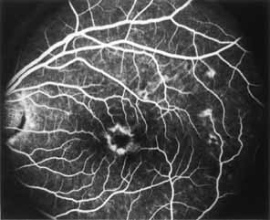
Fig. 31 Dominant slowly
progressive macular dystrophy. Fluorescein angiography in mid arteriovenous
phase of patient seen in Fig.
29 demonstrates central hypofluorescence with surrounding
hyperfluorescent corona similar to that seen in foveomacular vitelliform
dystrophy: adult type. Additional hyperfluorescence relates to the flecks
within the superotemporal and temporal posterior pole. |