Chapter 113B
Fluorescein Angiography of the Hereditary Chorioretinal Dystrophies*
KENNETH G. NOBLE
Main Menu   Table Of Contents

Search

GENERALIZED CHORIORETINAL DYSTROPHIES
VITREOTAPETORETINAL DYSTROPHIES
RETINAL VASCULAR DYSTROPHIES
MACULAR DYSTROPHIES
REFERENCES

The hereditary chorioretinal dystrophies for the most part affect visual processing at various loci. As such., they are best studied by diagnostic modalities that evaluate visual function, such as the electroretinogram, electro-oculogram, visual-evoked potential, dark adaptation, and visual fields. Fluorescein angiography highlights abnormalities in the vascular system and within the pigmented layers, and even in its relatively limited role, it has been helpful in diagnostic evaluation and pathogenesis.

Back to Top
GENERALIZED CHORIORETINAL DYSTROPHIES

RETINITIS PIGMENTOSA (RP)

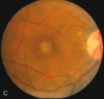
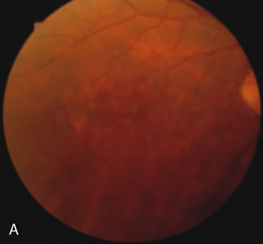
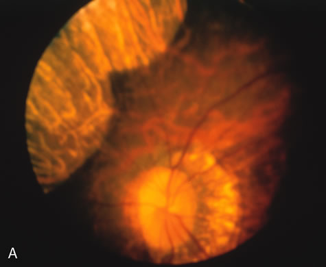
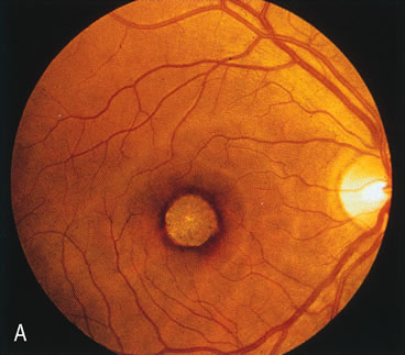
In retinitis pigmentosa (RP), the pigment abnormalities of atrophy, migration, and clumping are made apparent by transmitted hyperfluorescence and blocked hypofluorescence (Fig. 1A). Patients who have very minimal pigmentary alterations (pauci pigmentary RP) or no pigment abnormalities (RP sine pigmento) may show the abnormalities on fluorescien angiography (FA). It is uncommon to see choriocapillaris atrophy except in the late stages. This finding corresponds to the histopathology, which shows that the earliest abnormalities are in the photoreceptors and that the choroid is normal.1

Fig. 1. Retinitis pigmentosa. A. A typical area of bone spicule pigmentation. B. Diffuse dye leakage is apparent throughout the posterior pole. C. The early angiogram shows dilated and irregular retinal radial peripapillary capillaries and perifoveal retinal capillaries. D. Leakage from these vessels are evident in the late angiogram.

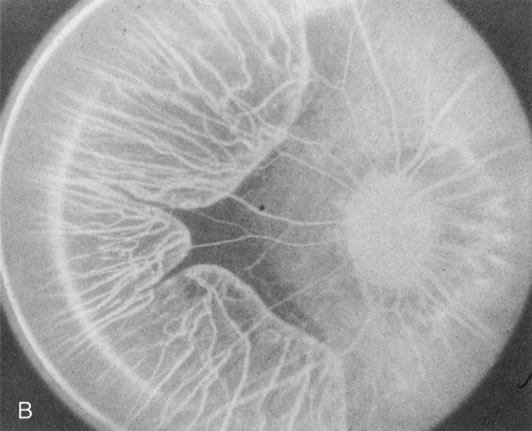
Dye leakage in RP may occur from the retinal vessels or at the level of the retinal pigment epithelium (Fig. 1B).2–4 The leakage may be seen in the macula and posterior pole, along the vascular arcades in the distribution of the radial peripapillary capillaries, and in the periphery (where an exudative vasculopathy resembling Coats' disease is suggested).

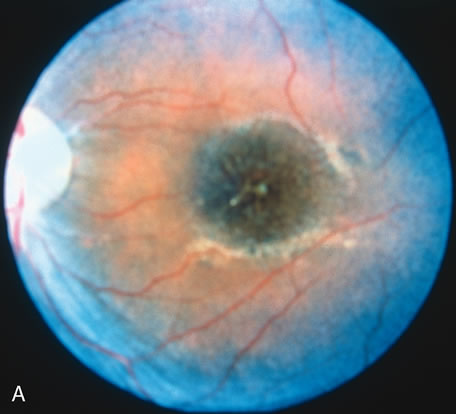
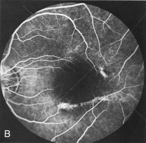
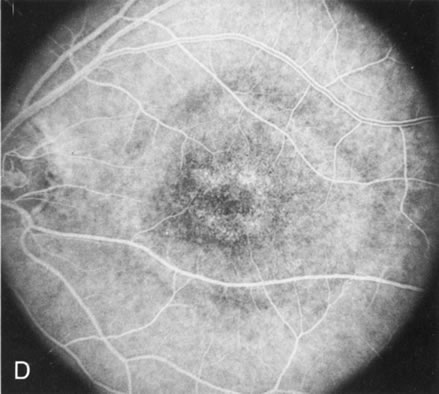
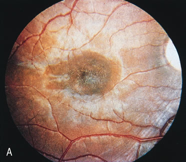
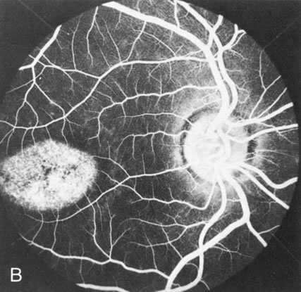
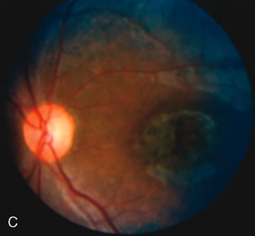
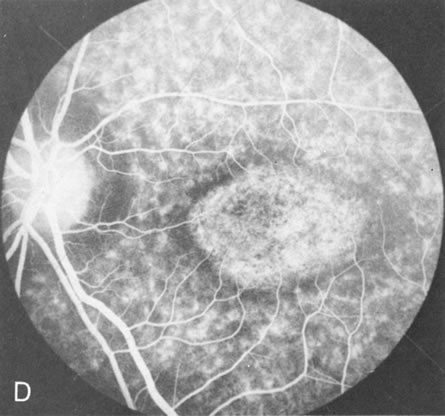
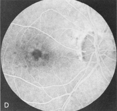
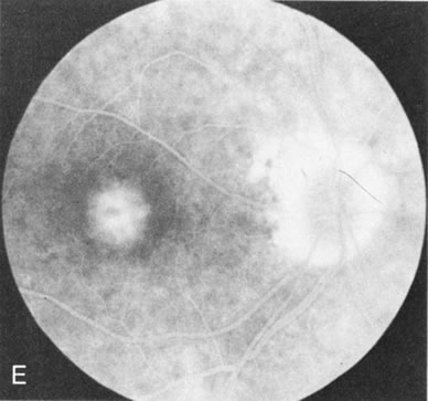
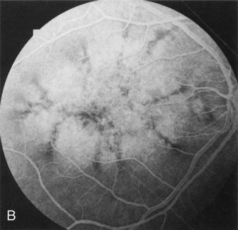
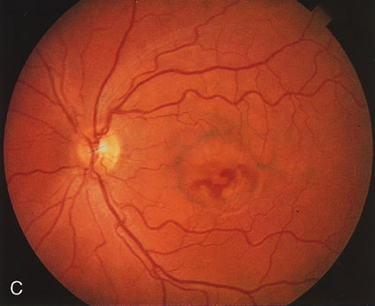
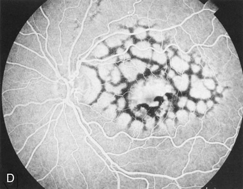
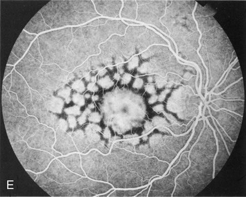
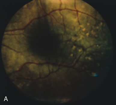
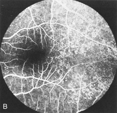
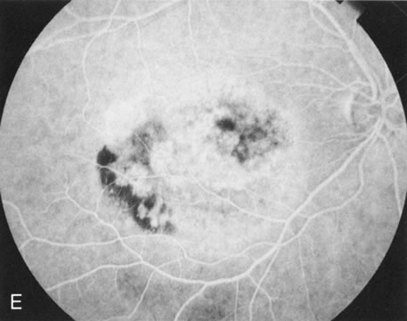
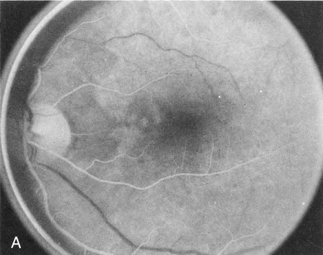
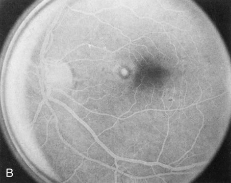
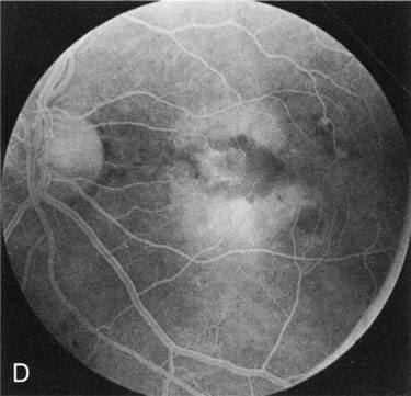
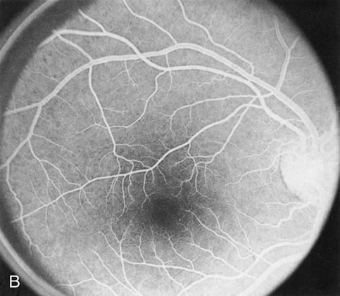
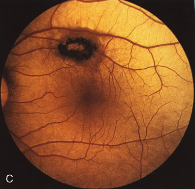
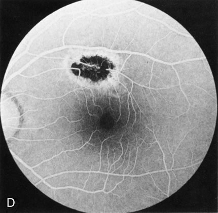
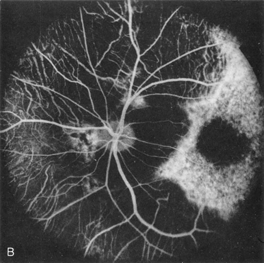
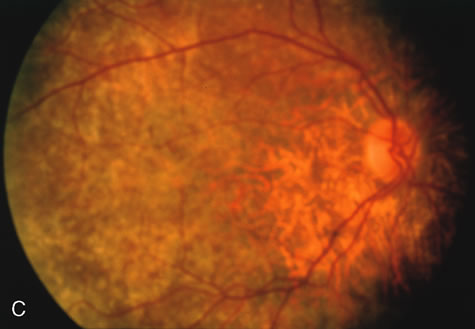
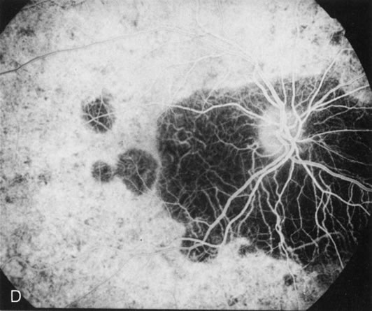
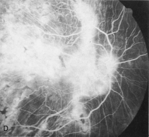
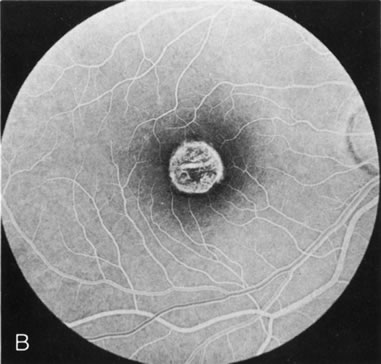
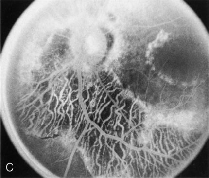
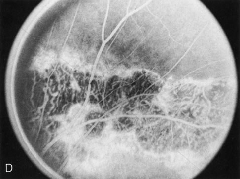
Of more clinical importance is the role of FA in the diagnosis and treatment of cystoid macular edema (CME) (Fig. 1C and D). Stereoscopic FA indicates that the leakage, which may be diffuse or have the typical petaloid stellate appearance of CME, can come from the perifoveal retinal capillaries, from the choroid through the RPE, or from a combination of both sources.4 With the recent suggestion that CME in RP may be successfully treated with acetazolamide,5, 6 FA is thus important to document the diagnosis of CME, establish the origin(s) of leakage, and follow patients during and after therapy.

The carrier female with XLR RP, who has the golden tapetal sheen reflex, has normal FA findings. This suggests that the abnormal reflex is not due an abnormal pigment layer or deposition7 (Fig. 2A and B).

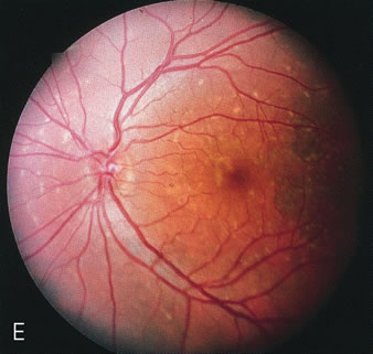
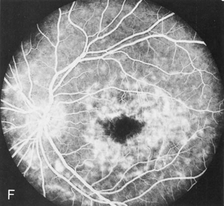
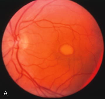
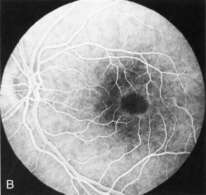
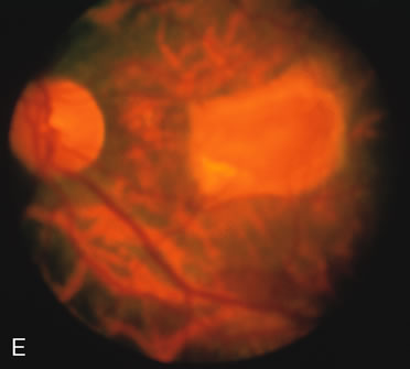
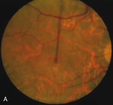
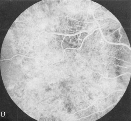
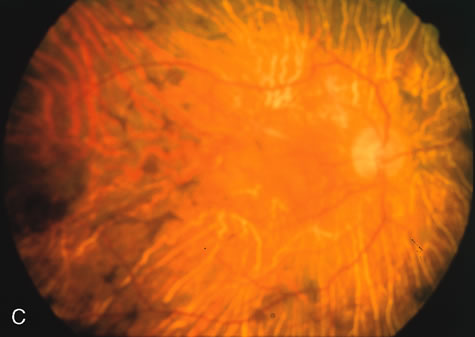
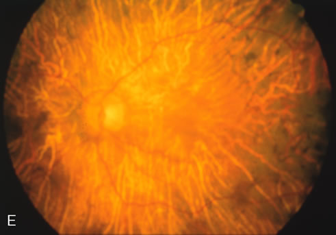
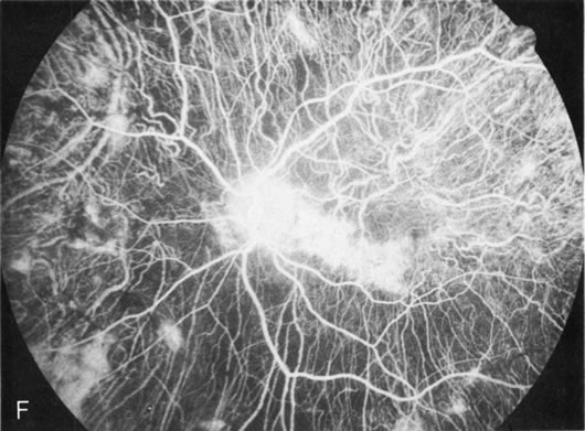
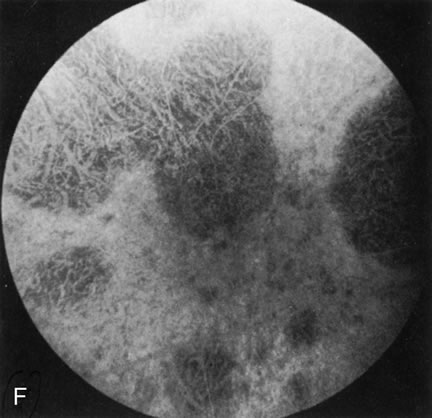
Fig. 2. Golden tapetal reflexes. There are three diseases in which there is an unusual golden reflex to the fundus: X-linked recessive RP in the carrier female, Oguchi's disease, and cone dystrophy. In all these diseases the angiogram is essentially normal, suggesting that this reflex is probably not related to pigment abnormalities. A, B. Carrier female of XLR retinitis pigmentosa. A golden scintillating reflex radiates from the macula. C, D. Oguchi's disease. A diffuse yellow metallic sheen is seen in the posterior pole (a pigmented chorioretinal lesion is an incidental finding). E, F. Progressive cone dystrophy. The typical bull's eye maculopathy is associated with a golden orange reflex.

CONGENITAL STATIONARY NIGHT BLINDNESS

FA is normal in congenital stationary night blindness (CSNB) with a normal fundus. However, in the two types of CSNB with an abnormal fundus (Oguchi's disease, fundus albipunctatus) FA does provide some interesting information.

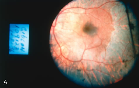
In Oguchi's disease the hallmark fundus finding is a yellow metallic sheen (Fig. 2C). A similar sheen has been seen in progressive cone dystrophies and juvenile macular degeneration.7 In all these diseases the abnormal reflex does not affect the normal transmission of fluorescein dye (Fig. 2D). The normal FA suggests that, like findings in the carrier female in XLR RP, the abnormal retinal reflex in this disease is unrelated to pigment concentration or distribution.

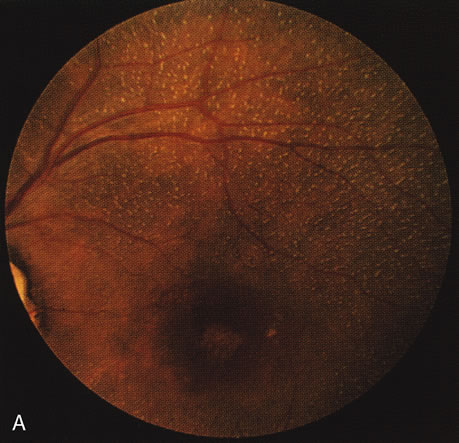
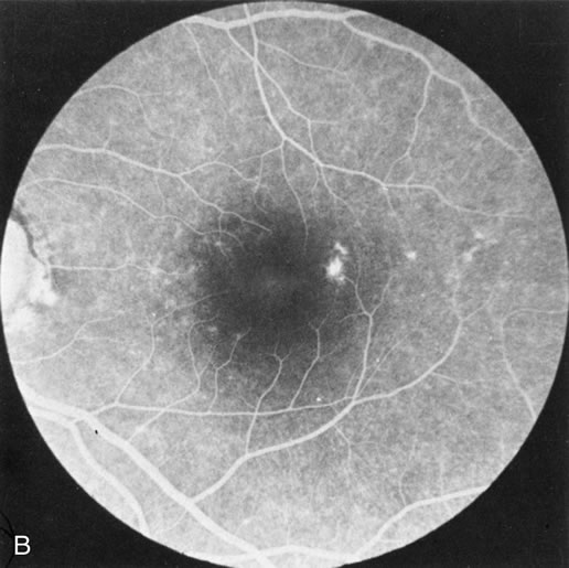
Fundus albipunctatus typically shows multiple, small white dots, which are deep in the retina, regular, and monotonous in their similar size, shape, and color and which involve the entire posterior pole into the equator while sparing the macula (Fig. 3A). These multiple dots are not apparent on FA. There may be a mottling of the background choroidal fluorescence and small areas of irregular transmission hyperfluorescence (especially surrounding the macula), but neither of these findings corresponds to the observed white dots8,9 (Fig. 3B).

Fig. 3. Fundus albipunctatus. The few small transmission defects on angiography (A) do not correspond to the multiple small white dots that are characteristic of this disease (B).

CONE DYSTROPHIES

Fluorescein angiography highlights observable fundus findings. In patients with a golden reflex the FA is normal (Fig. 2E, F) or shows a mild transmission hyperfluorescence.7

Choroidal Dystrophies

The choroidal dystrophies may be generalized (choroideremia, gyrate atrophy, generalized choroidal dystrophy) or localized to the posterior pole (central areolar choroidal dystrophy, peripapillary or pericentral choroidal dystrophy, Bietti's crystalline retinopathy). The hallmark finding in all these disorders is an atrophy of the choriocapillaris early in the course of the disease. Subtle loss of the choriocapillaris is documented and confirmed by FA in which there is persistent visualization of the mid-sized choroidal vessels.10 In this regard, FA helps to differentiate disorders that affect the choroid early in the course of the disease from disorders that initially affect the retinal pigment epithelium (RPE). It is also an excellent way to determine if there is progression in the course of the disease.

Choroideremia

In the early stages of choroideremia, before choroidal atrophy is funduscopically obvious and when the picture resembles RP, FA indicates diffuse choroidal atrophy throughout the entire retina. Only the macular area remains preserved (Fig. 4A and B).

Fig. 4. Choroideremia and choroideremia carrier. A. The choroidal atrophy in this affected male is not apparent in the fundus. B. However, the angiogram shows diffuse atrophy of the choriocapillaris with persistent visualization of the larger choroidal vessels. C. This carrier female has peripapillary choroidal atrophy and diffuse pigment mottling. D. The patchy areas of focal choroidal atrophy that occasionally occurs in carriers is evident on angiography.

The typical carrier female, with focal or diffuse pigment mottling, does not show choroidal atrophy. However, a few carrier females have a more severe form with focal areas of choroidal atrophy. The presence of these areas, and possible progression, can be documented by FA (Fig. 4C and D). These carriers exhibit a mosaicism, which is explained by the Lyon hypothesis of random X-chromosome inactivation.

Gyrate Atrophy

Unlike choroideremia, gyrate atrophy funduscopically has well-demarcated scalloped areas of choroidal atrophy. A hyperpigmented border separates the normal and abnormal tissue. These lesions begin as isolated areas in the midperiphery, which then merge to form a garland wreath, with progression peripherally and centrally, sparing only the macula.

FA demonstrates the sharp demarcation between normal and abnormal tissue, the former showing normal background fluorescence, the latter atrophy of the choriocapillaris (Fig. 5). Thus, the normal choriocapillaris background fluorescence in the early stages of gyrate atrophy is in contradistinction to the diffuse choriocapillaris atrophy in the early stages of choroideremia.

Fig. 5. Gyrate atrophy. The areas of choroidal atrophy (A) show choriocapillaris atrophy on the angiogram. (B) Adjacent areas of normal-appearing retina have a normal background choroidal flush.

Generalized Choroidal Dystrophy

Generalized choroidal dystrophy is usually noted in middle-aged mildly symptomatic individuals who show a predominantly peripapillary or pericentral distribution of choroidal atrophy. Gradually, over the years these areas enlarge to eventually involve the entire retina. These changes are vividly seen on FA (Fig. 6).

Fig. 6. Generalized choroidal dystrophy. This 65-year-old woman gradually developed enlarging, progressive areas of choroidal atrophy over a 20-year period. When initially seen, the abnormalities were confined to the peripapillary and macular region (A,B). In a recent examination, the generalized choroidal atrophy is vividly demonstrated on angiography (C–F).

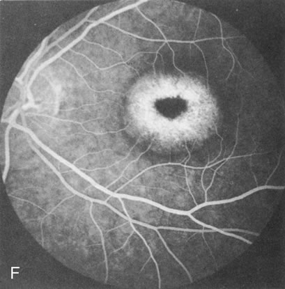
Central Areolar Choroidal Dystrophy

Although central areolar choroidal dystrophy (CACD) and peripapillary (pericentral) choroidal dystrophy are not generalized, but rather localized disorders, they are discussed here as part of the choroidal dystrophies because the angiographic findings are similar.

In CACD the bilateral macular lesions are solitary, circumscribed, and circular or ovoid in shape. They are unassociated with other findings such as drusen or flecks. FA will confirm the well-circumscribed area of choriocapillaris atrophy and further document that there are no associated findings that would lead to secondary choroidal atrophy in disorders such as age-related macular degeneration, Stargardt's fundus flavimaculatus, or dominant drusen of Bruch's membrane11 (Fig. 7 A and B).

Fig. 7. Central areolar choroidal dystrophy. The presence of choroidal atrophy in this well-circumscribed macular lesion (A) is confirmed by persistent visualization of the larger choroidal vessels as seen on angiography (B). Peripapillary (pericentral) choroidal dystrophy. The areas of choroidal atrophy are well-demarcated and contrast with the areas of normal choroid (C, D). Crystalline retinopathy (of Bietti). The areas of choroidal atrophy correspond to areas of the retina where crystals are not present (E, F).

Peripapillary (Pericentral) Choroidal Dystrophy

Peripapillary choroidal atrophy radiates from the optic nerve along the temporal vascular arcades. The macula is affected later in the course, and this is usually responsible for the onset of visual symptoms. FA shows the choroidal atrophy and the early macular changes (Fig. 7C and D).

Crystalline Retinopathy (of Bietti)

Crystalline retinopathy (of Bietti) is usually localized to the posterior pole but can be progressive. Glistening yellow crystals appear scattered throughout the retina (Fig. 7E). The angiogram has a typical and unique appearance. In the area of the crystals there is a transmitted hyperfluorescence; adjacent to these areas, where there are no crystals, choriocapillaris atrophy is evident (Fig. 7F). Progression of the disease is documented by FA,12 which shows enlarging areas of choriocapillaris atrophy.

Back to Top
VITREOTAPETORETINAL DYSTROPHIES
The vitreotapetoretinal dystrophies are a heterogeneous group of diseases that share the association of vitreous abnormalities with a tapetoretinal dystrophy. The initial triad of diseases included under this heading were juvenile retinoschisis, Goldmann-Favre disease, and Wagner's dystrophy. Additional disorders that qualify for inclusion include Stickler's syndrome (hereditary progressive arthro-ophthalmopathy), snowflake degeneration, and autosomal dominant vitreoretinochoroidopathy (ADVIRC).

When the macula has typical superficial linear plications radiating from the fovea seen with schisis (e.g., juvenile retinoschisis, Goldmann-Favre disease), FA is normal (Fig. 8A and B). This indicates that the pathology is in the inner retina (probably Henle's fiber layer, considering the spokewheel pattern). However, when the schisis flattens, it is not uncommon to see a transmitted hyperfluorescence suggesting a secondary atrophy to the RPE (Fig. 8C and D).

Fig. 8. Juvenile XLR retinoschisis. The superficial macula schisis (A) does not affect the angiogram (B) except in a few areas where it has flattened and resulted in some pigment dispersion. When the macular schisis has entirely flattened (C) there is a mild transmission hyperfluorescence (D). At this stage the diagnosis can be suspected by the presence of an inferior retinoschisis (present in half) and confirmed by the typical electroretinographic finding of a scotopic electronegative response (present in all affected males).

The periphery may show schisis or vascular sheathing. Histopathology of the peripheral schisis in juvenile retinoschisis documents a splitting of the nerve fiber layer.13 FA shows retinal vascular abnormalities, including capillary nonperfusion, focal and diffuse vascular leakage, and intraretinal neovascularization, in many of these disorders.14 These may or may not be associated with schisis.

As with generalized tapetoretinal dystrophy (RP), FA clearly delineates retinal vascular decompensation in some of these patients. What remains to be answered is whether this is part of the basic disease process or a secondary reaction to it.

Back to Top
RETINAL VASCULAR DYSTROPHIES
There are a few uncommon hereditary dystrophies in which the retinal vascular system seems to be the primary abnormality. In these cases FA is helpful, and sometimes essential, in making the diagnosis and determining the pathogenesis.

The most common disorder in this group is familial exudative vitreoretinopathy (FEVR), which clinically resembles retinopathy of prematurity, although it is dominantly inherited and unassociated with prematurity or perinatal supplemental oxygen therapy. The mildest changes, documented by FA, are similar to those of the retinopathy of prematurity, namely peripheral retinal capillary nonperfusion and arteriovenous anastomoses. In family members at risk who have a normal-appearing periphery, FA will reveal abnormalities in affected family members and is therefore a sensitive indicator of disease.15

Perifoveal retinal capillary obliteration16 (which may occur throughout the retina and involve the brain)17 requires FA to define this unusual inherited disorder. Inherited retinal venous beading, reported in two dominant pedigrees,18,19 has focal areas of retinal capillary nonperfusion in the posterior pole associated with retinal capillary leakage and intraretinal neovascularization.

Back to Top
MACULAR DYSTROPHIES

STARGARDT'S DISEASE (FUNDUS FLAVIMACULATUS)

In Stargardt's disease, thes most common hereditary macular dystrophy, FA has a variety of functions. In the mildest fundus presentation, with visual symptoms and visual function out of proportion to the observed maculopathy, FA can confirm or even make the diagnosis and can avoid a mistaken diagnosis of malingering, hysteria, or central nervous system disease (Fig. 9A and B). When a maculopathy is present without surrounding parafoval flecks, FA may show patchy areas of transmission hyperfluorescence in the posterior pole, indicating a more diffuse involvement (Fig. 9C and D).

Fig. 9. Stargardt's disease–fundus flavimaculatus. The mild maculopathy (without parafoveal flecks) (A) is confirmed by the angiogram (B). The relative absence of the underlying choroidal flush, resulting in an easier visualization of the overlying retinal capillary circulation, has been referred to as the “silent” or “dark” choroid, and is considered a common finding in this disease. The diagnosis is confirmed in an individual with a pigmentary maculopathy without flecks (C). Here the angiogram demonstrates widespread transmission hyperfluorescence and a “silent” or “dark” peripapillary area (D). When the posterior pole shows multiple yellowish-white flecks (E), the angiographic findings do not necessarily correspond to the flecks (F). It should also be noted that despite the widespread abnormalities, the background choroidal fluorescence is normal.

In a large majority of patients (86% in one study),20 there is an absence or decrease in the background choroidal fluorescence (which is referred to as the “silent” or “dark” choroid) (see Fig. 9B). This warrants special attention because it occurs so frequently, is rarely found in other retinal disorders,21 and may be related to histopathology that shows an increase in lipofuscin in the RPE.22

BEST'S VITELLIFORM MACULAR DYSTROPHY

The intact solid yellow egg yolk lesion in Best's vitelliform macular dystrophy (BVMD) typically shows hypofluorescence on FA due to blockage of the underlying choroidal fluorescence with the overlying retinal vasculature visible (Fig. 10A and, B). This finding places the lesion in front of the choroid, possibly within the RPE, but probably not within the neurosensory retina. There is no histopathology of the intact lesion to confirm this.


Fig. 10. Best's vitelliform macular dystrophy. The most characteristic angiographic finding in the solid yellow egg-yolk stage is blocked hypofluorescence (A, B). A morphologically mimicking lesion (pseudovitelliform degeneration) may be the result of leakage from the underlying choroid (C–E).

Histopathology in later stages of BVMD has shown an increase in lipofuscin in the RPE throughout the retina.23 If lipofuscin were within the intact egg-yolk lesion, this would block fluorescence. However, it is surprising that the “silent,” “dark” choroid seen in another disease with diffuse RPE lipofuscin accumulation (Stargardt's disease) does not occur in BVMD.

There are several acquired macular degenerations of different etiologies that have a macular lesion similar to the vitelliform stage of BVMD. FA may be helpful in identifying these cases of pseudovitelliform degeneration by showing leakage from the retinal capillaries or through the RPE and late staining of the lesions (Fig. 10CE). However, the electro-oculogram remains the most discriminating test because it is always abnormal in BVMD and usually normal in pseudovitelliform macular degeneration.

PIGMENT PATTERN DYSTROPHIES

The fundus abnormalities in pigment pattern dystrophies may be subtle, especially when the pigmentary changes are orange and yellow in color. However, FA is most dramatic in highlighting these abnormalities. Thus, FA will confirm, and in some circumstances provide, the diagnosis (Fig. 11AE). Although there is no histopathologic confirmation, the fundus and angiogram suggest that an abnormal deposition of lipofuscin in the RPE is responsible for the clinical picture.


Fig. 11. Pigment pattern dystrophies. Subtle changes of the fundus in this family member with a pattern dystrophy (A) are highlighted on angiography (B). Equally dramatic is the fluorescein angiography of this young woman who presented with poor vision in association with a retinal hemorrhage (C). The angiogram reveals a bilateral, symmetric reticular pattern of the posterior pole (D, E).

DOMINANT DRUSEN OF BRUCH'S MEMBRANE

There is no clear agreement as to what constitutes dominantly inherited drusen and what constitutes age-related degenerative drusen. There is a distinct group of younger individuals with bilateral, symmetric, uniformly small, round, discrete, yellow and white drusen. Histopathology demonstrates a nodular thickening of the RPE basement membrane, and these drusen have therefore been referred to as basal laminar, or cuticular, drusen.24

FA shows a characteristic pinpoint area of transmission hyperfluorescence corresponding to the drusen and reveals many more than are appreciated clinically (Fig. 12). Secondary changes such as pigment atrophy and dispersion, exudative and nonexudative detachments, and occasionally a neovascular membrane, will be revealed by FA (Fig. 12E).


Fig. 12. Dominant drusen of Bruch's membrane. The drusen appear as yellowish-white “blisters” predominantly in the temporal posterior pole (A). The angiogram shows many pinpoint areas of transmission hyperfluorescence typical of drusen, some of which have coalesced to form broader areas of hyperfluorescence (B). These drusen occasionally result in ingrowth of a choroidal neovascular membrane, such as occurred in this case over a 5-year period (C–E).

HEREDITARY HEMORRHAGIC MACULAR DYSTROPHY (PSEUDOINFLAMMATORY MACULAR DYSTROPHY OF SORSBY)

FA plays an important role in the understanding of this rare disorder. In the earliest manifestations of the disease, FA shows a single, isolated choroidal neovascular membrane unassociated with other causes of a membrane, such as drusen or angioid streaks (Fig. 13).25 Presumably this membrane is the cause of the subsequent hemorrhagic maculopathy with secondary disciform scarring, occurring initially in the macula but often extending throughout the retina.


Fig. 13. Hereditary hemorrhagic macular dystrophy (pseudoinflammatory macular dystrophy of Sorsby). Fluorescein angiography in this family member demonstrates that the earliest finding is an isolated choroidal neovascular membrane B). Despite laser photocoagulation, a hemorrhagic maculopathy developed in this eye (C, D) and eventually a disciform scar (E) formed, as it had in the fellow eye, and in the eyes of other affected family members.

Abnormal choroidal perfusion, manifested on FA as a delay in choriocapillaris filling, has been seen in the restudy of some of Sorsby's initial pedigrees.26 This unusual pattern has been related to the histopathologic finding of a “confluent, lipid-containing, amorphous deposit found between the basement membrane of the RPE and the inner collagenous layer of Bruch's membrane.”27

Back to Top
REFERENCES

1. Flannery JG, Farber DB, Bird AC et al: Degenerative changes in a retina affected with autosomal dominant retinitis pigmentosa. Invest Ophthalmol Vis Sci 30:191, 1989.

2. Fetkenhour CL, Choromokos E, Weinstein J et al: Cystoid macular edema in retinitis pigmentosa. Trans Am Acad Ophthalmol Otolaryngol 83:515, 1977.

3. Spalton DJ, Bird AC, Cleary PE: Retinitis pigmentosa and retinal oedema. Br J Ophthalmol 62:174, 1978.

4. Newsome DA: Retinal fluorescein leakage in retinitis pigmentosa. Am J Ophthalmol 101:354, 1986.

5. Cox SN, Hay E, Bird AC: Treatment of chronic macular edema with Acetazolamide. Arch Ophthalmol 106:1190, 1988.

6. Fishman GA, Gilbert LD, Fiscella RG et al: Acetazolamide for treatment of chronic macular edema in retinitis pigmentosa. Arch Ophthalmol 107:1445, 1989.

7. Noble KG, Margolis S, Carr RE: The golden tapetal sheen reflex in retinal disease. Am J Ophthalmol 107:211, 1989.

8. Carr RE, Margolis S, Siegel IM: Fluorescein angiography and vitamin A and oxalate levels in fundus albipunctatus. Am J Ophthalmol 82:549, 1976.

9. Marmor MF: Fundus albipunctatus: A clinical study of the fundus lesions, the physiologic deficit, and the vitamin E metabolism. Doc Ophthalmol 43:277, 1977.

10. Noble KG, Carr RE, Siegel IM: The role of fluorescein angiography in the hereditary choroidal dystrophies. Br J Ophthalmol 61:43, 1977.

11. Noble KG: Central areolar choroidal dystrophy. Am J Ophthalmol 84:310, 1977.

12. Yuzawa M, Mae Y, Matsui M: Bietti's crystalline retinopathy. Ophthalmol Paediatr Gen 7:9, 1986

13. Yanoff M, Rahn EK, Zimmerman LE: Histopathology of juvenile retinoschisis. Arch Ophthalmol 79:49, 1968.

14. Green JL, Jampol LM: Vascular opacification and leakage in X-linked (juvenile) retinoschisis. Br J Ophthalmol 63:368, 1979.

15. Ober RR, Bird AC, Hamilton AM et al: Autosomal dominant exudative vitreoretinopathy. Br J Ophthalmol 64: 112, 1980.

16. Ehlers N, Jensen VA: Hereditary central retinal angiopathy. Acta Ophthalmol 51:171, 1973.

17. Grand MG, Kaine J, Fulling K et al: Cerebroretinal vasculopathy: A new hereditary syndrome. Ophthalmology 95:649, 1988.

18. Meredith TA: Inherited retinal venous beading. Arch Ophthalmol 105:949, 1987.

19. Stewart MW, Gitter KA: Inherited retinal venous beading. Am J Ophthalmol 106:675, 1988.

20. Fishman GA, Farber F, Patez BS et al: Visual acuity in patients with Stargardt's macular dystrophy. Ophthalmology 94:809, 1987.

21. Uliss AE, Moore AT, Bird AC: The dark choroid in posterior retinal dystrophies. Ophthalmology 94:1423, 1987.

22. Eagle RC Jr , Lucier AC, Bernardino VB Jr et al: Retinal pigment epithelial abnormalities in fundus flavimaculatus: A light and electron microscopic study. Ophthalmology 87:1189, 1980.

23. Weingeist TA, Kobrin JL, Watzke RC: Histopathology of Best's macular dystrophy. Arch Ophthalmol 100:1108, 1982.

24. Gass JDM, Jallow S, Davis B: Adult vitelliform macular detachment occurring in patients with basal laminar drusen. Am J Ophthalmol 99:445, 1985.

25. Carr RE, Noble KG, Nasaduke I: Hereditary hemorrhagic macular dystrophy. Am J Ophthalmol 85:318, 1978.

26. Polkinghorne PJ, Capon MRC, Berninger T et al: Sorsby's fundus dystrophy: A clinical study. Ophthalmology 96:1763, 1989.

27. Capon MRC, Marshall J, Krafft JI et al: Sorsby's fundus dystrophy: A light and electron microscopic study. Ophthalmology 96:1769, 1989.

Back to Top

* Supported in part by a grant from the Retinitis Pigmentosa Foundation, the Research to Prevent Blindness and The Kirby Eye Institute.