|
|
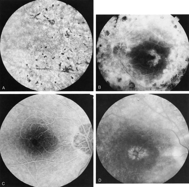
| Fig. 1. Retinitis pigmentosa. A. A typical area of bone spicule pigmentation. B. Diffuse dye leakage is apparent throughout the posterior pole. C. The early angiogram shows dilated and irregular retinal radial peripapillary capillaries and perifoveal retinal capillaries. D. Leakage from these vessels are evident in the late angiogram. |