|
|
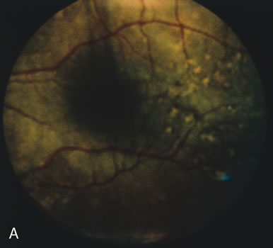
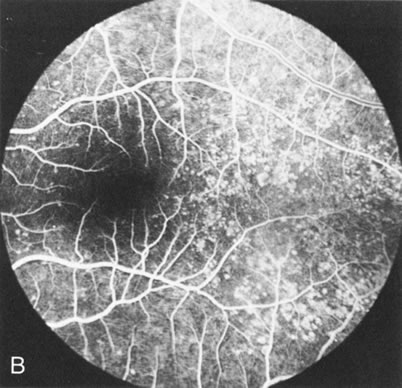
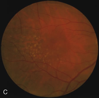
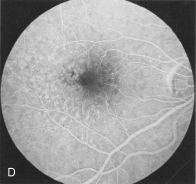
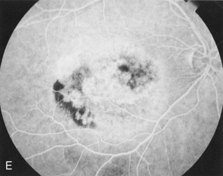
| Fig. 12. Dominant drusen of Bruch's membrane. The drusen appear as yellowish-white “blisters” predominantly in the temporal posterior pole (A). The angiogram shows many pinpoint areas of transmission hyperfluorescence typical of drusen, some of which have coalesced to form broader areas of hyperfluorescence (B). These drusen occasionally result in ingrowth of a choroidal neovascular membrane, such as occurred in this case over a 5-year period (C–E). |