|
|
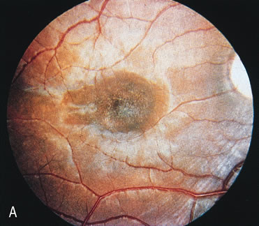
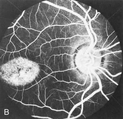
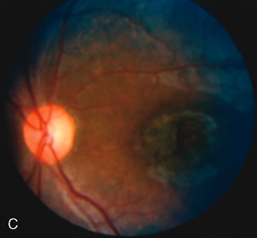
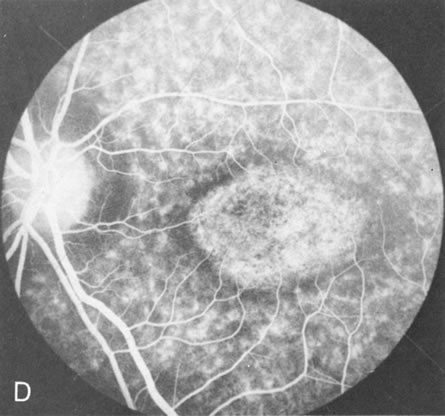
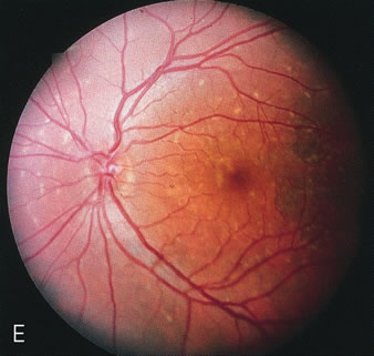
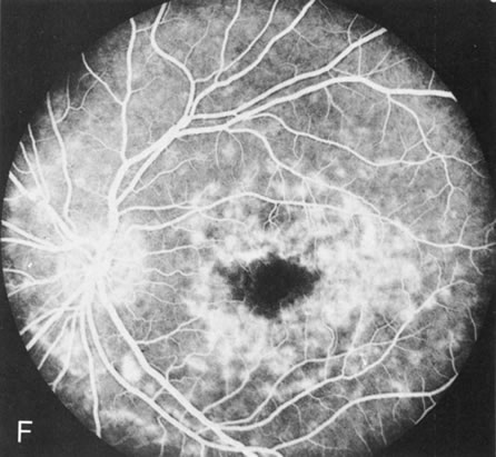
| Fig. 9. Stargardt's disease–fundus flavimaculatus. The mild maculopathy (without parafoveal flecks) (A) is confirmed by the angiogram (B). The relative absence of the underlying choroidal flush, resulting in an easier visualization of the overlying retinal capillary circulation, has been referred to as the “silent” or “dark” choroid, and is considered a common finding in this disease. The diagnosis is confirmed in an individual with a pigmentary maculopathy without flecks (C). Here the angiogram demonstrates widespread transmission hyperfluorescence and a “silent” or “dark” peripapillary area (D). When the posterior pole shows multiple yellowish-white flecks (E), the angiographic findings do not necessarily correspond to the flecks (F). It should also be noted that despite the widespread abnormalities, the background choroidal fluorescence is normal. |