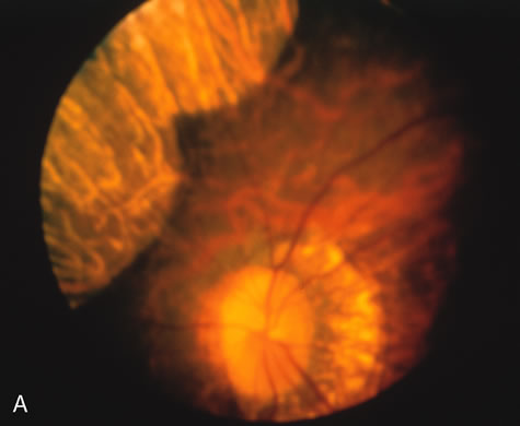
Fig. 5.
Gyrate atrophy. The areas of choroidal atrophy
(A)
show choriocapillaris atrophy on the angiogram.
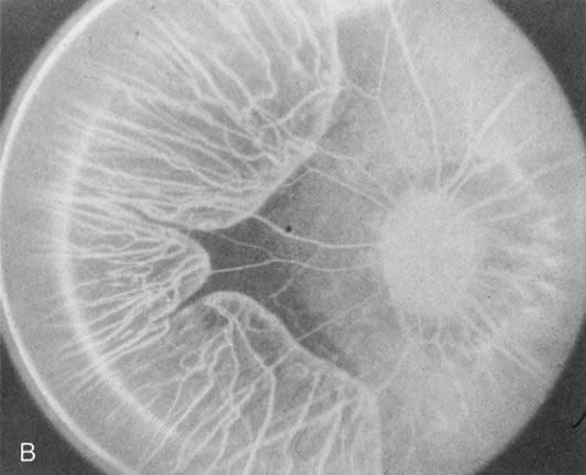
(B)
Adjacent areas of normal-appearing retina have a normal background choroidal flush.