|
|
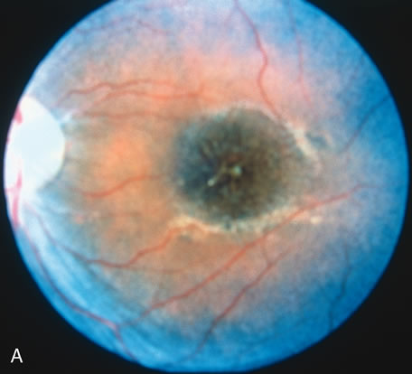
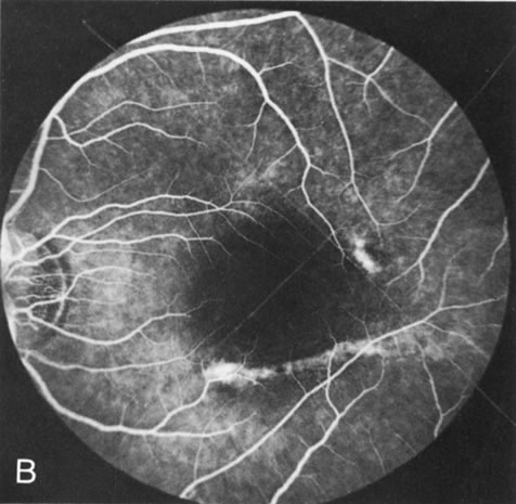
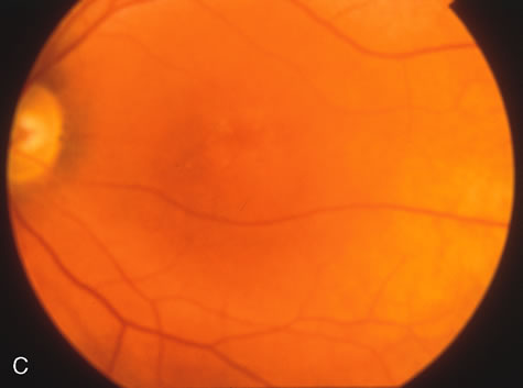
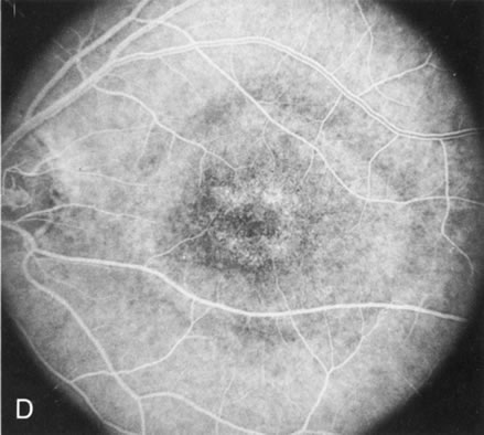
| Fig. 8. Juvenile XLR retinoschisis. The superficial macula schisis (A) does not affect the angiogram (B) except in a few areas where it has flattened and resulted in some pigment dispersion. When the macular schisis has entirely flattened (C) there is a mild transmission hyperfluorescence (D). At this stage the diagnosis can be suspected by the presence of an inferior retinoschisis (present in half) and confirmed by the typical electroretinographic finding of a scotopic electronegative response (present in all affected males). |