|
|
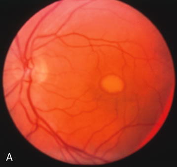
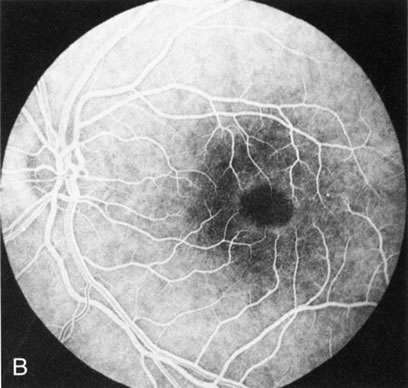
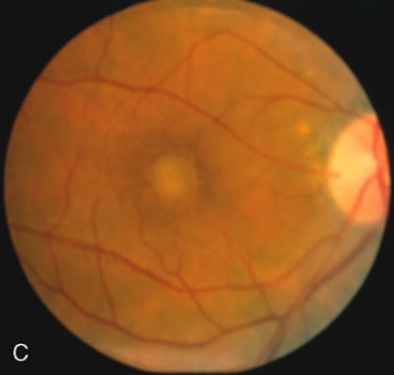
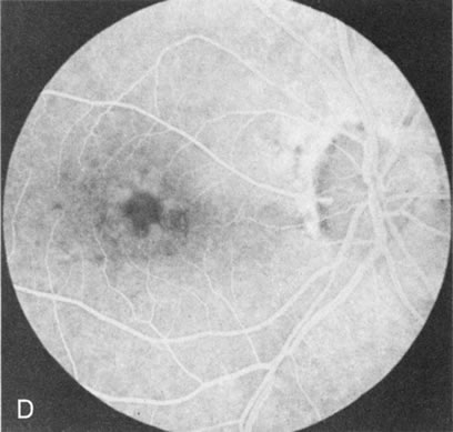
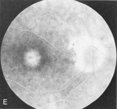
| Fig. 10. Best's vitelliform macular dystrophy. The most characteristic angiographic finding in the solid yellow egg-yolk stage is blocked hypofluorescence (A, B). A morphologically mimicking lesion (pseudovitelliform degeneration) may be the result of leakage from the underlying choroid (C–E). |