Chapter 14
Anatomy of the Zonular Apparatus
BARBARA W. STREETEN
Main Menu   Table Of Contents

Search

DEVELOPMENT AND SYNTHESIS OF THE ZONULE
COMPOSITION AND MOLECULAR GENETICS
ORIGIN OF THE ZONULE
CILIARY COURSE OF THE ZONULE
ZONULAR CIRCUMFERENTIAL GIRDLE FIBERS
CILIARY-LENS COURSE OF THE ZONULE
ZONULAR INSERTION ON THE LENS
ZONULAR COMPLICATIONS IN CATARACT SURGERY
CHANGES IN THE ZONULE WITH AGING
ZONULAR ROLE IN ACCOMMODATION
ACKNOWLEDGMENTS
REFERENCES

The zonular apparatus is a three-dimensional complex of microfibrils identical to the microfibrils of the elastic system in the rest of the body. These microfibrils are noncollagenous 10-nm beaded microfibrils composed predominantly of the protein fibrillin, and aggregate into fibers passing from the inner surface of the ciliary body to the outer surface of the lens capsule (Fig. 1). They are the only structural component directly holding the lens in place and are also designed to mediate movement forces of the ciliary muscle to the lens for accomodation. By attachments to the anterior hyaloid membrane, the posterior zonules may help to maintain this membrane and the anterior vitreous in apposition to the ciliary body and lens, which may play an additional role in accomodation.

Fig. 1. Insertion of the zonules on the anterior lens capsule. Cornea and iris have been removed and the eye has been stained with Gomori's hematoxylin. Ciliary processes are visible peripherally. Lens shrinkage during staining makes the perilenticular space wider than normal.

Because of great difficulties in understanding and even describing the elusive zonule, many terms have been used for it since the first complete description by the anatomist Zinn in 1753,1 including the zonule of Zinn, the annular ligament of Zinn, the suspensory ligament of the lens, the ciliary zonule, and the tertiary vitreous.

Back to Top
DEVELOPMENT AND SYNTHESIS OF THE ZONULE
Zonular fibers are first seen at the end of the third fetal month, lying within the secondary vitreous at its base and passing through the matrix of the tunica vasculosa lentis to attach to the lens capsule.2 The mechanisms and factors guiding their highly oriented and predictable attachment to sites on the lens capsule are still unknown. In late fetal life the adult pattern is already apparent, with silvery zonular fibers passing over the tops of the low ciliary processes while others dip into the valleys between the processes, and some change direction by running up along the sides of the processes (Fig. 2). The primary candidate for synthesis of the zonule is the ciliary nonpigmented epithelium. Autoradiographic studies on fetal and young mammalian eyes showed an early accumulation of labeled carbohydrate components and amino acids including cys-teine (present in high concentration in the zonule)on zonular fibrils close to the epithelium of the pars plana and posterior pars plicata.3,4 This proximity suggests that new protein is added at the ciliary end of the zonule and that the ciliary nonpigmented epithelium is its source. In culture, these cells as well as lens and ciliary pigment epithelial cells are capable of producing fibrillin microfibrils,5,6 as do many basement membrane-secreting epithelial cells. By in situ hybridization, the ciliary nonpigmented epithelium of the fetal mouse is indeed positive for fibrillin-1, the main protein of zonular and other elastic system microfibrils.7 However, the lens epithelium in the metabolically most active equatorial region has some fibrillin-1 positivity at the same developmental stages and may contribute as well to the early zonule.8 It is not known whether zonular synthesis is continuous postnatally through at least the most active period of ocular growth, or whether a continuous low-grade renewal process occurs.

Fig. 2. Silvery layer of delicate zonular fibers lies over the pars plana and low processes of the infantile eye. Some fibers pass over the tops of the processes (open arrow), some deep into the valleys, others higher on the sides of the processes (arrowheads) (unstained, × 45).

Back to Top
COMPOSITION AND MOLECULAR GENETICS
Because the zonular fibers arise within the vitreous, they were for many years assumed to be collagenous like the vitreous, whose small type II collagen fibers are similar in size to those in the zonule, resulting in the zonule being termed the tertiary vitreous.2 In recent decades they were shown to be composed of noncollagenous glycoprotein,9–12 consistent with their positivity to the periodic acid-Schiff (PAS) stain and association with both O- and N-linked oligosaccharides.13

Electron microscopy allowed the first significant insight into the nature of the zonule. In 1971 Raviola noted that the 10-nm size and tubular ultrastructure of the zonular fibers and their susceptibility to α-chymotrypsin digestion were similar to characteristics of the newly described microfibrils of elastic tissue.14,15 These two types of microfibrils were found to be strongly cross-reactive immunologically16 and to have similar biochemical profiles, including an unusually high cysteine content.11,12

With the advent of the molecular era, the primary component in all elastic system microfibrils was identified as a 350-kDa glycoprotein and named fibrillin,7 coded for by a gene on chromosome 15 q21.1.17,18 Immunoreactivity of zonular fibers for fibrillin is strong7,19 (Fig. 3). Classic Marfan syndrome with ectopia lentis was quickly linked to mutations on the fibrillin gene,17,20 usually of the missense type. More than 70 such mutations have now been reported, including some mutations in cases of simple dominant ectopia lentis.21 Another fibrillin protein, fibrillin-2, was found at the same time on chromosome 5, causing congenital contractural arachnodactyly when mutated, but no ocular involvement.17 More than 10 proteins besides fibrillin-1 have since been found associated with the elastic system microfibrils, many of them also with zonular microfibrils, from which one of the new proteins was isolated.22,23 It is unclear how many of these proteins are intimately involved in the structure of the microfibril, or what functions they may have. Most are known by acronyms referring to their microfibril glycoprotein nature, such as MAGP-1 and -2, MFAP-1 to -4, MAP, GP115/emilin, and LTBP-2,but also included are more familiar ubiquitous proteins including fibronectin and vitronectin.24

Fig. 3. Insertion of the anterior zonular fibers, strongly reactive with fibrillin-1 antibody on a completely nonstaining lens capsule.

The fibrillin-1 molecule has a large number of repeating domains resembling epidermal growth factor (EGF-like), all but two of the calcium-binding type, where most of the Marfan mutations have occurred.24 This is consistent with evidence that the presence of calcium is important for the stability of this unique beaded microfibril.25,26 The stability of the zonular bundles and their resistance to solubilization imply the presence also of strong cross-linkage sites, besides the great capacity for disulfide linkages between the many cysteines. Transglutaminase-induced glutamyl-lysine cross-linkages have also been identified in elastic microfibrils from several tissues.27 Sites in the molecule for RGD-integrin binding to cell membranes andmatrix are also recognizable.28 The adhesion mechanisms used by the zonular fibers are still incompletely known.

FUNCTION OF THE ZONULAR MICROFIBRILLAR SYSTEM

The elastic system is composed of three units,29 the first composed entirely of microfibrils, as far as can be determined identical to the zonular microfibrils. These are the first units to be synthesized during elastogenesis,15 serving as a template on which the completely different elastomeric protein, elastin, may be deposited. Many bundles of microfibrils in the body like the zonular type never become elasticized, and these are sometimes called oxytalan (acid-resistant) fibers.30 Like zonular bundles, they attach particularly to basement membranes of surfaces under stress, connecting them to each other just as zonular fibers connect the ciliary nonpigment epithelial basement membrane to the lens epithelial basement membrane. In deeper tissues these microfibrils can become partially elasticized bundles called elaunin fibers.29 These then connect to the larger fully elasticized elastic fibers, completing the third unit of the system. The long-recognized elasticity or stretchability of the zonular bundles clearly has a different basis than that of the elastomer elastin and appears to reside in reversible relationships of the fibrillin filaments to each other and to their associated beads, as well as to adjacent microfi-brils, when stretched. As part of the widespreadelastic system, the zonule and fibrillin-1 appear toshare in relatively few other systemic diseases be-sides the Marfan syndrome, including the Weil-Marchesani syndrome31 and the pseudoexfoliation syndrome.19,32,33

ZONULAR BUNDLE, FIBER, AND FIBRIL ARCHITECTURE

When seen during life through an iridectomy or under the dissecting microscope, the zonular fibers appear as glassy, straight rods of 5 to 30 μm in diameter (Fig. 4), occurring in diverging groups that appear as individual glassy rods when cut (Fig. 5). These groups of fibers are sticky when handled with instruments and show moderate elastic recoil after stretching by forceps. In one study of human eyes age 48 and 56 years, zonules attached to the lens capsule could be stretched for four times their normal length from capsule to ciliary body without apparent breakage or disinsertion.34 Because ofthis extensibility, zonular fibers and bundles canvary considerably in diameter, depending on processing methods.

Fig. 4. Unstained zonules seen in situ in a whole eye under the dissecting microscope. Zonular bundles stretch between the dark ciliary processes below to the lens above (× 150).

Fig. 5. When cut, the zonular bundles in Figure 4 appear as individual glassy rods (× 300).

Understanding of the three-dimensional architecture of this almost invisible zonular system has been slow to build up by use of many types of microscopy and special stains,35–38 but the combination of transmission and scanning electron microscopy has been the best tool to prove that the zonule is a system of fibers with a course that can be fairly accurately defined.

By light microscopy, zonular fiber bundles are eosinophilic, PAS-positive structures, often appearing to be ribbons connected by a paler interbundle “membrane” (Fig. 6). By scanning electron microscopy, the ribbons appear as groups of striated fiber bundles held together by a loose fibrogranular meshwork. Each fiber is composed of fibrils 10 nm in diameter, highly oriented and closely aggregated. Ultrastructurally, these fibrils have a tubular profile in cross section and a microperiodicity of 12 to14 nm seen along the fibril (Fig. 7). Dyes that precipitate and bind to polysaccharides show irregular granules and rod-like associated material on and between the fibrils.

Fig. 6. By scanning electron microscopy the ribbons of fiber bundles appear striated (× 2,200). Inset A. The flat ribbons of zonular bundles have a paler-staining “membrane” holding them together (arrow) (H&E, × 220). Inset B. The fibers are composed of highly oriented and tightly aggregated fibrils (× 23,000).

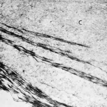
Fig. 7. Zonular fibrils average 10 nm in diameter. Most show a microperi-odicity of 12 nm (arrow) and they frequently appear hollow in cross section (small arrows). (TEM, × 81,600) Inset. C is lens capsule. The dark dots and filaments adherent to each zonular fibril are polysaccharide components preserved by fixatives containing Alcian blue (TEM, × 100,000).

The remarkably fine structural details of these zonular fibrils, or microfibrils as the identical fibrils are called in the rest of the elastic system, are shown best by rotary shadowing and electron microscopy of freshly homogenized zonular fibers.39,40 The individual zonular fibril is a beaded structure (“beads on a string”) consisting of 29- to 32-nm electron-dense beads held together by filaments that may be four in number and 2 to 3 nm in diameter (Fig. 8). The filaments can be seen more clearly when the fibrils are in a looser conformation. When they are in tighter conformation, many show cross-banding across the filaments between two beads. The beads' usual periodicity is 50 to 55 nm, but it can vary from 35 to 57 nm or more with different methods of preparation and 80 to 100 nm when stretched, without visible breakage of the filaments.41 Several studies suggest that changes in periodicity of the beads cannot fully explain the high degree of elasticity exhibited by fibrillin-containing microfibrils in tissue such as the zonular fibers, or in arteries of lower forms, perhaps requiring some reorientation of the fibrils.42,43 The nature of the bead is still not known, but the 148-nm filaments, the predicted length of a molecule of fibrillin, are thought to be arranged in a parallel staggered pattern with about one third overlap of adjacent molecules,27 although other patterns have also been suggested.44,45 The structure of elastic-system microfibrils throughout the body shows identical beaded fibrils.

Fig. 8. Rotary shadowing of a clump of human zonular fibrils in a 19-year-old patient. Darker straight fibril on right (large arrow) is in tight conformation (TEM, × 67,800). Top inset. Interbead filamentous subunits are seen (small arrows). Some bow outward, appearing to connect to every third bead (TEM, × 107,700). Bottom inset. A double-banded crosslinking region is visible across filaments between the beads (arrowhead). Three straight drumstick forms, probably interconnecting adhesion fibrils, extend down from beads or interbead areas (TEM, × 107,700).

Back to Top
ORIGIN OF THE ZONULE
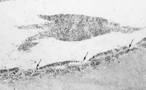
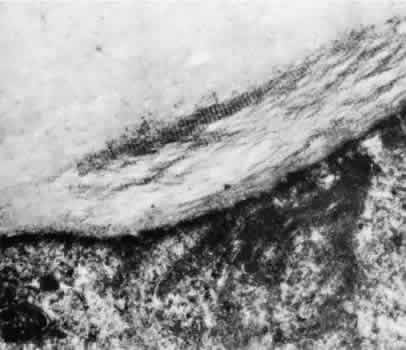
The ciliary zonular fibers originate in the pars plana as fine fiber bundles on the nonpigment epithelial basement membrane from 0.5 to 1 mm anterior to the ora serrata. They stain incompletely in gross specimens due to the overlying vitreous gel, narrowing to thin fibers as they near the ora (Fig. 9). By electron microscopy most appear to originate as small fibers containing 5 to 10 or more fibrils, adherent to the basement membrane of the epithelial cells, some becoming incorporated in the redundant infolding of the multilaminar basement membrane that develops with aging (Fig. 10). Small bundles are sometimes found superficially between these cells with no intervening basement membrane. Microfibrillar bundles found between some pigmented epithelial cells near their bases and in and under their basement membranes37,46 have been suggested to be zonular but are more likely a mixture of the abnormal elastic microfibrils found in the pigment epithelium's aging basement membrane and/or related to inserting microfibrils from elastic fibers of the underlying ciliary body stroma.

Fig. 9. Zonules (Z) on the pars plana taper toward the ora serrata (OR) between two retinal teeth (Gomori's hematoxylin, × 45).

Fig. 10. Attachment of zonular fibers to the ciliary epithelium of the pars plana. Small bundles (arrows) adhere to the surface and infoldings of the multilayeredepithelial basement membrane (TEM, × 17,000).

Back to Top
CILIARY COURSE OF THE ZONULE
Over the pars plana, the zonular fibers aggregate into bundles with the same pale membrane between them as illustrated in Figure 6, with multiple rows of these ribbons seen intermittently as new bundles are added (Fig. 11). The layer closest to the ciliary epithelium continues to have fine 0.1- to 0.5-μm fibers arising from the ciliary epithelium to join the main zonular stream over the rest of the pars plana, seen best in coronal sections, and sometimes is associated with small nodules of proliferated epithelium, called cap cells, appearing to be pulled inward by the fine attachments. These zonular bundles lie within the vitreous up to the mid-pars plana, where the anterior hyaloid membrane separates from the ciliary body to become an independent structure, and the zonules lie free in the posterior chamber (Fig. 12).

Fig. 11. Fine zonular fibers connect the main zonular ribbons to the ciliary epithelium over the whole pars plana. All sections were cut coronally. A. Intermittent double rows of zonular bundles (cm, ciliary muscle) (H&E, × 110). B. Elevated nodules of proliferated nonpigmented epithelium (cap cells) are often associated with these attachments (H&E, × 220). C. Fine zonular attachments at the posterior end of the pars plicata (PAS, × 220).

Fig. 12. Zonular fibers (Z) lie free in the posterior chamber from the point where the anterior hyaloid membrane (asterisk) begins to form in the mid-pars plana. B, elastic remnant of Bruch's membrane. (Toluidine blue, × 300)

The course of the zonule from the beginning of the pars plicata has been more difficult to elucidate and depends to some extent on the modality used to visualize it, because all have their artifacts and limitations. Scanning electron microscopy was a great step forward in understanding the three-dimensional nature of the zonule,46–55 although defects in methodology and low resolution in the early years led to erroneous impressions. A longtime controversy of importance in accomodative theory is whether some zonular fibers originate on the pars plicata and have a different function from those that are continuous from the pars plana to the lens.47 Recent investigators of both human and primate zonules have tended to conclude that most zonularfibers are continuous from pars plana to the lens,53,55 but they differ in estimating the number that may originate or terminate on the pars plicata and the degree of zonular adherence to this tissue. The following description is a synthesis of views about zonular anatomy in the pars plicata, obtained by several different methods and diagramed in Figure 13.

Fig. 13. Diagram showing the course of the zonular fibers from the pars plana of the ciliary body (right of figure) to their insertion on the lens (left of figure) as anterior (A), posterior (P), equatorial (E), and meridional (M). Almost all have a further attachment to the ciliary processes. The posterior zonular fibers include fibers (1) closely associated with the anterior hyaloid membrane throughout, (2) adherent to the top or sides of the ciliary crests, and (3) adherent to the valleys or minior plicae. The equatorial fibers (4) branch from others on the sides of the processes, and the anterior fibers (5) are adherent in the valley region.

With increasing height and complexity of the ciliary processes, the zonular bundles from their first entry between the posterior ciliary processes and minor plicae show considerable variation in distribution, but some general principles are apparent. Most of the bundles destined to insert on the posterior lens capsule become somewhat elevated above the level of the main zonular stream in the middle to anterior pars plana region. The one to three large bundles that pass over the crests of each ciliary process are the most highly elevated, especially over the prominent ciliary processes often found nasally. These bundles usually remain attached to the anterior hyaloid membrane up to the level of the ciliary processes. They then have some attachments to the crests and sides of the processes by direct adhesion or by joining branch bundles from below before passing to the posterior lens capsule at about a 30-degree angle, some still maintaining an attachment to the anterior hyaloid membrane. By the same direct and indirect attachments, the remaining posterior zonules become strongly adherent in the valleys between the ciliary processes or on the sides of the minor plicae, at intervals of 0.2 to 0.4 mm in from the posterior end of the pars plicata. These posterior zonulesin the valleys exit from the ciliary body at an angle of 50 degrees or more (Fig. 14). There is a continual exchange of fibers between bundles throughout their attachments to the valleys and walls of the pars plicata. Fibers from the deeper group of posterior zonules become associated with the anterior hyaloid membrane again only as it lies over their attachments on the lens posterior capsule.

Fig. 14. A. Overview of ciliary body, zonular fibers, and iris. The lens was removed after drying of the specimen. The angle between the anterior zonular fibers (AZ) and posterior fibers (PZ) has been preserved in some areas. VB, remnant of the vitreous base. (SEM, × 20) B. Zonular fibers continually change from one bundle to another (asterisk) (SEM, × 250).

When the anterior hyaloid membrane is removed for viewing or microscopy of this region, as it usually is, many of the posterior zonules are torn away or broken,54 which can give the erroneous impression that no zonules pass over the ciliary crests or that a number terminate on the crests.50 Fibers destined for the anterior and equatorial lens capsule are also strongly adherent to the valleys, between the minor plicae, or along the lower sides of the ciliary processes. They part company from the posterior zonules by continuing in an almost straight course to their insertion. Rohen has referred to the strong adherence of anterior and posterior zonules in the valleys and on the process walls as zonular “plexi” and the zone of divergence for these two main streams as the zonular “fork.”55 The anterior zonules are extensively embedded in infoldings of ciliary epithelium in the valleys, so it is difficult to peel them back from this region without breakage. A single zonular bundle from the pars plana may send fibers to all three lens regions. This fact, plus the constant interbundle exchange, guarantees the interrelatedness of the whole zonular complex. Both anterior and posterior zonular fibers exit from the pars plicata in ribbon-like swaths, lining up parallel to the ciliary processes (Fig. 15). The few zonules passing to the equator of the lens arise from the midsides of the processes or from the valleys and usually derive from anterior zonular bundles (see Fig. 13).

Fig. 15. Coronal section of zonular bundles attached to multiple sites (arrows) along sides and close to top of the ciliary process in the pars plicata (PAS, × 400). Inset. One bundle attachesthe anterior hyaloid membrane to the top (arrow) of a process(H&E, × 200).

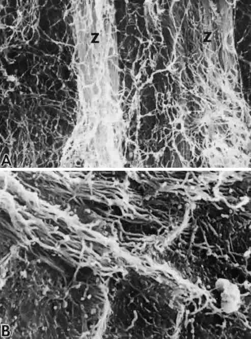
The close attachment of almost all zonular fibers to the ciliary processes or to the plicae during their course is an important one. It occurs by direct adhesion of zonular fibers to the ciliary epithelial basement membrane at the edges and base of large bundles, and by fine strands from the larger bundles appearing to splay out over the epithelium (Fig. 16A), of importance to accomodation theory (see later discussion of accomodative mechanisms).47,55On scanning microscopy, such bundles fan out in acharacteristic pattern at their attachment sites, partially embedded, or perhaps sometimes ending, in the rich fibrogranular matrix of the ciliary basement membrane (Fig. 16B) before regrouping to continue. Larger zonular bundles are attached here than in any other site, and they even become enmeshed in the early thickening of basement membrane occurring here (Fig. 17). Thus, between the attachment of small zonular fibers to the main zonular stream over the pars plana and the firm attachments of the main zonular fibers to the pars plicata, most of the system is closely bound to the ciliary body. This prevents the zonular fibers from forming a cord between their origin and their lens insertion that would interfere with their ability to transmit accomodative forces to the lens during accomodation.

Fig. 16. Zonular fibers and fibrogranular surface of ciliary nonpigmented epithelial basement membrane. A. Small fibrillar zonules on the sides of the ciliary processes. Whether they are continuous or terminating cannot always be determined (SEM, × 250). B. Higher-power view from boxed area in A, showing zonules merging into fibrogranular basement and adhesive surface matrix (SEM, × 5,000).

Fig. 17. Coronal section through a valley of the pars plicata shows superficial blending of zonular fibers and bundles (Z) into the multilaminar, already thickened basement membrane of the ciliary epithelium in a 19-year-old patient (TEM, × 26,200). Inset. Large zonular bundles (Z) are embedded in the even thicker basement membrane of the older eye (50-year-old patient). A dark cap cell (CAP) lies on the surface (TEM, × 5,000).

The “crossing” of anterior and posterior zonular fibers seen histologically as they pursue their different paths has interested microscopists for years.Salzmann noted in 1912 that “this double anchorage brings about a stronger fixation of the entire zonular mass at the posterior border of the corona ciliaris, and this is the reason why with few exceptions the zonular fibers break away at this place.”57

Back to Top
ZONULAR CIRCUMFERENTIAL GIRDLE FIBERS
Early anatomists noted the presence of some zonular-like bundles passing “the wrong way” (that is, perpendicular to the main zonular stream) in a circumferential (coronal) girdle-like pattern over the ciliary processes, the pars plana, and the posterior zonular lens insertion.57–59 Because the girdlefibers lie within the anterior hyaloid membrane, they are best seen when this membrane has not been completely removed. Eisner60 has found them by slit-lamp examination in postmortem eyes, and other observers have noted them on one or another of these three sites using a variety of microscopic and staining methods in humans and other primates.36–38,50,53–55,61 These girdle fibers are often seen to branch from underlying zonular fibers and are fibrillin-positive, so there is no question that they have the same nature.

POSTERIOR CILIARY GIRDLE AND ABERRANT ZONULAR FIBERS

The posterior girdle of zonular bundles lies within the vitreous just above the main zonular stream 1 to 2 mm anterior to the ora serrata (Fig. 18). This is also the area in which the median vitreous tract inserts, to use Eisner's terminology.60 Vitreous collagen fibrils in the immediate vicinity of the zonular girdle fibers have a similar circumferential orientation. This suggests that the orientation of both types of fibers may be a response to the same mechanical stresses, such as vitreous traction during ocular movement and accommodative excursions.

Fig. 18. Posterior girdle zonules on the pars plana in the vitreous base. A. Arrows indicate fibers passing circumferentially in vitreous base remnants (vb) on the pars plana. Ciliary processes and minor plicae are on the upper left (SEM, × 65). B. High-power view of above area, showing 1.5-μm circumferential zonular bundle (cz) among the collagen fibers of the cortical vitreous that are also oriented circumferentially. Round particles are melanin granules (SEM, × 9,000).

Short bundles that stain like zonular fibers may pass back over the ora serrata into the vitreous at all ages, either from the posterior girdle or separately without any apparent attachment to the ciliary body. Aberrant zonular fibers are commonly found posterior to the ora serrata in association with peripheral abnormalities such as zonular traction tufts62 and also adjacent to meridional folds, enclosed ciliary bays, and peripheral retinal rosettes or “granulations.” These aberrant fibers generally do not join the main zonular stream but end close to the ciliary surface a few millimeters anterior to the ora serrata.

ANTERIOR CILIARY AND LENTICULAR GIRDLE

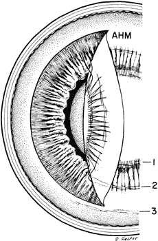
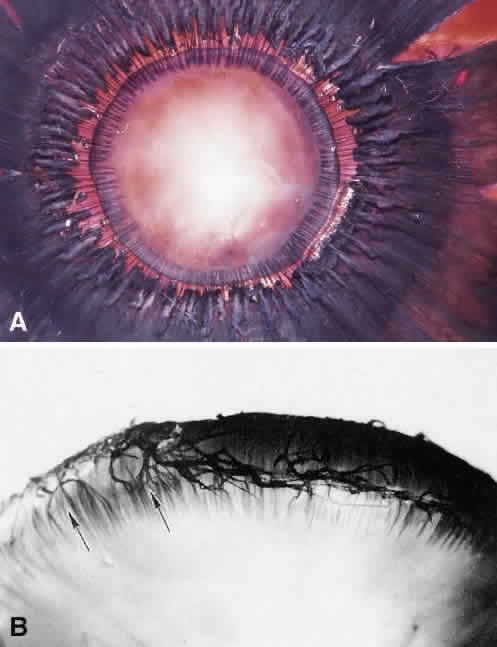
Prominent circumferential zonular fibers lie over the crests of the ciliary processes (Fig. 19), arising from branches of the posterior zonular bundles as they pass over the crests or from others attaching in the ciliary valleys. This anterior girdle lies superficially on and within the anterior hyaloid membrane, helping to bind the membrane close to the ciliary processes and resisting the pull of the coronary vitreous tract, which inserts here.60 These fibers usually remain on the hyaloid membrane when the latter is torn away, along with some posterior zonular fibers. In young eyes the girdle fibers are very fine, but in old eyes they are stout cords. Some vitreous collagen fibers here also have a circumferential orientation. As diagrammed in Figure 20, another group of circumferential zonular girdle fibers is lenticular. These can also be seen in Figure 19B in the anterior hyaloid membrane above the anterior ciliary girdle. Most of the lenticular girdle fibers lie on the flattened and fanning posterior zonular bundles near the end of their lens insertions at the approximate site of Wieger's ligament. Eisner'shyaloid vitreous tract also inserts here.60 Thesethree fibrillin-positive girdles represent sites where changes in tractional forces affect both the vitreous and the zonular system. The girdles may help maintain the posterior chamber and support the vitreous gel.

Fig. 19. Anterior and lenticular girdle zonular fibers. A. Circumferential zonular fibers (arrows) lying over the crests of the ciliary processes are attached to the anterior hyaloid membrane, which has not been removed in this intact eye. Lens and posterior zonular fibers are visible above (Gomori's hematoxylin, × 15). B. Anterior hyaloid membrane removed as a sheet from the same specimen. Many circumferential zonular fibers (horizontal) and posterior zonules (vertical) remain adherent to the membrane. The horizontal fibers include scanty lenticular (above) and more plentiful anterior ciliary girdle fibers below (Gomori's hematoxylin, × 60). C. Anterior hyaloid membrane with attaching posterior zonules on peripheral lens capsule and associated horizontal lenticular girdle fibers (flat preparation, immunostained for fibrillin-1, × 60).

Fig. 20. Distribution of the circumferentially oriented zonules as lenticular (1), anterior (2), and posterior (3) girdles. When the anterior hyaloid membrane (AHM) is reflected, many of the girdle fibers will remain adherent to it.

Back to Top
CILIARY-LENS COURSE OF THE ZONULE
No zonular fibers pass across the anterior heads or beaks of the ciliary processes, because the latter curve forward out of the plane of the zonular fibers (Fig. 21). The zonular bundles associated with each of the major ciliary processes form a unit as they pass along the sides of the process and over its head on their way to the lens. They emerge on either side of the process as flat ribbons of bundles 30 to 60 μm in diameter, about 6 to 10 deriving from each ciliary unit (see Fig. 4). If the method of preparation involves drying, the bundles may separate or clump together as their interfibrillar matrix dehydrates and be quite variable in number. An empty V-shaped area is left in the ciliary unit over the beaks of the anterior ciliary processes. The concept that pigmentary glaucoma might involve rubbing of zonular bundles against an abnormally bowed-back peripheral iris in this region, with loss of peripheral iris pigment and pigment epithelium, is a reasonable one.63 Bundles from adjacent zonular units continue to interchange, so that evidence of segmental origin and vacant areas is much reduced as the zonular fibers approach their very regular multilayered insertion on the lens capsule (see Fig. 1). There had been a long-standing question about whether the zonule forms a continuous membrane around the lens. The early anatomists discovered that air bubbles were easily trapped among the zonular bundles and could be made to dissect a channel between the anterior and posterior layers of zonular fibers (Hanover's canal) or between the posterior zonular fibers and the anterior hyaloid membrane (canal of Petit).64 This phenomenon is probably due to surface tension of the sticky zonular bundles. Modern studies show the zonule as a discontinuous fiber bundle system allowing inflammatory cells and pigment to percolate among the ribbon-like zonular bundles back to the anterior hyaloid membrane. It is unknown whether the sticky mucoid characteristics of the zonular ribbons and zonular lamella might have any influence on diffusion through the lens capsule in the insertional region.

Fig. 21. Scanning micrograph of the anterior zonular insertion after removal of cornea and iris. The anterior heads of the ciliary processes are free of zonules, leaving an empty V as the zonular bundles enter on each side of the processes. Lens is 25% smaller than normal owing to processing shrinkage (× 25).

Back to Top
ZONULAR INSERTION ON THE LENS
The region of zonular insertion on the lens can be difficult to analyze even with use of several methods of microscopy, due to difficulty in keeping all of the zonular complex attached when isolating the lens and in recognizing artifactual losses. The presentation here summarizes conclusions drawn from light microscopy, transmission and scanning electronmicroscopy, and immunostaining, particularly for fibrillin-1. By scanning electron microscopy the zonular insertional region shows complete covering by a layer different from the surface of the bare lens capsule, including the zonular bundles and an apparently similar background material between them (Fig. 22).

Fig. 22. View of the whole zonular insertional region and broad lens equator of an elderly patient, appearing different from the dark surface of the rest of the bare capsule because of its coating of zonular fibers and matrix. Anterior (A), posterior (P), and a few small equatorial (E) zonular bundles are visible. Some posterior zonular bundles were lost when the vitreous was removed (SEM, × 64). (Streeten BW: The zonular insertion: A scanning electron microscopic study. Invest Ophthalmol Vis Sci 16:364, 1977)

ANTERIOR ZONULAR INSERTION AND ZONULAR LAMELLA

The zonular fibers inserting on the lens form a layer that extends 1.5 mm onto the anterior surface of the lens capsule and 1.25 mm onto the posterior capsule in the adult. With increasing size of the lens, the inserting bundles appear flatter, with fewer underlying layers of attaching fibers visible, but at least two main layers are usually apparent. These rapidly spread out on the capsule as 5- to 10-μm bundles, presenting a remarkably symmetric appearance, with only occasional bundles passing ahead of the main insertion (Fig. 23). They continue to decrease in size, tapering over a flat insertional area whose length depends on the size and age of the lens, ending as 0.3- to 0.4-μm small bundles whose fibrils blend into the surface fibrillar layer of the capsule in a basket-weave fashion. For 1 mm central to the tips of the zonular inserting fibers, the surface fibrillar meshwork becomes gradually thinner until only the pebbly surface of the bare lens capsule is visible. All along the anterior insertion, the zonular bundles join a surface layer of more loosely arranged fibrils of similar size that often have a horizontally rippled appearance. When the capsular surface is abraded, as in Figure 24, by pulling away of some inserting zonular bundles, it is seen that the rumpled surface layer has fibrils like those of the meshwork around the inserting anterior zonular fibers, some of which remain adherent to the fibrogranular surface layer still on the anterior capsule. A rolling up and retraction of the distal abraded layer indicates that it has some elasticity and contains a few small linear zonular fibers within it.

Fig. 23. Zonular bundles insertingon the anterior lens capsule. A. Theinserting ends taper to thin points (SEM, × 700). B. Terminating zonu-lar fibrils (Z) end by a blendingof their fibrils into the superficialcapsule in a basket-weave fashion (SEM, × 21,000). Inset shows pebbly surface of bare lens capsule, cen-tral to the zonular insertion (SEM, × 16,000). (Streeten BW: The zonular insertion: A scanning electron microscopic study. Invest Ophthalmol Vis Sci 16:364, 1977)

Fig. 24. The nature of the superficial zonular insertion. A. A dark area of surface abrasion in the anterior end of the zonular insertion shows variable rippling and retraction of the remaining layer (boxed area) (SEM, × 645). B. At the edge of the abrasion in the boxed area of A, individual fibrils blend into the fibrogranular matrix of the superficial lens capsule (SEM, × 10,700). (Streeten BW: The zonular insertion: A scanning electron microscopic study. Invest Ophthalmol Vis Sci 16:364, 1977)

The layer of lenticular zonular fibers and the superficial lens capsule into which they insert was named the zonular lamella by Berger.65 This superficial 0.6 to 0.9 μm of the capsule in the insertional area is more loosely arranged than the rest of the capsule and has a higher content of glycosaminoglycans with Alcian blue staining, also shown by the zonule (Fig. 25). A high content of proteoglycans is found in this junctional region, with a large chondroitin sulfate proteoglycan (chondroitinase ABC-sensitive) surrounding bundles of zonular fibers and their junction with the capsule, and extensive hep-aran sulfate (heparitinase-sensitive) in the adjacent superficial capsule. Evidence of chondroitin sulfate associated with the zonule was found earlier biochemically9,11 and more recently by impressive immunohistochemistry and synthesis studies.66,67 Other moieties related to adhesion of zonular and other elastic microfibrils to basement membranes, such as fibronectin,68,69 vitronectin,70 and integrin αvβ3 are being further investigated.

Fig. 25. Proteoglycans of the zonular lamella and zonule. A. Zonular fiber layer (Z) and superficial lens capsule (S) stain more deeply for glycosaminoglycan than the rest of the capsule. LE, lens epithelium. (Alcian blue, × 1,000) B. Lens from a 6-month-old infant stained only with cuprolinic blue and uranyl acetate. The superficial capsule (S) shows heavy staining of thin stick-like aggregates of heparan sulfate glycosaminoglycan. Larger aggregates and a few smaller ones (arrows), primarily chondroitin sulfates, are seen on the capsular surface and around the periphery of zonular bundles (Z) (TEM, × 53,900).

By transmission electron microscopy, the zonular lamella of the superficial lens into which the zonule inserts has a loose fibrogranular vacuolated appearance, the latter perhaps related to water held by its proteoglycan. Its fibrils when visible are 1 to 3 nm in diameter (Fig. 26) compared with the 10-nm zonular fibrils. The zonular fibers insert by sending individual fibrils and small 0.07- to 0.5-μm fibers into the superficial capsule to a depth of 0.6 to 1.6 μm. The horizontal surface rippling seen between the anterior inserting zonular bundles is associated with rippling of the underlying superficial capsule as well, where a small number of their zonular fibers insert, emphasizing the malleability of this zonular lamellar capsule.

Fig. 26. Anterior zonular insertion. A. Coronal section of anterior zonular fiber bundle (above) inserting into the lens capsule. The loose superficial capsule (CAP) shows 10-nm banded zonular fibrils (arrows) to a depth of 1.6 μm. Fibrils of the capsular matrix at 1 to 3 nm are barely visible (TEM, × 65,000).B. Behind the main anterior inserting bundles, a few small zonular fibers join others in the superficial capsule (Z IN S. CAP). Both tissues participate in the capsular rippling (asterisk) (TEM, × 42,400). Inset. Rippled superficial layer fibrils merging with fibrils of the attaching zonular bundles (Z) (SEM, × 6,400).

Staining with fibrillin-1 gives a new view of the familiar “capsular inclusions” that are said to begin accumulating in the superficial and deep capsule from the beginning of the second decade.71,72 Anteriorly they are seen dramatically in flat preparations as fibrillin-positive, comma-shaped forms, slightly deeper than the inserting zonular fibers themselves (Fig. 27), and especially clear where anterior zonules are torn away. In some capsules they can be very profuse even at age 16, extending halfway back over the insertional area, having no clear connection to zonular fibers. In tangentially cut sections of the insertion they are curved, thin fibrogranular forms about 1 to 6 μm long, mostly 2 to 3 μm, but a few up to 9 μm long when joined together. Besides immunoreacting for fibrillin-1, they are also posi-tive for vitronectin and another binding protein,HNK-1. Whether they have a normal adhesivefunction or are to be considered degenerative isuncertain. Studies of intact fibrillin microfibrilshave shown that they can be quickly degraded by nuetrophil elastase, chymotrypsin, and trypsin,73 but what enzymes might be involved in any normal turnover processes is not known.

Fig. 27. Anterior capsular superficial inclusions. A. Small comma-shaped fibrillin-positive bodies are seen in the superficial capsule from which zonules have torn away (immunostained for fibrillin-1,flat preparation, original magnification × 132). B. Tangential cuts across the insertion show the inclu-sions as slightly less regular curved forms of 1 to 8 μm in the capsular matrix (immunostained forfibrillin-1, TEM, × 8,100).

MERIDIONAL ZONULAR FIBERS

The zonular layer thickens from 1.0 to 1.7 μm behind the anterior zonular insertion, where it shows a marked radial ribbing produced by the deep layer of meridional zonular fibers (Fig. 28A). These small fibers vary from 3 to 10 μm in width and are covered by the loose surface meshwork fibrils seen elsewhere. They are intimately involved in the insertion of the regional zonular fibers, as seen in Figure 28B, where many branches of an equatorial zonular bundle appear to merge onto the surfaces of the meridionals or enter between them for their own insertions. Zonular bundles inserting directly on the equator are quite infrequent and vary in number but can be seen at all ages, even into the tenth decade. Although some are up to 60 μm in diameter, most are 10 to 15 μm and attach almost perpendicularly, quickly merging with the meridional fibers so they cannot be individually distinguished. By transmission electron microscopy the midequatorial meridional bundles insert superficially in the capsule as small bundles (Fig. 28C).

Fig. 28. Meridional zonular fibers. A. Ribbing of the lens equator by the meridional zonular fibers between the anterior (above) and posterior (below) zonular bundles (SEM, × 195). B. Equatorial zonular bundle at its junction with the meridional zonular fibers (SEM, × 470). C. Coronal section of two 0.5-μm meridional zonular fibers (m) within the superficial capsule (TEM, × 51,000). (A & B from Streeten BW: The zonular insertion: A scanning electron microsopic study. Invest Ophthalmol Vis Sci 16;364, 1977)

The meridionals appear to extend over the entire zonular insertion as an initial zonular layer into which the larger bundles merge (Fig. 29), best seen when there is good preservation of the larger zonular bundles. Most of this zonular complex is torn from the lens during an intracapsular cataract extraction or separation of lenses from donor eyes. What typically remains is only the inserting tips of the anterior zonular fibers and occasional remnants of meridional fibers (Fig. 30A). Pulling forward on some of the zonular complex, as seen in Figure 30B, shows how easy it is to remove the whole complex like a blanket, leaving bare capsule visible.

Fig. 29. Whole zonular insertion on a flat preparation of lens capsule with careful preservation of entire zonular complex, showing inserting anterior zonular bundles above and some posterior zonular bundles below, connected by the meridional zonule fibers (immunosained for fibrillin-1, original magnification × 14).

Fig. 30. The zonule and cataract extraction. A. Lens after removal intracapsularly by cryoprobe (round probe mark above). Only a few tips of anterior zonular fibers and a sector of meridional zonules remain (Gomori's hematoxylin, × 10). B. Experimental stripping of zonular complex at level of posterior (P) and meridional zonular fibers, with a few deeper inserting zonules revealed (arrows) (SEM, × 600). (Streeten BW: The zonular insertion: A scanning electron microscopic study. Invest Ophthalmol Vis Sci 16:364, 1977)

The most unusual feature of the meridionals is their strong attachment in the postequatorial region, approximately over the terminal meridional nuclear rows of lens epithelial cells (Fig. 31), as seen in flat preparations of whole lens capsules immunostained for fibrillin-1.74 The very regular outline of these10-μm columns of meridional zonular insertion may remain when most of the zonular complex has been completely removed. The insertional columns are seen to have small zonular fibers inserting on them as larger bundles are pulled away, sometimes with paler spaces across them forming a partial “waffle” pattern. The meridional zonules that attach strongly to this area can be seen to continue both above and below this attachment region. This pattern is obscured when more of the meridional zonules remain attached and may even be hidden if large numbers of posterior zonules cover the area. When the zonular fibers have been mostly removed, the base of their attachments is visualized as profuse aggregates of small fibrillin-positive filaments. Coronal cross sections through this meridional insertion show three features. Two rows of fibrillin-positive zonular material lie on the lens capsule, the outer row segmented into 3.3-μm ellipses of zonular material and the innermost layer appearing somewhat more irregular but continuous (Fig. 32). The overlying zonular bundle seen in this figure is 9.9 μm in toto. This complex appears to represent a single meridional insertion made up of three small zonular fibers with an overlying more loosely attached bundle.

Fig. 31. The meridional zonular fiber system in flat capsular preparations, staining for fibrillin-1. A. Artifactual pulling aside of inserting zonules reveals the vertical posterior meridional insertion site (arrows) (original magnification × 14). B. Dense fibrillin staining seen in a “waffle” pattern of vertical lines consisting of 10-μm zonular fibers to which small bundles attach, as seen on lower right (original magnification × 66). C. In areas where the inserting zonules have been pulled away, their base consists of profuse small zonular microfibrils (original magnification × 330).

Fig. 32. Coronal sections through the meridional insertion region show two layers of inserted zonular material on the capsule, the innermost having a less organized fibrillar pattern, and the outermost well-delineated regular elliptical 3.3-μm bundles (arrows), with an overlying loosely attached 10-μm zonular bundle (TEM, × 6100).

In young eyes, some evidence of this radial pattern can extend up almost to the anterior zonular insertions, but it becomes increasingly restricted to the equatorial region. This intriguing site may be related to the 10-μm vertical bands of fibrillin staining reported by Wheatley and colleagues75,76 in postequatorial frozen sections of lens capsule cut coronally. Interpretation in these studies was hindered by incomplete preservation of the zonular insertional region, such that no connection was found between the anterior inserting zonular tips and the equatorial inserting zonules, which were thought to be the only region of zonular insertion on the lens. The great regularity and persistence of this area overlying the most metabolically active site for new lens fiber formation is of interest, also the fact that the peripheral lens fibers average 10.2 μm in diameter.77 Perhaps this postequatorial region is established in an early period of zonular aggregation on the lens capsule to help direct a radial zonular pattern, rather than a haphazard coating of the insertional region by fibrillin matrix. Regulatory and adhesive factors from the underlying lens cells might well be required.

POSTERIOR ZONULAR INSERTION

The posterior zonular bundles insert in two or three layers over a 0.4- to 0.5-mm area. The superficial bundles have a slightly more fanned insertion than do the anterior zonular bundles, with fewer interbundle connections (Fig. 33). Their insertion begins at the posterior edge of the lens equator, just beyond the terminus of the subcapsular lens epithelial cells. The innermost layer of posterior zonules begins to insert earlier, with some adhesion to the meridional zonular fibers. All are covered by a denser layer of loosely arranged meshwork fibrils than over the anterior zonular fibers (Fig. 34). When exposed,however, the insertions show the usual pattern of fibrils blending into a fibrogranular capsular matrix. The superficial capsule or zonular lamella into which they insert is especially loose in its outer2 μm, and the zonular fibers penetrate in multilay-ers for almost the whole 2-μm thickness (Fig. 35).No penetration of any zonular fiber deeper than2.6 μm into the lens capsule has been found.

Fig. 33. Posterior zonular insertion. A. In intact eye (Gomori hematoxylin, × 7). B. Posterior lens and equator of an extracted lens. The posterior zonular insertion begins over the end of the meridional nuclear rows of lens epithelium (arrows) (Gomori's hematoxylin, × 15). (Streeten BW, Pulaski JP: Posterior zonules and lens extraction. Arch Ophthalmol 96:132, 1978. Copyright 1978, American Medical Association)

Fig. 34. Insertion of the posterior zonular fibers. A. A thick layer of looser fibrils covers the inserting zonular fibrils (Z) (SEM, × 9,200). B. When exposed, the deep insertion shows the usual blending of zonular fibrils into the fibrogranular matrix of the superficial capsule (SEM, × 15,000). (Streeten BW, Pulaski JP: Posterior zonules and lens extraction. Arch Ophthalmol 96:132, 1978. Copyright 1978, American Medical Association)

Fig. 35. Posterior zonule fibers behind the equator insert deeply into the very loose superficial matrix of the lens capsule (C) (Ruthenium red, TEM, × 29,000).

The presence of sporadic bundles of fibrils in the equatorial and midperipheral lens capsule close to the lens epithelium was once thought to represent deep zonular insertions, but no direct connection to surface zonules has been found, and these inclusions were not seen before the second decade.72,73 Some are prominently banded at 45 to 50 nm (Fig. 36), which is the periodicity of fibrillin fibrils, and stain for fibrillin-1. Others found in midcapsule in increasing numbers with age are fibrogranular with tubular profiles. They are fibrillin- and vitronectin-positive, like the anterior ones, and show no zonular fiber connection. In the exfoliation syndrome, similar capsular inclusions are much larger and very profuse. All types appear to represent aberrant synthesis by the lens epithelium, but some relation to a normal lens secretory function is not ruled out.

Fig. 36. Deep capsular inclusions. Fibrils deep in the lens capsule adjacent to the epithelium showing banding at 50 nm, characteristic of material often found in aging and abnormal basement membranes (TEM, × 24,000).

POSTERIOR ZONULAR ATTACHMENT TO THE ANTERIOR HYALOID MEMBRANE

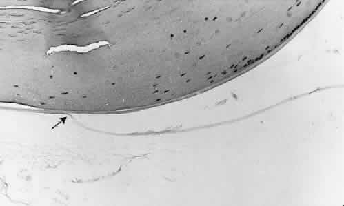
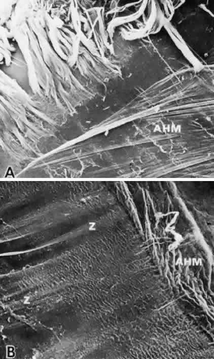
The posterior zonular insertion is closely related to the attachment of the anterior hyaloid membrane on the lens capsule. This anterior hyaloid attachment was defined by Wieger (cited by Vail)1 as an 8- to 9-mm circular area at the edge of the posterior lens, which he called the hyaloideocapsular ligament (Fig. 37). This junctional area is also known as Egger's line when seen at the slit lamp.60 By microscopy the attachment site is found at variable distances behind the equator, depending on the method of preparation and the age of the patient. In childhood, the anterior hyaloid membrane is normally closely attached to the whole posterior zonular insertion and lens (Fig. 38). In the first two or three decades, removal of the intact lens is accompanied by the vitreous due to the strength of this adhesion. In later adult life, the anterior hyaloid membrane more readily peels back to the end of the zonular insertion and separates completelywith slight traction. The reason for the strength of Wieger's “ligament” is not evident, because no special differentiation of capsule or zonule has been recognized here, except for some circumferential girdle zonular fibers. During intracapsular cataract extraction it was not uncommon for a superficial flap of either capsule or anterior hyaloid to be torn from Wieger's area, often with vitreous loss. Many of the avulsed posterior zonules and circumferential zonular fibers remain adherent to the anterior hyaloid membrane after an uneventful intracapsular extraction (Fig. 39).54

Fig. 37. Junction of the anterior hyaloid membrane and the lens capsule: site of Wieger's “ligament” (arrow) (H&E, × 220).

Fig. 38. Posterior zonular bundles and the anterior hyaloid membrane (AHM). A. Irregular retraction of the AHM from the posterior zonular insertion in a 3-year-old patient. Note layering of posterior zonular bundles (SEM, × 90). B. Complete retraction of the AHM to the end of the posterior zonular fibers (Z) in a 71-year-old patient (SEM, × 400).

Fig. 39. Anterior hyaloid membrane after an intracapsular cataract extraction showing adherent zonular bun-dles fanning out as though attachingto the posterior lens capsule (SEM,× 400). Inset. Extensive zonular fibersremain on the hyaloid in this regionof Wieger's ligament. (Streeten BW,Pulaski JP: Posterior zonules andlens extraction. Arch Ophthalmol 96:3132, 1978. Copyright 1978, American Medical Association)

Back to Top
ZONULAR COMPLICATIONS IN CATARACT SURGERY
The unpleasant complication of zonular dialysis or dehiscence from the capsule during extracapsular cataract extraction continues to focus interest on zonular attachments to the lens. About 5% of cases show zonular dehiscences recognizable at the time of surgery,78 with a larger number demonstrable by special stains postmortem.79 Some dialyses may be extensive enough to modify the plans for lens evacuation or placement of an intraocular lens. Dehiscences with vitreous presentation appear to occur by posterior zonular or hyaloid tearing from the capsule at the same sites as described for intracapsular extraction. Most commonly, dehiscences develop during the removal of the lens nucleus or cortical material78 as traction is exerted on the zonular attachments. In experimental zonular extraction for a previous study, it appeared that backward tipping of the lens was especially apt to result in vitreous presentation from circumferential tearing in Wieger's attachment region. When specific disease affects the strength of the zonular system, such as in the pseudoexfoliation syn-drome, the fragile fibers are four times more likely to break78 or may already have ruptured before surgery, with an occult subluxation.80
Back to Top
CHANGES IN THE ZONULE WITH AGING
In fetal and infantile eyes, the zonular fibers are finer and less aggregated than in adult eyes. There is also considerably more proteoglycan staining with Alcian blue or cuprolinic blue in the young zonule (see Fig. 25), which is also noted in some animals.81 This decrease in subsequent years may indicate either that this material was mostly necessary during the developmental stage or that its loss is a degenerative phenomenon. In the elderly, the zonules appear thinner and fewer and are more easily ruptured than in the young adult, although this fragility with age is variable.82 Ultrastructurally, their attachment to the ciliary body basement membrane was found less close and their network disorganized, with shorter microfibrils and dispersed beads by scanning microscopy.83 This decrease in microfibril size was suggested to result from decreased fibrillin synthesis with age, as noted in the aging human aorta.84 Equatorial zonules become especially scanty. In the first two decades of life, the flat insertional areas are narrow, grasping the thin lens equator at its periphery, with the zonular bundles appearing thick and closely packed (Fig. 40). As the lens continues to grow in diameter and thickness, the insertional areas are widened and displaced relatively more centrally.85,86 Because of this radial lens growth and the great expansion of the equator, the distance between the anterior and posterior zonular insertions is considerably increased while their distances from the ciliary valleys are decreased, and might tend to lessen their mechanical advantage. For uncertain reasons, there is also a gradual true narrowing of the zonule-free area in the center of the anterior capsule, from approximately 8 mm in diameter at age 20 to 6.5 mm in the eighth decade and rarely to as little as 5.5 mm.86 This narrowing should be considered when planning anterior capsulectomy to avoid direct injury to the zonular nsertion.

Fig. 40. Widening of the anterior zonular insertion and relative inward displacement accompany growth of the lens equator with age. The central zonule-free capsule also becomes narrower. Perilenticular space is minimally widened by lens shrinkage in older eyes (Gomori's hematoxylin). (Stark WJ, Streeten BW: The anterior capsulotomy of extracapsular cataract extraction. Ophthalmic Surg 15:911–917, 1984)

It is likely that none of these zonular changes contributes to the development of presbyopia as a primary factor, but rather reflects the continuous lens growth with later aging pathology.

Back to Top
ZONULAR ROLE IN ACCOMMODATION
Because the zonule is the effector arm of the accommodative mechanism, its anatomy must directly reflect the workings of this mechanism. There is little question that the zonule in the nonaccommodated state maintains tension on the lens capsule, decreasing the naturally more spherical shape of the lens. During accommodation the ciliary processes move forward and inward, relaxing zonular tension, according to most views since Helmholtz,87 and shown dramatically by videotapes of centrally stimulated accommodation in rhesus monkey eyes after removal of the iris.88 The lens decreases in circumference and increases in thickness, with the nucleus thickening more than the cortex.89 The anterior surface of the lens becomes more curved and displaced anteriorly, increasing its refractive power. Failure of the posterior lens surface to undergo comparable changes in curvature and displacement has been somewhat puzzling. Explanations include support from a relatively incompressible vitreous.90 The mechanical disadvantage of the more obliquely inserted posterior zonular fibers might also be relevant, especially as the lens thickens with growth. Further evidence for a vitreous support role was deduced from a mathematical model of accommodation.91 The configuration of the posterior zonules was calculated to be inadequate to raise the posterior forces necessary for accommodation, without vitreous support.

Rohen and colleagues47,55 proposed that there are two different types of zonules: the main zonular fibers passing directly from the pars plana to the lens, and “tension zonules” connecting the main stream to the ciliary processes. They suggested that during accommodation the tension fibers are placed on stretch, allowing the main zonular fibers to slacken. This theory assumes that the pars plana has no anterior movement during accommodation, conflicting with the traditional view and warranting further investigation. Others have not found these two separate systems of zonules.50,53 There is no doubt, however, that most zonules are extensively attached to the ciliary processes, especially on the ciliary valleys and walls where the large number of puncta adherens junctions between the epithelial cells is evidence of tractional stress, as described by Ober and Rohen.56 During extreme accommodation the inserting zonular fibers were observed to fold over on themselves by videography,88 suggesting that they are isolated at least partly functionally from the pars plana fibers by their strong ciliary attachments. Relaxation of the human zonule during near accomodation has also been noted by ultrasound biomicroscopy.92 A new theory of accomodation offered by Schachar and colleagues with a proposed physical model93,94 is based on the equatorial zonules being the active components in accomodation, where they are envisioned as being under tension, reducing tension on the anterior and posterior zonules and allowing the lens to round up. Anatomically the minimal number of equatorial zonules observed by most seems inadequate to allow them to exert such independent action. This theory is still being actively investigated.

Factors controlling the synthesis and turnover of zonular components are unknown. McKanna and Casagrande95 presented evidence that there is a feedback mechanism among the variables of zonular fiber development, accommodation, and lens growth during “emmetropization” in the infantile eye. In the elongated eyes of tree shrews with unilateral lid-suture myopia, they found the lens and zonule to be hypoplastic. They postulated that blurred vision induced excessive chronic accommodation, reducing tension on the zonular fibers and lens and leading to hypoplasia of both structures by feedback loops.

Back to Top
ACKNOWLEDGMENTS
Funded in part by research grant EY01602 from the National Eye Institute, National Institutes of Health. Acknowledgement is made with gratitude to Dr. Yue Qi, Dr. Zong-Yi Li, Robert Wallace, and Judith Strauss for outstanding research contributions and to Carolyn Buckbee for excellent manuscript preparation.
Back to Top
REFERENCES

1. Vail D: The zonule of Zinn and ligament of Wieger. Trans Ophthalmol Soc UK 77:441, 1957

2. Barber AN: Embryology of the Human Eye, pp 154–157. St. Louis, CV Mosby, 1955

3. Gloor BP: Zur Entwicklung des Glaskorpers und der Zonule: Vl. Autoradiographische Untersuchungen zur Entwicklung der Zonule der Maus mit 3H-markeiten Aminoauren und 3H-Glucose. Graefes Arch Clin Exp Ophthalmol 189:105–124, 1974

4. Rhodes RH, Mandelbaum SH, Minckler DS, Cleary PE: Tritrated fucose incorporation in the vitreous body, lens and zonules of the pigmented rabbit. Exp Eye Res 34:921–931, 1982

5. Briglin CA, Liz-Y, Streeten BW: Cultured lens epithelial cells synthesize matrix immunopositive for elastic microfibrillar proteins, including fibrillin and a zonular peptide. Invest Ophthalmol Vis Sci 32(suppl):777, 1991

6. Robb BW, Wachi H, Schaub T et al: Characterization of an in vitro model of elastic fiber assembly. Mol Biol Cell 10:3595–3605, 1999

7. Sakai LY, Keene DR, Engvall E: Fibrillin, a new 350-kd glycoprotein, is a component of extracellular microfibrils.J Cell Biol 103:2499–2509, 1986

8. Qi Y, Streeten BW, Wallace RN, Hoepner: Expression of fibrillin-1 mRNA in cells related to the zonular system of the developing mouse eye by in situ hybridization. Invest Ophthalmol Vis Sci 37(suppl):90, 1996

9. Buddecke E, Wollensak J: Zur Biochemie der Zonulafaser des Rinderauges. Z Naturforsch 216:337–341, 1966

10. Dische Z, Murty VLN: An insoluble structural glycoprotein: A major constituent of the zonula Zinni. Invest Ophthalmol Vis Sci 13:991–995, 1974

11. Streeten BW, Swann DA, Licari PA et al: The protein composition of the ocular zonules. Invest Ophthalmol Vis Sci 24:119–123, 1983

12. Streeten BW, Gibson SA: Identification of extractable proteins from the bovine ocular zonule: Major zonular antigens of 32 kd and 250 kd. Curr Eye Res 7:139–146, 1988

13. Streeten BW, Gibson SA, Li Z-Y: Lectin binding topseudoexfoliative material and the ocular zonules. Invest Ophthalmol Vis Sci 27:1516–1521, 1986

14. Raviola G: The fine structure of the ciliary zonule and ciliary epithelium. With special regard to the organization and insertion of the zonular fibrils. Invest Ophthalmol Vis Sci 10:851–869, 1971

15. Ross R, Bornstein P: The elastic fiber: 1. The separation and partial characterization of its macromolecular components. J Cell Biol 40:366–381, 1969

16. Streeten BW, Licari PA, Marucci AA, Dougherty RM: Immunohistochemical comparison of ocular zonules and the microfibrils of elastic tissue. Invest Ophthalmol Vis Sci 21:130–135, 1981

17. Lee B. Godfrey M, Vitale E et al: Linkage of Marfan syndrome and a phenotypically related disorder to two different fibrillin genes. Nature 352:330–334, 1991

18. Magenis RE, Maslen CL, Smith L et al: Localization of the fibrillin (FBN) gene to chromosome 15 and band 21.1.Genomics 11:346–351, 1991

19. Streeten BW, Gibson SA, Dark AJ: Pseudoexfoliative material contains an elastic microfibrillar-associated glycoprotein. Trans Am Ophthalmol Soc 84:304–320, 1986

20. Dietz HC, Cutting GR, Pyeritz RE et al: Marfan syndrome caused by a recurrent de novo missense mutation in the fibrillin gene. Nature 352:337–339, 1991

21. Tsipouras P, DelMastro R, Safarazi M et al: Linkage analysis demonstrates that Marfan syndrome, ectopia lentis, and congenital contractural arachnodactyly are linked to the fibrillin genes on chromosomes 15 and 5. N Engl J Med 326:905–909, 1991

22. Horrigan S, Rich C, Streeten BW et al: Characterization of a microfibril-associated protein through recombinant DNA techniques. J Biol Chem 267:10087–10095, 1992

23. Yeh H, Chow M, Abrams WR et al: Structure of the hu-man gene encoding the associated microfibrillar protein(MFAP-1) and localization to chromosome 15q21. Genomics 23:443–449, 1994

24. Kielty CM, Shuttleworth CA: Microfibrillar elements of the dermal matrix. Microscopy Res Tech 38:413–427, 1997

25. cmReinhardt DP, Mechling DE, Boswell BA et al:Calcium determines the shape of fibrillin. J Biol Chem 272:7368–7373, 1997

26. Cardy CM, Handford PA: Metal ion dependency of microfibrils supports a rod-like conformation for fibrillin-1 calcium-binding epidermal growth factor-like domains. J Mol Biol 276:855–860, 1998

27. Qian R-Q, Glanville RW: Alignment of fibrillar molecules in elastic microfibrils is defined by transglutaminase-derived cross-links. Biochemistry 36:15841–15847, 1997

28. Fleischmajer R, Contard P, Schwartz et al: Elastin-associated microfibrils (10 nm) in a three-dimensional fibroblast culture. J Invest Dermatol 97:639–643, 1991

29. Cotta-Pereira G, Rodrigo FG, Bittencourt-Sampaio S: Oxytalan, elaunin and elastic fibers in the human skin. J Invest Dermatol 66:143–148, 1976

30. Fullmer HM, Sheetz JH, Narkates AJ: Oxytalan connective tissue fibers: A review. J Oral Pathol 3:296–316, 1975

31. Wirtz MK, Samples JR, Kramer PL et al: Weill-Marchesani syndrome: possible linkage of the autosomal dominant form to 15q21.1Am J Med Genetics 65:68–75, 1996

32. Schlötzer-Schrehardt U: Pseudoexfoliation syndrome: Ocular manifestation of a systemic disorder? Arch Ophthalmol 110:1752–1756, 1992

33. Streeten BW, Li ZY, Wallace RN et al: Pseudoexfoliative fibrillopathy in visceral organs of a patient with pseudoexfoliation syndrome. Arch Ophthalmol 110:1757–1762, 1992

34. Canals M, Costa-Vila J, Potau JM et al: Scanning electron microsocopy of the human zonule of the lens (zonula ciliaris). Acta Anatomica 157:309–314, 1996

35. Wislocki GB: The anterior segment of the eye of the rhesus monkey investigated by histochemical means. Am J Anat 91:233–255, 1952

36. Garzino A: Le fibre della zonula di Zinn studiate a fresco con il microscopio a contralto di fase. Rass Ital Ottalmol 22:3, 1953

37. McCulloch C: The zonule of Zinn: Its origin, course and insertion, and its relation to neighboring structures. Trans Am Ophthalmol Soc 52:525–585, 1954Üc

38. Kaczurowski Ml: I. Zonular fibers of the human eye. Am J Ophthalmol 58:1030–1047, 1964; II. The surface of the vitreous. Am J Ophthalmol 63:419–428, 1967

39. Wallace RN, Streeten BW, Hanna RB: Rotary shadowing of elastic system microfibrils in the ocular zonule, vitreous, and ligamentum nuchae. Curr Eye Res 10:99–109, 1991

40. Ren ZX, Brewton RG, Mayne R: An analysis by rotary shadowing of the mammalian vitreous humor and zonular apparatus. J Ultrastruct Biol 106:57–63, 1991

41. Keene DR, Maddox BK, Kuo H-J et al: Extraction of extendable beaded structures and their identification as fibrillin-containing extracellular matrix microfibrils. J Histochem Cytochem 39:441–449, 1991

42. Wess TJ, Purslow PP, Kielty CM: Fibrillin-rich microfibrils: an x-ray diffraction study of the fundamental axial periodicity. FEBS Letters 413:424–428, 1997

43. McConnell CJ, Demont ME, Wright GM: Microfibrils provide non-linear elastic behavior in the abdominal artery of the lobster Homarus americanus. J Physiol 499:513–526, 1997

44. Reinhardt DP, Keene DR, Corson GM et al: Fibrillin-1: Organization in microfibrils and structural properties. J Mol Biol 258:104–116, 1996

45. Downing AK, Knott V, Werner JM et al: Solution structure of a pair of calcium-binding epidermal growth factor-like domains: implications for the Marfan syndrome and other genetic disorders. Cell 85:597–605, 1996

46. Roll P. Reich M, Hofmann H: Der Verlauf der Zonulafasern. Graefes Arch Clin Exp Ophthalmol 195:41–47, 1975

47. Rohen JW, Rentsch FJ: Der konstructive Bau des Zonnula Apparates beim Menschen und dessen functionelle Bedeutung: Morphologische Grundlagen fur eine neue Akkomodationstheorie. Graefes Arch Clin Exp Ophthalmol 178:1–19, 1969

48. Bornfeld N, Spitznas M, Breipohl W: Scanning electron microscopy of the zonule of Zinn: 1. Human eyes. Graefes Arch Clin Exp Ophthalmol 192:117–129, 1974

49. Reich ME, Roll P, Hofmann H, Scher A: Rasterelektronen mikroskopische Untersuchungen des voreren Augenab-schnittes mit besonderer Berucksichtigung des Verlaufs der Zonula Zinnii. Graefes Arch Clin Exp Ophthalmol 1975; 195:271–283 19

50. Davanger M: The suspensory apparatus of the lens: The surface of the ciliary body: A scanning electron microscopic study. Acta Ophthalmol 53:19–33, 1975

51. Farnsworth PN, Mauriello JA, Burke-Gadomski P et al: Surface ultrastructure of the human lens capsule and zonular attachments. Invest Ophthalmol Vis Sci 15:36–40, 1976

52. Streeten BW: The zonular insertion: A scanning electron microscopic study. Invest Ophthalmol Vis Sci 16:364–375, 1977

53. Farnsworth PNB, Burke P: Three-dimensional architecture of the suspensory apparatus of the lens of the rhesus monkey. Exp Eye Res 25:563–576, 1977

54. Streeten BW, Pulaski JP: Posterior zonules and lens extraction. Arch Ophthalmol 96:132–138, 1978

55. Rohen JW: Scanning electron microscopic studies of the zonular apparatus in human and monkey eyes. Invest Ophthalmol Vis Sci 18:133–144, 1979

56. Ober M, Rohen JW: Regional differences in the fine structure of the ciliary epithelium related to accommodation. Invest Ophthalmol Vis Sci 18:655–664, 1979

57. Salzmann M: The Anatomy and Histology of the Human Eyeball in the Normal State, EVL Brown (trans). Chicago, University of Chicago Press, 1912:155–162

58. Gamier RV: Uber den normalen und pathologischen Zustand der Zonula Zinnii. Arch Augenheilkd 24:32, 1892

59. Graf Spee H: Ueber den Bau der Zonulafasern und ihre Anordinung im menschlichen Aug. Anat Anz 21(suppl):236, 1892

60. Eisner G: Clinical examination of the vitreous. Trans Ophthalmol Soc UK 95:360–363, 1975

61. Streeten BW: A method for studying the zonule in gross specimens. Presented at the Spring Meeting of the Association for Research in Vision and Ophthalmology, Sarasota, FL, 1974, p 10

62. Foos RY: Zonular traction tufts of the peripheral retina in cadaver eyes. Arch Ophthalmol 82:620–632, 1969

63. Campbell DG: Pigmentary dispersion and glaucma: A new theory. Arch Ophthalmol 97:1667–1972, 1979

64. Duke-Elder S, Wybar KC: The Anatomy of the Visual System. London, Henry Kimpton, 1961:324–338

65. Berger E: Beitrage zur Anatomie der Zonula Zinnii. Graefes Arch Clin Exp Ophthalmol 28:28, 1882

66. Kielty CM, Whittaker SP, Shuttleworth CA: Fibrillin: Evidence that chondroitin sulphate proteoglycans are components of microfibrils and associate with newly synthesised monomers. FEBS Letters 386:169–173, 1996

67. Chan FL, Choi HL, Underhill CB: Hyaluronan and chondroitin sulfate proteoglycans are colocalized to the ciliary zonule of the rat eye: a histochmeical and immunocytochemical study. Histochem Cell Biol 107:289–301, 1997

68. Goldfischer S, Coltoff-Schiller B, Goldfischer M: Microfibrils, elastic anchoring components of the extracellular matrix, are associated with fibronectin in the zonule of Zinn and aorta. Tissue Cell 17:441–450, 1985

69. Inoue S, Leblond CP, Rico P, Grant D: Association of fibronectin with the microfibrils of connective tissue. Am J Anat 186:43–54, 1989

70. Li Z-Y, Streeten BW, Wallace RN: Vitronectin localizes to pseudoexfoliative fibers in ocular and conjunctival sites by immunoelectron microscopy. Invest Ophthalmol Vis Sci 32(suppl):777, 1991

71. Dark AJ, Streeten BW, Jones DB: Accumulation of fibrillar protein in the aging human lens capsule. Arch Ophthalmol 82:815–821, 1969

72. Seland JH: Ultrastructural changes in the normal human lens capsule from birth to old age. Acta Ophthalmol 52:688–706, 1974

73. Kielty CM, Wooley DE, Whittaker SP et al: Catabolism of intact fibrillin microfibrils by neutrophil elastase, chymotrypsin and trypsin. FEBS Letters 351:85–89, 1994

74. Qi Y, Streeten BW, Wallace RN: The pattern of zonular and matrix attachment proteins on the normal human lens capsule. Invest Ophthalmol Vis Sci 35(suppl):2211, 1994

75. Wheatley HM, Traboulsi EI, Flowers BE et al: Immunohistochemical localization of fibrillin in human ocular tissues. Relevance to th Marfan syndrome. Arch Ophthalmol 113:103–109, 1995

76. Mir S, Wheatley HM, Maumenee Hussels IE et al: A comparative histologic study of the fibrillin microfibrillar system in the lens capsule of normal subjects with Marfan syndrome. Invest Ophthalmol Vis Sci 39:84–93, 1998

77. Brown NAP, Bron AJ, Sparrow JM: An estimate of the size and shape of the human lens fiber in vivo. Br J Ophthalmol 71:916–922, 1987

78. Guzek JP, Holm M, Cotter J: Risk factors for intraoperative complications in 1000 extracapsular cataract cases. Ophthalmology 94:461–466, 1987

79. Wilson DJ, Jaeger MJ, Green WR: Effects of extracapsular cataract extraction on the lens zonules. Ophthalmology 94:467–470, 1987

80. Skuta GL: Zonular dialysis during extracapsular cataract extraction in pseudoexfoliation syndrome. Arch Ophthalmol 105:632–634, 1987

81. Bock P: The distribution of disulfide groups in Descemet's membrane, lens capsule, and zonular fibers. Acta Histochem 63:127–136, 1978

82. Buschmann W, Linnert D, Hofmann W, Gross A: The tensile strength of human zonule and its alteration with age. Graefes Arch Clin Exp Ophthalmol 206:183–190, 1978

83. Hanssen E, Franc S, Garrone R: Fibrillin-rich microfibrils: structural modifications during ageing in normal human zonule. Submicrosc Cytol Pathol 30:365–369, 1998

84. Godfrey M, Nejezchleb PA, Schaeffer GB et al: Elastin and fibrillin on RNA and protein levels in the ontogeny of normal human aorta. Connect Tissue Res 29:61–69, 1993

85. Farnsworth PN, Shyne SE: Anterior zonular shifts with age. Exp Eye Res 28:291–297, 1979

86. Stark WJ, Streeten BW: The anterior capsulotomy of extracapsular cataract extraction. Ophthalmic Surg 15:911–917, 1984

87. Duke-Elder A, Abrams D: Ophthalmic optics and refraction. In: Duke-Elder S, ed. System of Ophthalmology, vol 5, pp 180–183. St. Louis, CV Mosby, 1970

88. Neider MW, Crawford K, Kaufman PL et al: In vivo videography of the rhesus monkey accommodative apparatus: Age-related loss ofciliary muscle response to central stimulation. Arch Ophthalmol 108:69–74, 1990

89. Brown N: The change in shape and internal form of the lens of the eye on accommodation. Exp Eye Res 15:441–459, 1973

90. Coleman DJ: Unified model for accommodative mechanism. Am J Ophthalmol 69:1063–1079, 1970

91. Koretz JF, Handelman GH: Model of the accommodative mechanism in the human eye. Vision Res 22:917–927,1982

92. Ludwig K, Wegscheider E, Hoops JP et al: In vitro imaging of the human zonular apparatus with high-resolution ultrasound biomcroscopy. Graefes Arch Exp Ophthalmol 237:361–371, 1999

93. Schachar RA, Cudmore DP, Torti R et al: A physical model demonstrating Schachar's hypothesis of accommodation. Ann Ophthalmol 26:4–9, 1994

94. Schachar RA: Zonular function: A new hypothesis with clinical implications. Ann Ophthalmol 26:36–38, 1994

95. McKanna JA, Casagrande VA: Reduced lens development in lid-suture myopia. Exp Eye Res 26:715–723, 1978

Back to Top