|
|
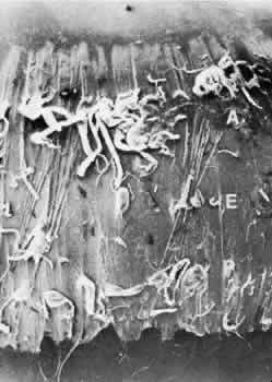
| Fig. 22. View of the whole zonular insertional region and broad lens equator of an elderly patient, appearing different from the dark surface of the rest of the bare capsule because of its coating of zonular fibers and matrix. Anterior (A), posterior (P), and a few small equatorial (E) zonular bundles are visible. Some posterior zonular bundles were lost when the vitreous was removed (SEM, × 64). (Streeten BW: The zonular insertion: A scanning electron microscopic study. Invest Ophthalmol Vis Sci 16:364, 1977) |