|
|
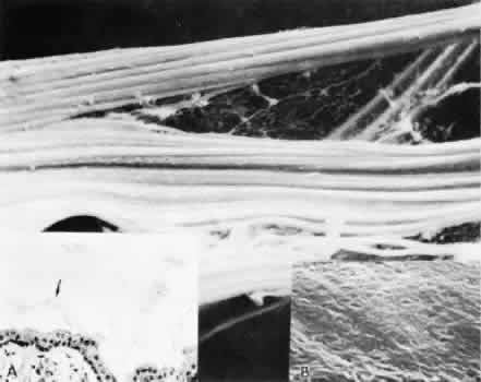
| Fig. 6. By scanning electron microscopy the ribbons of fiber bundles appear striated (× 2,200). Inset A. The flat ribbons of zonular bundles have a paler-staining “membrane” holding them together (arrow) (H&E, × 220). Inset B. The fibers are composed of highly oriented and tightly aggregated fibrils (× 23,000). |