|
|
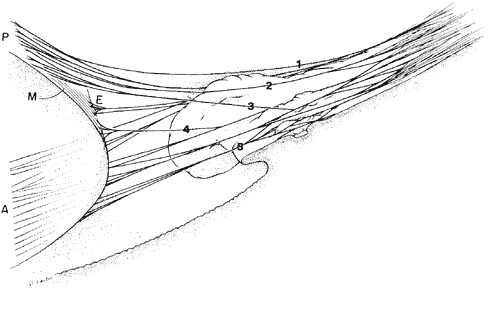
| Fig. 13. Diagram showing the course of the zonular fibers from the pars plana of the ciliary body (right of figure) to their insertion on the lens (left of figure) as anterior (A), posterior (P), equatorial (E), and meridional (M). Almost all have a further attachment to the ciliary processes. The posterior zonular fibers include fibers (1) closely associated with the anterior hyaloid membrane throughout, (2) adherent to the top or sides of the ciliary crests, and (3) adherent to the valleys or minior plicae. The equatorial fibers (4) branch from others on the sides of the processes, and the anterior fibers (5) are adherent in the valley region. |