|
|
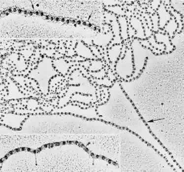
| Fig. 8. Rotary shadowing of a clump of human zonular fibrils in a 19-year-old patient. Darker straight fibril on right (large arrow) is in tight conformation (TEM, × 67,800). Top inset. Interbead filamentous subunits are seen (small arrows). Some bow outward, appearing to connect to every third bead (TEM, × 107,700). Bottom inset. A double-banded crosslinking region is visible across filaments between the beads (arrowhead). Three straight drumstick forms, probably interconnecting adhesion fibrils, extend down from beads or interbead areas (TEM, × 107,700). |