|
|
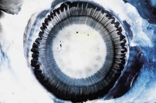
| Fig. 1. Insertion of the zonules on the anterior lens capsule. Cornea and iris have been removed and the eye has been stained with Gomori's hematoxylin. Ciliary processes are visible peripherally. Lens shrinkage during staining makes the perilenticular space wider than normal. |