|
|
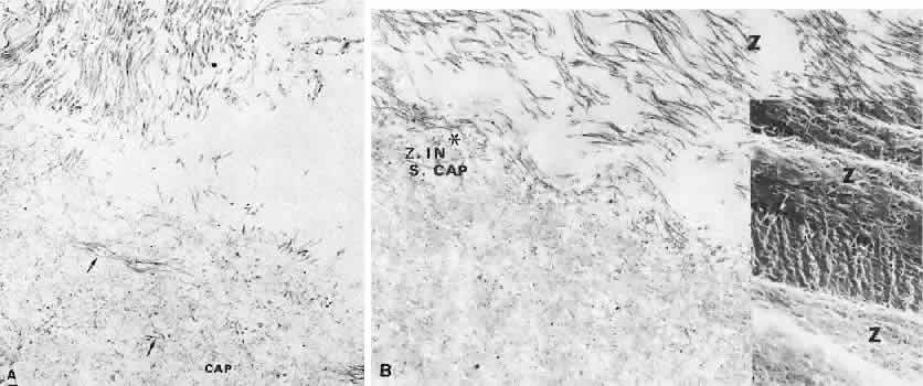
| Fig. 26. Anterior zonular insertion. A. Coronal section of anterior zonular fiber bundle (above) inserting into the lens capsule. The loose superficial capsule (CAP) shows 10-nm banded zonular fibrils (arrows) to a depth of 1.6 μm. Fibrils of the capsular matrix at 1 to 3 nm are barely visible (TEM, × 65,000).B. Behind the main anterior inserting bundles, a few small zonular fibers join others in the superficial capsule (Z IN S. CAP). Both tissues participate in the capsular rippling (asterisk) (TEM, × 42,400). Inset. Rippled superficial layer fibrils merging with fibrils of the attaching zonular bundles (Z) (SEM, × 6,400). |